. 2016:2016:5741518.
doi: 10.1155/2016/5741518. Epub 2016 Dec 25.

Urinary Extracellular Vesicles: Potential Biomarkers of Renal Function in Diabetic Patients

Affiliations

Urinary Extracellular Vesicles: Potential Biomarkers of Renal Function in Diabetic Patients

Agnieszka Kamińska et al. J Diabetes Res. 2016.

Abstract

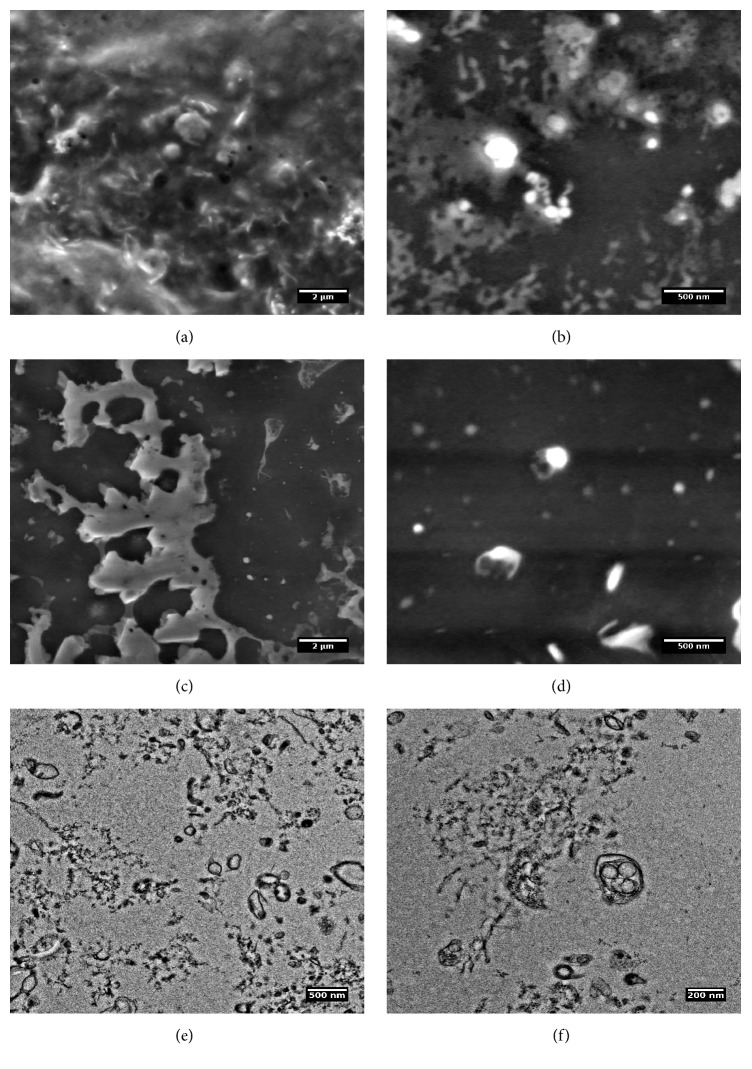
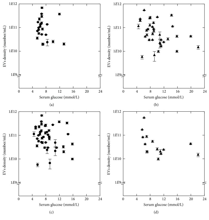
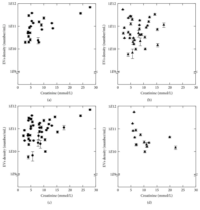
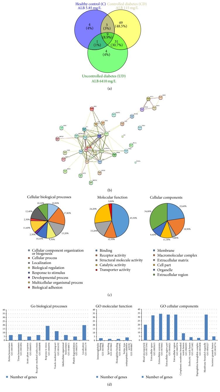
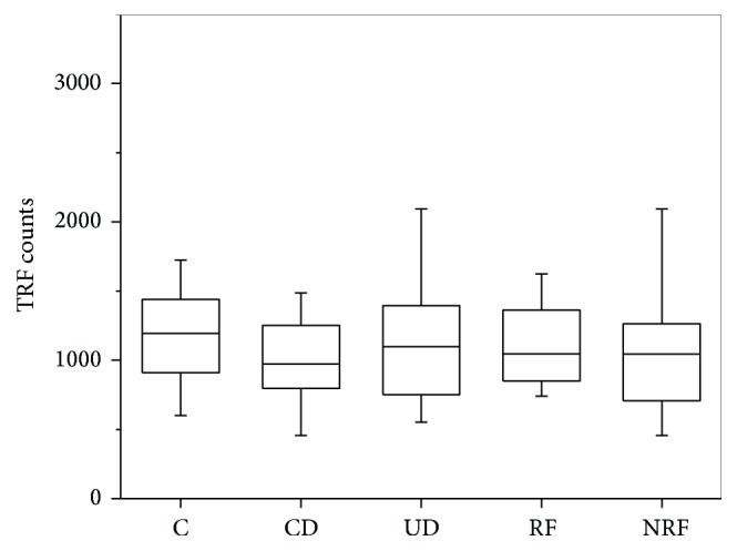
The aim of this study was to check the relationship between the density of urinary EVs, their size distribution, and the progress of early renal damage in type 2 diabetic patients (DMt2). Patients were enrolled to this study, and glycated hemoglobin (HbA1c) below 7% was a threshold for properly controlled diabetic patients (CD) and poorly controlled diabetic patients (UD). Patients were further divided into two groups: diabetic patients without renal failure (NRF) and with renal failure (RF) according to the Glomerular Filtration Rate. Density and diameter of EVs were determined by Tunable Resistive Pulse Sensing. Additionally, EVs were visualized by means of Transmission and Environmental Scanning Electron Microscopy. Nano-liquid chromatography coupled offline with mass spectrometry (MALDI-TOF-MS/MS) was applied for proteomic analysis. RF had reduced density of EVs compared to NRF. The size distribution study showed that CD had larger EVs (mode) than UD (115 versus 109 nm; p < 0.05); nevertheless the mean EVs diameter was smaller in controls than in the CD group (123 versus 134 nm; p < 0.05). It was demonstrated that EVs are abundant in urine. Albumin, uromodulin, and number of unique proteins related to cell stress and secretion were detected in the EVs fraction. Density and size of urinary EVs reflect deteriorated renal function and can be considered as potential renal damage biomarkers.


Conflict of interest statement

The authors declare that they have no competing interests.

Figures

Figure 1
Figure 1
Environmental Scanning Electron Microscopy (ESEM) (a–d) and Transmission Electron Microscopy (e, f) images of urinary extracellular vesicles (EVs) isolated from a urine sample. ESEM images show that EVs form aggregates and they are clustered on the surface. TEM analysis visualizes the variety of different vesicle-like objects in diameter mostly around 130–160 nm. Interestingly, multivesicle objects were also present in urine that confirms integrity of EVs during preparation.
Figure 2
Figure 2
Relationship between EVs density and serum glucose level in study groups: CD (a), UD (b), NRF (c), and RF (d). EVs density values are given as mean (SD). Spearman's rank correlation coefficient, p < 0.05.
Figure 3
Figure 3
Relationship between EVs density and urine creatinine concentration in study groups: CD (a), UD (b), NRF (c), and RF (d). EVs density values are given as mean (SD). Spearman's rank correlation coefficient, p < 0.05.
Figure 4
Figure 4
Proteomic analysis of urinary extracellular vesicles: mass spectrometry results from representative samples. (a) Venn diagram shows that in EVs from controlled diabetic patient with microalbuminuria there is a large number of unique proteins (n = 49), in a healthy control and in an uncontrolled diabetic patient with macroalbuminuria the number of unique proteins is very low (n = 4) [20]. (b) Protein-to-protein interaction analysis of common 45 proteins selected from Venn diagram shows the central role of albumin among urinary EV-related proteins; the list of submitted proteins is available in a supplementary data file (Supplementary Table 2) [22]. (c, d) Gene Ontology analysis showed that most of identified proteins are related to extracellular region or they are related to membrane organelles (exosomes); their localization corresponds with molecular function (receptors and transport proteins) [19]. A1BG: alpha-1-B glycoprotein; A2M: alpha-2-macroglobulin; ACTB: actin, beta; AFM: afamin; ALB: albumin; AMBP: alpha-1-microglobulin/bikunin precursor; ANPEP: alanyl (membrane) aminopeptidase; APOD: apolipoprotein D; AZGP1: alpha-2-glycoprotein 1, zinc-binding; CD59: CD59 molecule, complement; regulatory protein; CP: ceruloplasmin; GC: group-specific component (vitamin D binding protein); HBA1: hemoglobin, alpha 1; HBB: hemoglobin, beta; HP: haptoglobin; HPX: hemopexin; HSPG2: heparan sulfate proteoglycan 2; IGJ: immunoglobulin J polypeptide; IGLL5: immunoglobulin lambda-like polypeptide 5; KRT1: keratin 1; KRT2: keratin 2; KRT9: keratin 9; KRT10: keratin 10; LRG1: leucine-rich alpha-2-glycoprotein 1; MASP2: mannan-binding lectin serine peptidase 2; MUC1: mucin 1; ORM1: orosomucoid 1; ORM2: orosomucoid 2; PLG: plasminogen; PODXL: podocalyxin-like; POTEE: POTE ankyrin domain family member E; SERPINA1: serpin peptidase inhibitor, clade A, member 1; SERPINA3: serpin peptidase; inhibitor, clade A, member 3; TF: transferrin; UMOD: uromodulin.
Figure 5
Figure 5
Urine CD81 level. Results from Time Resolved Fluorescence assay. Kruskal-Wallis test: median TRF counts, p = 0.5 at α < 0.05 significance level.

References

    1. Tuttle K. R., Bakris G. L., Bilous R. W., et al. Diabetic kidney disease: a report from an ADA consensus conference. Diabetes Care. 2014;37(10):2864–2883. doi: 10.2337/dc14-1296. - DOI - PMC - PubMed
    1. Leehey D. J., Kramer H. J., Daoud T. M., Chatha M. P., Isreb M. A. Progression of kidney disease in type 2 diabetes—beyond blood pressure control: an observational study. BMC Nephrology. 2005;6:1471–2369. doi: 10.1186/1471-2369-6-8. - DOI - PMC - PubMed
    1. National Kidney Foundation. Diabetes—a major risk factor for kidney disease, https://www.kidney.org/news/newsroom/factsheets/FastFacts/
    1. Yuana Y., Sturk A., Nieuwland R. Extracellular vesicles in physiological and pathological conditions. Blood Reviews. 2013;27(1):31–39. doi: 10.1016/j.blre.2012.12.002. - DOI - PubMed
    1. Ranghino A., Dimuccio V., Papadimitriou E., Bussolati B. Extracellular vesicles in the urine: markers and mediators of tissue damage and regeneration. Clinical Kidney Journal. 2015;8(1):23–30. doi: 10.1093/ckj/sfu136. - DOI - PMC - PubMed