Review
. 2016 Sep 14;2(6):733-749.
doi: 10.1016/j.jcmgh.2016.09.002. eCollection 2016 Nov.

The Hepatic Lymphatic Vascular System: Structure, Function, Markers, and Lymphangiogenesis

Affiliations
Review

The Hepatic Lymphatic Vascular System: Structure, Function, Markers, and Lymphangiogenesis

Masatake Tanaka et al. Cell Mol Gastroenterol Hepatol. .

Abstract

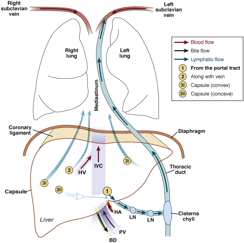
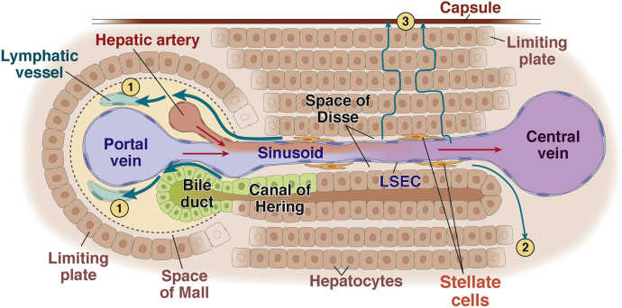
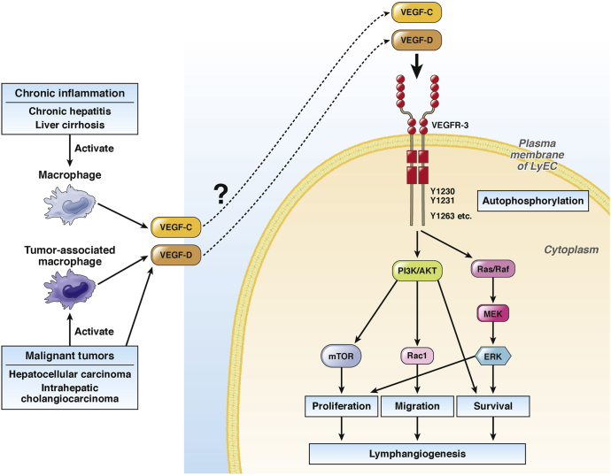
The lymphatic vascular system has been minimally explored in the liver despite its essential functions including maintenance of tissue fluid homeostasis. The discovery of specific markers for lymphatic endothelial cells has advanced the study of lymphatics by methods including imaging, cell isolation, and transgenic animal models and has resulted in rapid progress in lymphatic vascular research during the last decade. These studies have yielded concrete evidence that lymphatic vessel dysfunction plays an important role in the pathogenesis of many diseases. This article reviews the current knowledge of the structure, function, and markers of the hepatic lymphatic vascular system as well as factors associated with hepatic lymphangiogenesis and compares liver lymphatics with those in other tissues.

Keywords: CCl4, carbon tetrachloride; Cirrhosis; EHE, epithelioid hemangioendothelioma; HA, hyaluronan; HBx Ag, hepatitis B x antigen; HCC, hepatocellular carcinoma; IFN, interferon; IL, interleukin; Inflammation; LSEC, liver sinusoidal endothelial cell; LYVE-1, lymphatic vessel endothelial hyaluronan receptor 1; LyEC, lymphatic endothelial cell; NO, nitric oxide; Portal Hypertension; Prox1, prospero homeobox protein 1; VEGF; VEGF, vascular endothelial growth factor; VEGFR, vascular endothelial growth factor receptor; mTOR, mammalian target of rapamycin.


Figures

Figure 1
Figure 1
Schematic diagram of macro-anatomy of hepatic lymphatic vascular system. (1) Lymphatic capillaries in the portal tract coalesce into collecting vessels, which drain to lymph nodes at the hepatic hilum and the lesser omentum. Efferent lymphatic vessels (LV) from these lymph nodes connect to celiac lymph nodes, which drain to the cisterna chyli, the enlarged origin of the thoracic duct. Lymphatic fluid through the thoracic duct drains to the left subclavicular vein and returns to the systemic blood circulation. (2) Lymphatic vessels along the central vein (CV) converge into large lymphatic vessels along the hepatic vein (HV), which then traverse along the inferior vena cava (IVC) through the diaphragm toward mediastinal lymph nodes. (3) Lymphatic fluid running underneath the capsule of the convex surface of the liver (3i) drains to mediastinal lymph nodes through the coronary ligament, whereas that of the concave surface (3ii) drains to lymph nodes of the hepatic hilum and regional lymph nodes. BD, bile duct; HA, hepatic artery; LN, lymph node; PV, portal vein.
Figure 2
Figure 2
Schematic diagram of the micro-anatomy of the hepatic lymphatic vascular system. Blood flow (red arrows) from the portal vein (PV) and hepatic artery (HA) enters the liver. Plasma components are filtered through LSECs into the space of Disse, the interstitial space between LSECs and hepatocytes, and are regarded as the source of lymphatic fluid. Lymphatic fluid in the space of Disse mostly flows through the space of Mall, the space between the stroma of the portal tract and the outermost hepatocytes, into the interstitium of the portal tract and then into lymphatic capillaries (1). Some portion of the lymphatic fluid in the space of Disse flows into the interstitium around the central vein (2) or underneath the hepatic capsule (3).
Figure 3
Figure 3
Intracellular signaling pathways in lymphangiogenesis. Signaling via VEGF-C/D and VEGFR-3 is the most well-known pathway for lymphangiogenesis. VEGF-C or VEGF-D binds to its receptor VEGFR-3 in the plasma membrane of LyECs, which facilitates signal transduction through various intracellular signaling pathways, leading to lymphangiogenesis. In the liver, activated macrophages in chronic inflammatory conditions, such as chronic hepatitis and liver cirrhosis, secrete VEGF-C and/or VEGF-D. Malignant liver tumors, such as HCC and intrahepatic cholangiocarcinoma, also secrete VEGF-C and/or VEGF-D. Furthermore, these malignant tumors activate tumor-associated macrophages, which also secrete VEGF-C and/or VEGF-D. Secreted VEGF-C and VEGF-D are likely related to lymphangiogenesis in liver diseases through VEGFR-3–mediated pathways.

References

    1. Chung C., Iwakiri Y. The lymphatic vascular system in liver diseases: its role in ascites formation. Clin Mol Hepatol. 2013;19:99–104. - PMC - PubMed
    1. Tammela T., Alitalo K. Lymphangiogenesis: molecular mechanisms and future promise. Cell. 2010;140:460–476. - PubMed
    1. Schulte-Merker S., Sabine A., Petrova T.V. Lymphatic vascular morphogenesis in development, physiology, and disease. J Cell Biol. 2011;193:607–618. - PMC - PubMed
    1. Koltowska K., Betterman K.L., Harvey N.L. Getting out and about: the emergence and morphogenesis of the vertebrate lymphatic vasculature. Development. 2013;140:1857–1870. - PubMed
    1. Kaipainen A., Korhonen J., Mustonen T. Expression of the fms-like tyrosine kinase 4 gene becomes restricted to lymphatic endothelium during development. Proc Natl Acad Sci U S A. 1995;92:3566–3570. - PMC - PubMed