Review
. 2017 Apr;8(2):227-241.
doi: 10.1007/s13244-016-0539-9. Epub 2017 Jan 19.

Mature cystic teratoma of the ovary: a cutting edge overview on imaging features

Affiliations
Review

Mature cystic teratoma of the ovary: a cutting edge overview on imaging features

Hilal Sahin et al. Insights Imaging. 2017 Apr.

Abstract

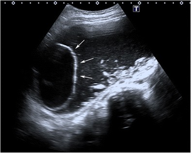
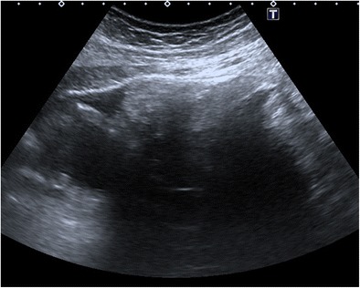
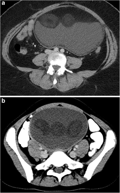
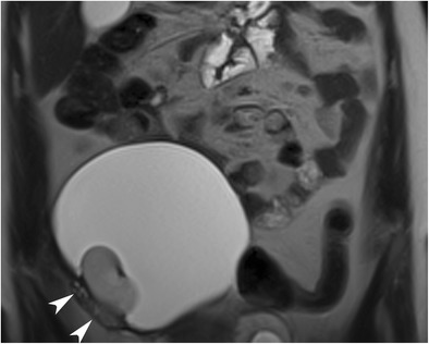
Mature cystic teratoma (MCT) is the most common neoplasm of the ovary and includes at least two well- differentiated germ cell layers. Different combinations of mature tissue derivatives with varying arrangements in the tumour cause a wide spectrum of radiological presentation ranging from a purely cystic mass to a complex cystic mass with a considerable solid component. In different imaging modalities, each radiological feature reflects a specific pathologic equivalent that forms because of diverse compositions of histological components. Understanding uncommon findings as well as the classic signs with basic knowledge of pathological equivalents permits a more accurate diagnosis and guides adequate treatment. In this review, radiological features of MCT in different imaging modalities (US, CT, MR imaging) including specific signs and useful radiological artefacts with brief emphasis on pathological basics are discussed. Teaching points • Ovarian mature cystic teratomas (MCTs) have a wide spectrum of radiological presentation.• Each radiological feature of MCT reflects a specific pathologic equivalent.• Understanding radiological signs with basic knowledge of pathology can permit a more accurate diagnosis.

Keywords: Cyst; Dermoid; Neoplasm; Ovary; Teratoma.


Conflict of interest statement

The authors declare that they have no conflicts of interest concerning this article.

Figures

Fig. 1
Fig. 1
Transverse sonogram of a 20-year-old female with a mature cystic teratoma. A densely echogenic protuberance on the left side of the wall projecting into the cystic lumen is consistent with a Rokitansky nodule
Fig. 2
Fig. 2
Transverse sonogram of an 18-year-old female with mature cystic teratoma. Shadowing echodensity projecting into the cystic lumen is seen, which was proved to contain fat and hair in the pathological specimen
Fig. 3
Fig. 3
Transverse sonogram of a 14-year-old female with mature cystic teratoma demonstrates a cystic Rokitansky nodule (arrows) in a cystic mass. Note that there is echogenic debris at the gravity-dependent portion of the cyst
Fig. 4
Fig. 4
Transverse sonogram of a 16-year-old female with mature cystic teratoma (a) demonstrates high-amplitude echoes in two different regions (arrows). Axial computed tomography image (b) shows that those regions contain fat (arrowheads). In the macroscopic specimen (c), whitish sebaceous material with fat and hair is seen in the opened cystic lumen
Fig. 5
Fig. 5
Transverse sonogram of a 16-year-old female with a mature cystic teratoma. An amorphous echogenic region is seen in the near field that causes posterior shadowing and obscures the posterior portion of the lesion and any structures behind it
Fig. 6
Fig. 6
Transverse sonogram of a 51-year-old female with mature cystic teratoma. Echogenic lines and dots, making the dot-dash sign, are seen in a left adnexal mass regarding hair arranged in different orientations. B bladder
Fig. 7
Fig. 7
Transverse sonogram of an 18-year-old female with fat-fluid level in a mature cystic teratoma. Supernatant hyperechoic sebum forms a layer with dependent hypoechoic aqueous fluid
Fig. 8
Fig. 8
Transverse sonogram of a 14-year-old female with mature cystic teratoma shows multiple floating spherical echogenic structures that differ in size and shape
Fig. 9
Fig. 9
Transverse sonogram of an 18-year-old female with a mature cystic teratoma. There are three dark comet tail appearances (arrows) floating in the interface of the fat-fluid level consistent with hair balls pathologically
Fig. 10
Fig. 10
Axial CT image of a 25-year-old female with mature cystic teratoma (MCT). Two round fatty structures (arrows) are seen in the left adnexal mass (a). In another case with the fat-fluid level in an MCT, the axial CT image (b) shows a thin fat layer covering the inner side of the wall (arrowheads)
Fig. 11
Fig. 11
Axial CT image of a 37-year-old female with a mature cystic teratoma shows a rounded Rokitansky nodule (arrowheads) made up of fat and a tooth-like structure in a high-density cystic mass
Fig. 12
Fig. 12
Axial CT image of an 18-year-old female with mature cystic teratoma shows a tooth-like high-density structure in the centre of a Rokitansky nodule (arrowheads)
Fig. 13
Fig. 13
Axial CT image of an 18-year-old female with mature cystic teratoma (MCT) (a) shows two rounded floating balls in the interface of the fat-fluid level. The bigger one has a low density core and a low density outer shell. In another case with MCT (b), floating balls have relatively high density cores
Fig. 14
Fig. 14
Coronal T1-weighted MR image of a 40-year-old female with mature cystic teratoma (a) shows a heterogeneous mass with high signal intensity areas in the right adnexa. In the fat-saturated T1-weighted image (b), the major fat component of the lesion is suppressed regarding the diagnosis of teratoma. In the macroscopic specimen (c), the fat component is seen as a yellow area at the centre of the tumour (arrows)
Fig. 15
Fig. 15
Axial T1-weighted in-phase MR image of a 56-year-old female with mature cystic teratoma (a) shows a large mass with a high signal intensity part creating an interface (arrowheads) with a low signal intensity part. T1-weighted opposed-phase image (b) demonstrates the fat component with decreased signal intensity in the supernatant layer regarding the presence of sebum
Fig. 16
Fig. 16
Coronal T2-weighted MR image of a 42-year-old female with mature cystic teratoma shows chemical shift artefact both inside the tumour and in the borders. A low signal intensity band (arrow) is seen on the cranial border of the cyst with a high intensity band on the opposite side
Fig. 17
Fig. 17
Axial T1-weighted MR image of a 31-year-old female with mature cystic teratoma (a) shows high signal intensity of the supernatant fatty layer creating an interface with a low signal intensity aqueous layer. Fat-saturated T1-weighted image (b) demonstrates a major fat component with decreased signal intensity in the supernatant layer and free-floating hypointense fat particles in the aqueous layer. A Rokitansky nodule with cystic components is seen in the posterior part of the cyst. Cut section of the tumour (c) shows yellowish sebaceous material leaking from the cystic lumen. Note the black hairs in that material
Fig. 18
Fig. 18
Sagittal T2-weighted MR image of a 56-year-old female (same case as in Fig. 15) with mature cystic teratoma shows chemical shift artefact (arrowheads) in the interface of two different hyperintense layers. Note that the complete gravity-dependent layering of the fatty component in the cranial part is not formed regarding the high viscosity. A floating rounded structure is also seen in the cystic lumen (arrow)
Fig. 19
Fig. 19
Axial T1-weighted MR image of a 31-year-old female with mature cystic teratoma (a) (same case as in Fig. 17). A floating ball is seen in the interface of the high signal intensity fat and low signal intensity aqueous fluid layer (arrow). Axial T2-weighted image (b) shows chemical shift artefact in and around the ball (arrow). In the macroscopic specimen (c), the floating ball corresponds to whitish creamy material containing hair and keratin
Fig. 20
Fig. 20
Coronal T2-weighted MR image of a 21-year-old female with mature cystic teratoma. A bean-shaped Rokitansky nodule is seen lying on the right side of the wall of the cystic mass. Ovarian parenchyma with normal signal intensity (arrowheads) can be seen near the cyst wall where the Rokitansky nodule arises
Fig. 21
Fig. 21
Sagittal T2-weighted MR image of a 22-year-old female with mature cystic teratoma (a). A hyperintense structure (arrows) with an obvious chemical shift artefact lying in the interface of the fat-fluid level is seen in a cystic mass. It is consistent with matted hair and keratinoid material in the macroscopic specimen (b). Note the Rokitansky nodule at the posterior wall with hyperintense parts that were proven to be fat and linear hair shafts (arrowhead) arising from the nodule in the T2-weighted image. Cut-section photograph of the Rokitansky nodule is shown in Fig. 25d
Fig. 22
Fig. 22
Axial T1-weighted MR image of a 42-year-old female with mature cystic teratoma (a) shows a palm tree-like structure projecting into the high signal intensity mass (arrowheads). Fat-saturated contrast-enhanced T1-weighted image (b) demonstrates a suppressed fat component in the tumour (arrowheads)
Fig. 23
Fig. 23
Axial T2-weighted (a), fat-saturated T2-weighted (b), in-phase (c) and opposed-phase T1-weighted (d) MR images of a 56-year-old female with a floating ball (arrowheads) in a mature cystic teratoma. The central core of the ball contains minor fat, which is seen as decreased signal intensity in the opposed-phase image, and an outer layer with a major fat signal that decreases in the fat-saturated T2-weighted image
Fig. 24
Fig. 24
Axial fat-saturated T2-weighted (a), diffusion-weighted (b 800) image (b) and ADC map (c) images of a 14-year-old female with a mature cystic teratoma. Multiple floating structures (arrows) are seen in the anterior part of the cystic mass. Those structures show diffusion restriction regarding the keratin content, which was also proven pathologically
Fig. 25
Fig. 25
Axial T1-weighted (a), diffusion-weighted (b 800) image (b) and ADC map (c) images of a 22-year-old female with a mature cystic teratoma (same case as in Fig. 21). A hypointense cauliflower-like mass is seen near the interface of the fat-fluid level and a Rokitansky nodule is seen in the posterior part of the cystic lesion (a). The cauliflower-like mass shows diffusion restriction regarding the keratin content (b, c). Whitish cheese-like material regarding keratin (arrows) is seen in the lumen in the macroscopic specimen of the tumour (d). Cut section of the Rokitansky nodule (arrowheads) shows a yellowish fat component, creamy keratin and fine hair shafts arising from the nodule

References

    1. Rha SE, Byun JY, Jung SE, Kim HL, Oh SN, Kim H, et al. Atypical CT and MRI manifestations of mature ovarian cystic teratomas. AJR. 2004;183:743–750. doi: 10.2214/ajr.183.3.1830743. - DOI - PubMed
    1. Outwater EK, Siegelman ES, Hunt JL. Ovarian teratomas: tumor types and imaging characteristics. RadioGraphics. 2001;21:475–490. doi: 10.1148/radiographics.21.2.g01mr09475. - DOI - PubMed
    1. Weiss JR, Burgess JR, Kaplan KJ. Fetiform teratoma (homunculus) Arch Pathol Lab Med. 2006;130:1552–1556. - PubMed
    1. Saba L, Guerriero S, Sulcis R, Virgilio B, Melis G, Mallarini G. Mature and immature ovarian teratomas: CT, US and MR imaging characteristics. Eur J Radiol. 2009;72:454–463. doi: 10.1016/j.ejrad.2008.07.044. - DOI - PubMed
    1. Patel MD, Feldstein VA, Lipson SD, Chen DC, Filly RA. Cystic teratoma of the ovary: diagnostic value of sonography. AJR. 1998;171:1061–1065. doi: 10.2214/ajr.171.4.9762997. - DOI - PubMed

LinkOut - more resources