. 2016 Dec 22;17(Suppl 3):153.
doi: 10.1186/s12863-016-0461-7.

Genes of susceptibility to early neurodegenerative changes in the rat retina and brain: analysis by means of congenic strains

Affiliations

Genes of susceptibility to early neurodegenerative changes in the rat retina and brain: analysis by means of congenic strains

Elena E Korbolina et al. BMC Genet. .

Abstract

Background: There has been considerable interest in discovery of the genetic architecture of complex traits, particularly age-related neurodegenerative disorders. To predict disease risk and to understand its genetic basis in humans, it is necessary to study animal models. Our previous research on the accelerated-senescence OXYS strain has revealed two quantitative trait loci (QTLs) on rat chromosome 1 that are associated with early cataract and/or retinopathy as well as with behavioral abnormalities. Each locus was partially mapped within the introgressed segments in a certain congenic strain: WAG/OXYS-1.1 or WAG/OXYS-1.2. Retinal transcriptome profiling of 20-day-old congenic and OXYS rats by high-throughput RNA sequencing uncovered relevant candidate genes and pathways. Nonetheless, the question remained open whether the same genetic components simultaneously have effects on various manifestations of the accelerated-senescence phenotype in OXYS rats. The present study was designed to analyze the genes of susceptibility to early neurodegenerative processes taking place in the OXYS rat retina and brain and to assess their potential functional clustering. The study was based on the findings from recent publications (including mapping of quantitative trait loci) and on comparative phenotyping of congenic rat strains.

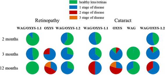
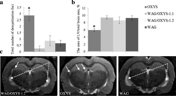
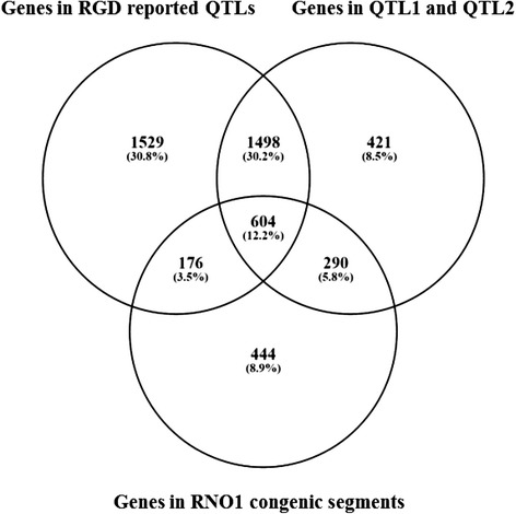
Results: The backcrossing of Wistar Albino Glaxo (WAG) and OXYS strains to generate the congenics resulted in two congenic strains with high susceptibility to cataract and retinopathy but with no obvious signs of Alzheimer's disease-like brain pathology that are specific for OXYS rats. Thus, the genes of susceptibility to brain neurodegeneration were not introgressed into the congenic strains or there is a strong effect of the genetic background on the disease phenotype. Moreover, the progression of retinopathy with age was relatively less severe in the WAG background compared to the OXYS background. A comparative analysis of previously defined QTLs and congenic segments led to identification of candidate genes with a suspected effect on brain neurodegeneration including the genes showing differential expression in the congenic strains.

Conclusion: Overall, our findings suggest that the cause of the cataract and the cause of retinopathy phenotypes in OXYS rats may be genetically linked to each other within the introgressed segments in the WAG/OXYS-1.1 and/or WAG/OXYS-1.2 congenic strains.

Keywords: Age-related macular degeneration; Alzheimer’s disease; Congenic strain; Genetic architecture of complex trait; OXYS rats; Quantitative trait locus.


Figures

Fig. 1
Fig. 1
The incidence of cataract and retinopathy in congenic (WAG/OXYS-1.1 and WAG/OXYS-1.2) and parental (OXYS and WAG) rat strains at the age of 2, 3, and 12 months. We did not observe any cases of retinopathy in WAG rats at any age. Data are presented as a distribution of rat eyes with the stages of cataract and retinopathy (n = 30 eyes). Colors are labeled with 0 through 2 and correspond to a certain stage of cataract or retinopathy, scoring 0–3, as explained in Methods
Fig. 2
Fig. 2
Retinas of 3-month-old WAG a and OXYS b c rats. a Open chorioretinal vessels with a small amount of blood cells (white dotted arrows); the cellular prismatic pigment epithelium with prominent microvilli (white arrows). b The ganglion neuron nucleus with pyknosis (black arrow); hyperchromic oligodendrocyte with nuclear pyknosis in the layer of nerve fibers (dashed black arrow); stasis of blood cells in the intraretinal vessel (white dashed arrow); hyperchromic pyknomorphic radial glial cells (white arrows); hyperchromic pyknomorphic associative neurons (red arrows). c Stasis and sludge of the blood cells in the choroidal vessels (black dashed arrows); a pyknotic nucleus of an RPE cell (black arrows); disorientation and disaggregation of the outer segments of photoreceptors (asterisk); the photoreceptors containing a nucleus with pyknosis (white dotted arrows). Abbreviations: OPL, outer plexiform layer; INL, inner nuclear layer; IPL, the inner plexiform layer; ONL, outer nuclear layer; GL, ganglion cell layer, RPE, retinal pigment epithelium, Ch, choroid. The scale bar is 10 μm
Fig. 3
Fig. 3
Retinas of 3-month-old WAG/OXYS-1.2 rats. a Migration of macrophages into the inner plexiform and ganglion layers (white arrows). Migration of the perikaryon of the associative neuron into the inner plexiform layer (dashed black arrow). b Advanced intraretinal capillary with phenomenon of the blood cells stasis (black arrows); lymphocytic infiltration (white arrows); NGC: the nucleus of the ganglion neuron. Abbreviations: OPL: outer plexiform layer; INL: inner nuclear layer; IPL: the inner plexiform layer; ONL: outer nuclear layer; GL: ganglion cell layer. The scale bar is 10 μm
Fig. 4
Fig. 4
Retinas of 10-month-old WAG/OXYS-1.2 a and WAG/OXYS-1.1 b rats. The thinning of the outer nuclear layer with a reduction in the number of the photoreceptors in the retina of a 16-month-old WAG/OXYS-1.2 rat c. a The open intraretinal vessels (red arrows). b Open intraretinal capillaries (red arrows); the flattening of the RPE cells, RPE cells containing a nucleus with pyknosis (white dashed arrows); migration of mononuclear phagocytes within the subretinal space with RPE detachment (black dashed arrow). c Open intraretinal capillaries (red arrows); the flattening of the RPE cells, RPE cells containing a nucleus with pyknosis (black arrows); the destructive changes in neurosensory and associative neurons (black dashed arrows). Abbreviations: OPL: outer plexiform layer, INL: inner nuclear layer; IPL: the inner plexiform layer; ONL: outer nuclear layer; GL: ganglion cell layer, RPE: retinal pigment epithelium, PhL: photoreceptor layer. The scale bar is 10 μm
Fig. 5
Fig. 5
Results for the two-dimensional principal component analysis (PCA) of rat behavioral parameters recorded in the 8-arm radial maze test. According to the results of PCA, the rats of two congenic strains, WAG/OXYS-1.1 and WAG/OXYS-1.2, proved to be much closer to the parameters of WAG rats, than to those of OXYS rats. Legend: PC1 and PC2: principal components 1 and 2, respectively
Fig. 6
Fig. 6
An MRI morphometric study of the brain of 12-month-old OXYS, WAG/OXYS 1.1, and WAG/OXYS-1.2 rats. a The total number of demyelinating foci is significantly greater in the OXYS brain than in the brain of the congenic and WAG rats. b The lateral ventricles in the congenic and WAG rats are enlarged when compared with OXYS rats. c Axial slices of the brain of 12-month-old WAG/OXYS-1.2, WAG, and OXYS rats. The foci of demyelination (white arrows) and the increase in the size of lateral ventricles in WAG and congenic rats (white dotted arrows) are visible. Not all congenic and WAG rats have the demyelination lesions. The data are shown as mean ± SEM. Abbreviation: LV, lateral ventricles; *P < 0.05 for differences between the strains
Fig. 7
Fig. 7
The Venn diagram showing overlaps among the three analyzed sets of RNO1 genes. The genes located within the previously determined QTL1 and QTL2 [23], the genes located within the mapped congenic segments in WAG/OXYS-1.1 and WAG/OXYS-1.2 rat congenic strains [24], and the genes located within rat QTLs, presented in RGD were analyzed. The number of genes in each group as reported by RGD is shown in the corresponding field

References

    1. Fushan AA, Turanov AA, Lee S-G, Kim EB, Lobanov AV, Yim SH, et al. Gene expression defines natural changes in mammalian lifespan. Aging Cell. 2015;14:352–365. doi: 10.1111/acel.12283. - DOI - PMC - PubMed
    1. Ackert-Bicknell CL, Anderson LC, Sheehan S, Hill WG, Chang B, Churchill GA, et al. Aging Research Using Mouse Models. Curr. Protoc. Mouse Biol. [Internet]. Hoboken, NJ, USA: John Wiley & Sons, Inc.; 2015. p. 95–133. Available from: http://doi.wiley.com/10.1002/9780470942390.mo140195. - PMC - PubMed
    1. Bertram L. The genetic epidemiology of neurodegenerative disease. J Clin Invest. 2005;115:1449–1457. doi: 10.1172/JCI24761. - DOI - PMC - PubMed
    1. Jellinger KA. Basic mechanisms of neurodegeneration: a critical update. J. Cell. Mol. Med. [Internet]. 2010; Available from: http://doi.wiley.com/10.1111/j.1582-4934.2010.01010.x. - PMC - PubMed
    1. Marciniuk K, Taschuk R, Napper S. Evidence for prion-like mechanisms in several neurodegenerative diseases: potential implications for immunotherapy. Clin Dev Immunol. 2013;2013:1–20. doi: 10.1155/2013/473706. - DOI - PMC - PubMed

Publication types

MeSH terms