. 2017 Jun 9;26(6):1001-1016.
doi: 10.3727/096368916X694391. Epub 2017 Jan 20.

Human Dental Pulp Stem Cells Are More Effective Than Human Bone Marrow-Derived Mesenchymal Stem Cells in Cerebral Ischemic Injury

Human Dental Pulp Stem Cells Are More Effective Than Human Bone Marrow-Derived Mesenchymal Stem Cells in Cerebral Ischemic Injury

Miyeoun Song et al. Cell Transplant. .

Abstract

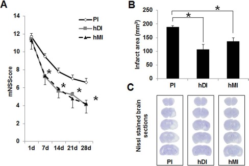
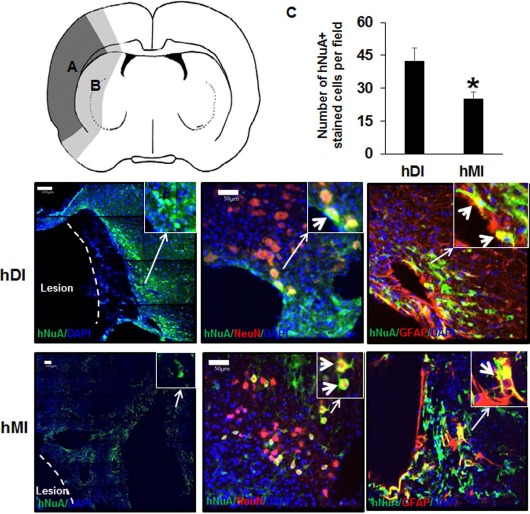
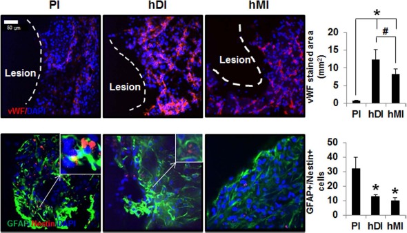
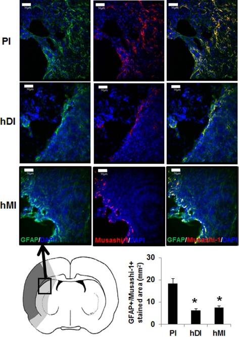
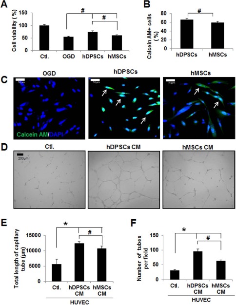
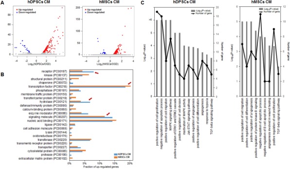
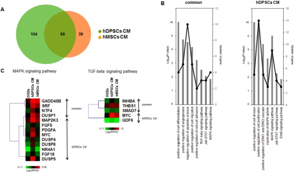
We compared the therapeutic effects and mechanism of transplanted human dental pulp stem cells (hDPSCs) and human bone marrow-derived mesenchymal stem cells (hBM-MSCs) in a rat stroke model and an in vitro model of ischemia. Rats were intravenously injected with hDPSCs or hBM-MSCs 24 h after middle cerebral artery occlusion (MCAo), and both groups showed improved functional recovery and reduced infarct volume versus control rats, but the hDPSC group showed greater reduction in infarct volume than the hBM-MSC group. The positive area for the endothelial cell marker was greater in the lesion boundary areas in the hDPSC group than in the hBM-MSC group. Administration of hDPSCs to rats with stroke significantly decreased reactive gliosis, as evidenced by the attenuation of MCAo-induced GFAP+/nestin+ and GFAP+/Musashi-1+ cells, compared with hBM-MSCs. In vivo findings were confirmed by in vitro data illustrating that hDPSCs showed superior neuroprotective, migratory, and in vitro angiogenic effects in oxygen-glucose deprivation (OGD)-injured human astrocytes (hAs) versus hBM-MSCs. Comprehensive comparative bioinformatics analyses from hDPSC- and hBM-MSC-treated in vitro OGD-injured hAs were examined by RNA sequencing technology. In gene ontology and KEGG pathway analyses, significant pathways in the hDPSC-treated group were the MAPK and TGF-β signaling pathways. Thus, hDPSCs may be a better cell therapy source for ischemic stroke than hBM-MSCs.


Figures

Figure 1.
Figure 1.
Effects of intravenous (IV) transplantation of human dental pulp stem cells (hDPSCs) and human bone marrow-derived mesenchymal stem cells (hBM-MSCs) on neurological function and infarct volume after middle cerebral artery occlusion (MCAo). (A) Functional recovery was assessed by a modified neurologic severity score (mNSS) test at days 7, 14, 21, and 28. PI, ischemia with PBS injection (n = 9); hDI, ischemia with hDPSC injection (n = 9); hMI, ischemia with hBM-MSC injection (n = 9). (B) Infarct size of rat brains at 28 days after MCAo. Infarct volume measurements were performed on Nissl-stained coronal sections. (C) Nissl-stained brain sections of the PI, hDI, and hMI groups, respectively. Data are expressed as mean ± standard deviation (SD). *p < 0.05 compared to the control group (Student's t-test).
Figure 2.
Figure 2.
Immunohistochemical staining of rat brains to evaluate targeting migration and neurogenic differentiation of injected hDPSCs (hDI) or hBM-MSCs (hMI) 28 days after MCAo. Schematic diagram depicting the regions of transplanted cells. hDPSCs and hBM-MSCs were investigated in the brain regions of the ischemic hemisphere: (A) ischemic core indicated by dark gray; (B) boundary zone indicated by bright gray. Representative image of double staining of nucleus [4′,6-diamidino-2-phenylindole (DAPI), blue], human nucleus (hNuA, green), astrocytes [glial fibrillary acidic protein (GFAP), red], and neurons (NeuN, red). The right upper windows of images show high-magnification images of the arrow area. Thick arrows in the right upper windows indicate hNuA+/NeuN+ and hNuA+/GFAP+ cells. Scale bars: 100 μm (hNuA/DAPI-stained section images), 50 μm (hNuA/GFAP or hNuA/NeuN-stained section images). These images are representative of three independent experiments. (C) Quantification of human stem cells by human antibody immmunoreactivity in the rat brain. Data are expressed as mean±SD. *p < 0.05 compared to the control group (Student's t-test).
Figure 3.
Figure 3.
Immunohistochemical staining of rat brains to evaluate angiogenesis and astrogliosis of hDI or hMI 28 days after MCAo. Representative image of double staining of nucleus (DAPI, blue), von Willebrand factor (vWF, red), GFAP (green), and nestin (red). Scale bar: 50 μm. Positive cells and areas in the brain from MCAo rats were analyzed using an image analyzer. The right upper windows of images show high-magnification images of the arrow area. The thick arrow in right upper windows indicates GFAP+/nestin+ cells. vWF+ (A) and double-labeled (GFAP and nestin; B) cells were counted in three to five coronal sections per animal (n = 5). Data are expressed as mean ± SD. *p < 0.05 compared to the control group (Student's t-test); #p < 0.05 compared to each group (Student's t-test).
Figure 4.
Figure 4.
Immunohistochemical staining of rat brains to evaluate astrogliosis using GFAP/Musashi-1 staining of injected hDPSCs (hDI group) or hBM-MSCs (hMI group) 28 days after MCAo. Schematic diagram depicting the regions of transplanted cells sampled for quantitative analyses. Ischemic core is indicated by dark gray, and boundary zone is indicated by bright gray. Representative image of double staining of nucleus (DAPI, blue), GFAP (green), and Musashi-1 (red). Scale bars: 50 μm. Positive stained areas (GFAP+/Musashi-1+ cells) in the brain from MCAo rats were analyzed using an image analyzer. GFAP and Musashi-1 double-stained areas were measured in three to five coronal sections per animal (n = 5). Data are expressed as mean ± SD. *p < 0.05 compared to the control group (Student's t-test).
Figure 5.
Figure 5.
Effect of hDPSCs and hBM-MSCs on cell viability (A), migration (B, C), and angiogenesis (D, E) in oxygen–glucose deprivation (OGD)-injured human astrocytes (hAs). (A) hA cell viability was measured by 3-(4,5-dimethylthiazol-2-yl)-2,5-diphenyltetrazolium bromide (MTT) assay. Data are representative of three independent experiments. Ctl., control. (B) Graph showing that hDPSCs or hBM-MSCs migrated to ischemic hAs in the Transwell system. (C) Representative image of migration of hDPSCs or hBM-MSCs to ischemic hAs. Calcein acetoxymethyl ester (calcein AM)/DAPI double-positive cells indicate migrated hDPSCs or hBM-MSCs. (D) Representative capillary formation with conditioned medium (CM) obtained from hDPSC- and hBM-MSC-treated human umbilical vein endothelial cells (HUVECs). The graph shows the average total length of capillary tube (μm) (E) and number of tubes per field (F) in three independent experiments. Data are expressed as mean ± SD (n = 5). *p < 0.05 compared to the control group (Student's t-test); #p < 0.05 compared to each group (Student's t-test).
Figure 6.
Figure 6.
Gene expression profile differences in OGD-injured hAs treated with hDPSCs and hBM-MSCs. (A) Differential gene expression in OGD-injured control hAs and posttreatment of hDPSCs (left) or hBM-MSCs (right). Primary cultured hDPSCs and commercial hBM-MSCs were obtained from 10 and 3 donors separately. The x-axis represents the log2 fold changes, and the y-axis is the –log10-adjusted p value. Upregulated and downregulated genes in each treatment are colored red and blue, respectively. (B) Protein classification of genes upregulated in hDPSCs and hBM-MSCs. Four red arrows indicate the following protein classes: “receptor,” “structural protein,” “hydrolase,” and “signaling molecule,” showing the differences in the composition of upregulated genes between the treatments. (C) Gene ontology (GO) term and Kyoto Encyclopedia of Genes and Genomes (KEGG) pathway enrichment in genes upregulated in hDPSC- and hBM-MSC-treated hAs. The p values of significantly enriched terms or pathways and the number of the genes in the GO term and KEGG pathways are shown as bar plots (–log10 p value) and line graphs (number of genes in the specific enriched terms or pathways).
Figure 7.
Figure 7.
Comparison of hDPSC- and hBM-MSC-treated hA gene regulation. (A) Venn diagram of upregulated genes in hDPSC- and hBM-MSC-treated hAs. Primary cultured hDPSCs and commercial hBM-MSCs were obtained from 10 and 3 donors separately. (B) GO term and KEGG pathway enrichment of genes commonly (left) and exclusively (right) upregulated between the hDPSC- and hBM-MSC-treated groups. The overrepresented pathways and the numbers of genes in the GO terms and KEGG pathways are shown as bar plots (–log10 p value) and line graphs, similar to Figure 6C. (C) Heatmaps of the mitogen-activated protein kinase (MAPK) and transforming growth factor-β (TGF-β) signaling pathways. The expression values (log2 RPKM) of upregulated genes in hDPSCs and hBM-MSCs were calculated and used to perform hierarchal clustering and for generation of heatmaps. On the right side of the heatmap, the labels show the genes upregulated in both hDPSCs and hBM-MSCs and those upregulated exclusively in hDPSCs.

Similar articles

Cited by

References

    1. Donnan G.A., Fisher M., Macleod M., Davis S.M. Stroke. Lancet 2008; 10: 1612–23. - PubMed
    1. Chen J., Li Y., Wang L., Zhang Z., Lu D., Lu M., Chopp M. Therapeutic benefit of intravenous administration of bone marrow stoma cells after cerebral ischemia in rats. Stroke 2001; 32: 1005–11. - PubMed
    1. Nomura T., Honmou O., Harada K., Houkin K., Hamada H., Kocsis J.D. IV infusion of brain-derived neurotrophic factor gene-modified human mesenchymal stem cells protects against injury in a cerebral ischemia model in adult rat. Neuroscience 2005; 136: 161–9. - PMC - PubMed
    1. Lindvall O., Kokaia Z. Stem cell research in stroke: How far from the clinic? Stroke 2011; 42: 2369–75. - PubMed
    1. Gronthos S., Mankani M., Brahim J., Robey P.G., Shi S. Postnatal human dental pulp stem cells (DPSCs) in vitro and in vivo. Proc Natl Acad Sci USA 2000; 97: 13625–30. - PMC - PubMed

MeSH terms

LinkOut - more resources