. 2017 Jan 20:7:40878.
doi: 10.1038/srep40878.

A conserved TLR5 binding and activation hot spot on flagellin

Affiliations

A conserved TLR5 binding and activation hot spot on flagellin

Wan Seok Song et al. Sci Rep. .

Abstract

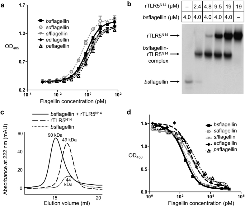
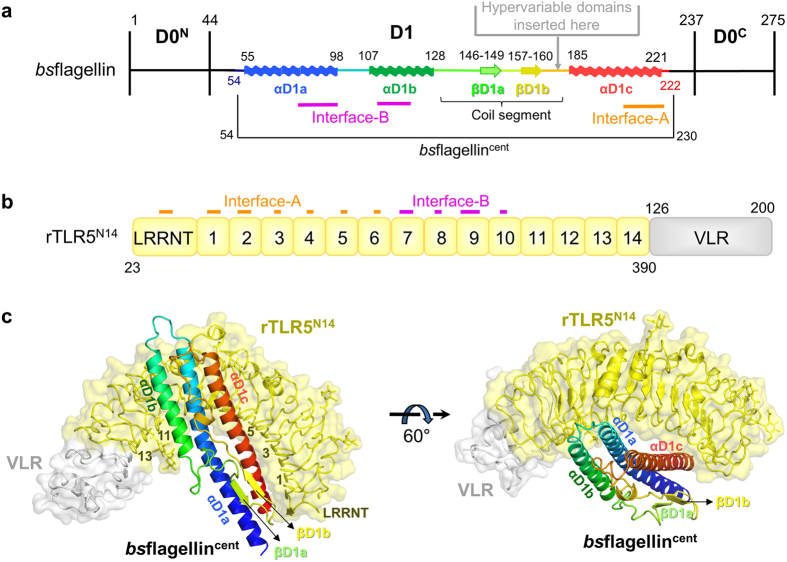
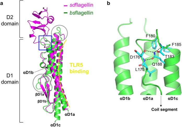
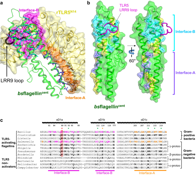
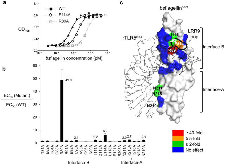
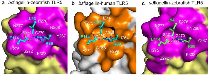
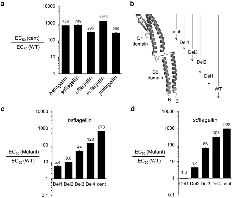
Flagellin is a bacterial protein that polymerizes into the flagellar filament and is essential for bacterial motility. When flagellated bacteria invade the host, flagellin is recognized by Toll-like receptor 5 (TLR5) as a pathogen invasion signal and eventually evokes the innate immune response. Here, we provide a conserved structural mechanism by which flagellins from Gram-negative γ-proteobacteria and Gram-positive Firmicutes bacteria bind and activate TLR5. The comparative structural analysis using our crystal structure of a complex between Bacillus subtilis flagellin (bsflagellin) and TLR5 at 2.1 Å resolution, combined with the alanine scanning analysis of the binding interface, reveals a common hot spot in flagellin for TLR5 activation. An arginine residue (bsflagellin R89) of the flagellin D1 domain and its adjacent residues (bsflagellin E114 and L93) constitute a hot spot that provides shape and chemical complementarity to a cavity generated by the loop of leucine-rich repeat 9 in TLR5. In addition to the flagellin D1 domain, the D0 domain also contributes to TLR5 activity through structurally dispersed regions, but not a single focal area. These results establish the groundwork for the future design of flagellin-based therapeutics.


Figures

Figure 1
Figure 1. TLR5 interaction and activation by flagellins.
(a) TLR5 signaling activities of bsflagellin, sdflagellin, paflagellin, sfflagellin, and ecflagellin. Activities were determined in duplicate using the HEK293TLR5 reporter cell assay. The data [means ± standard deviation (S.D.); n = 2] are representative of three independent experiments that yielded similar results (Table 1). (b) Native PAGE analysis of the direct interaction between bsflagellin and rTLR5N14. (c) Gel-filtration analysis of the complex formation between bsflagellin and rTLR5N14. The apparent molecular weight of each peak was estimated using the elution volumes of gel-filtration standards and is shown near the peak. Protein elution was monitored by optical absorbance at 222 nm instead of 280 nm because bsflagellin does not contain any tryptophan and tyrosine residues that are detectable with 280 nm absorbance. (d) rTLR5N14-binding capacity of bsflagellin, sdflagellin, paflagellin, sfflagellin, and ecflagellin determined with the competitive binding assay. The data shown are representative of at least three independent experiments that yielded similar results (Table 1).
Figure 2
Figure 2. Crystal structure of the bsflagellincent-rTLR5N14 complex.
(a) Domain organization of bsflagellin. bsflagellin consists of two domains, D0 and D1. The D0 domain is divided into D0N and D0C by the insertion of the D1 domain. In three- or four-domain flagellins, hypervariable domains are located between the coil segment and the αD1c helix of the D1 domain. bsflagellin residues 54–222 were built in the bsflagellincent-rTLR5N14 complex structure and are rainbow-colored from blue to red with secondary structure assignments. bsflagellin residues in binding interface-A and interface-B are schematically indicated by orange and magenta lines, respectively. (b) Schematic representation of the rTLR5N14 LRR modules (zebrafish TLR5 LRRNT-LRR14, yellow) fused to the C-terminal fragment of the hagfish variable lymphocyte receptor (VLR, gray). Binding interface-A and interface-B are delineated by orange and magenta lines, respectively. The LRR modules of rTLR5N14 are labeled accordingly. (c) Overall structure of the complex between bsflagellincent and rTLR5N14. rTLR5N14 is depicted in yellow ribbons with a transparent yellow surface representation, and its fusion partner, VLR, is colored in gray. bsflagellincent is shown in the rainbow-colored ribbon diagram from the N-terminus in blue to the C-terminus in red.
Figure 3
Figure 3. Flagellin structure in the bsflagellincent-rTLR5N14 complex.
(a) Structural comparison of bsflagellincent (green ribbons) and sdflagellinD1-D2 (magenta ribbons, PDB ID 3V47). The unique structural features of bsflagellincent are highlighted by a blue rectangle and gray circles. A sharp turn between the coil segment and αD1c helix in the bsflagellincent structure replaces the hypervariable D2 domain of sdflagellinD1-D2. (b) Close-up view of the blue rectangle in Fig. 3a.
Figure 4
Figure 4. bsflagellincent-rTLR5N14 interaction.
(a) Binding interfaces of the bsflagellincent-rTLR5N14 complex. rTLR5N14 is shown in yellow ribbons and a surface representation. Flagellin-binding TLR5 residues in interface-A and interface-B are represented by orange and magenta surfaces, respectively, and are also depicted with sticks. bsflagellincent is shown as green ribbons. TLR5-binding bsflagellin residues in interface-A and interface-B are depicted as light blue and cyan sticks, respectively. The LRR9 loop is represented by a black thick coil. (b) Comparison of the TLR5-binding interfaces of bsflagellin and sdflagellin. bsflagellin residues that are conserved with sdflagellin are shown as light blue (interface-A) and cyan (interface-B) surfaces with sticks. bsflagellin residues that are different from sdflagellin are colored in yellow. The TLR5 LRR9 loop is depicted as a magenta coil. (c) Alignment of the flagellin amino acid sequences at and around the primary binding interface. bsflagellin and sdflagellin residues in interface-A and interface-B are colored in orange and magenta, respectively, and are shown in bold. Residues of other flagellins that are identical to the bsflagellin residues are bolded. bsflagellin residues with significant effects on TLR5 activation are designated as ‘*’ color-coded as in Fig. 5c. bsflagellin R89 and its equivalent residues in other TLR5-activating flagellins are highlighted by a red box. The amino acid sequences are derived from Bacillus subtilis subspecies spizizenii strain W23, Clostridium tyrobutyricum, Listeria innocua serovar 6a, Salmonella enterica subspecies enterica serovar Dublin, Escherichia coli strain K-12, Pseudomonas aeruginosa, Shigella flexneri 2a strain 301, Serratia marcescens WW4, Bordetella bronchiseptica, Helicobacter pylori J99, Campylobacter jejuni subspecies jejuni, Bartonella bacilliformis, and Rhizobium meliloti strain 1021.
Figure 5
Figure 5. TLR5 signaling activities of bsflagellin alanine mutants.
(a) TLR5 signaling activities of bsflagellinWT and its mutants, R89A and E114A. Activities were determined using the HEK293TLR5 reporter cell assay. The data (means ± S.D.; n = 2) are representative of at least three independent experiments that yielded similar results. (b) The relative TLR5 signaling activities of bsflagellin alanine mutants, compared to bsflagellinWT. The data represent the means ± S.D. from at least three independent experiments. (c) Contributions of bsflagellin residues to TLR5 signaling activity that were color-coded by the effects of the mutations.
Figure 6
Figure 6. Flagellin-TLR5 interactions at and near bsflagellin R89A.
(a) bsflagellin R89, E114, and L93 (cyan sticks) observed in the cavity formed by the zebrafish TLR5 LRR9 loop (magenta surface) in the bsflagellincent-rTLR5N14 structure. Hydrogen bonds are represented by black (bsflagellin-to-TLR5 interactions) and cyan (bsflagellin-to-bsflagellin interactions) dashed lines. (b) bsflagellin R89, E114, and L93 (cyan sticks) observed in the cavity formed by the human TLR5 LRR9 loop (orange surface) in the bsflagellin-human TLR5 model. The complex model was generated by calculating a homology-based model of human TLR5 with the Modeller program, combining it with the bsflagellincent structure obtained from the bsflagellincent-rTLR5N14 structure, and applying structure idealization with the Refmac5 program. Hydrogen bonds are represented by black (bsflagellin-to-TLR5 interactions) and cyan (bsflagellin-to-bsflagellin interactions) dashed lines. (c) sdflagellin R90, E114, and L94 (green sticks) observed in the cavity formed by the zebrafish TLR5 LRR9 loop (magenta surface) in the sdflagellinD1-D2-rTLR5N14 structure (PDB ID 3V47). Hydrogen bonds are represented by black (sdflagellin-to-TLR5 interactions) and green (sdflagellin-to-sdflagellin interactions) dashed lines.
Figure 7
Figure 7. TLR5 signaling activity of bsflagellin and sdflagellin deletion mutants (means ± S.D.; n = 2).
(a) TLR5 signaling activities of bsflagellincent, sdflagellincent, paflagellincent, sfflagellincent, and ecflagellincent, compared to WT flagellins. Activities were determined using the HEK293TLR5 reporter cell assay. (b) Schematic representations of the flagellin deletion constructs (Del1, Del2, Del3, Del4, and cent). The N- and C-termini of flagellin are labeled as ‘N’ and ‘C’, respectively. (c) TLR5 signaling activities of bsflagellin-Del1, Del2, Del3, Del4, and cent, compared to bsflagellinWT. Activities were determined using the HEK293TLR5 reporter cell assay. (d) TLR5 signaling activities of sdflagellin-Del1, Del2, Del3, Del4, and cent, compared to WT sdflagellin.

References

    1. Yonekura K., Maki-Yonekura S. & Namba K. Growth mechanism of the bacterial flagellar filament. Research in microbiology 153, 191–197 (2002). - PubMed
    1. Macnab R. M. Genetics and biogenesis of bacterial flagella. Annual review of genetics 26, 131–158 (1992). - PubMed
    1. Ramos H. C., Rumbo M. & Sirard J. C. Bacterial flagellins: mediators of pathogenicity and host immune responses in mucosa. Trends in microbiology 12, 509–517 (2004). - PubMed
    1. Takeda K., Kaisho T. & Akira S. Toll-like receptors. Annu Rev Immunol 21, 335–376 (2003). - PubMed
    1. Franchi L., Munoz-Planillo R. & Nunez G. Sensing and reacting to microbes through the inflammasomes. Nat Immunol 13, 325–332 (2012). - PMC - PubMed

Publication types

LinkOut - more resources