. 2017 Jan 20:7:40660.
doi: 10.1038/srep40660.

Identification of LukPQ, a novel, equid-adapted leukocidin of Staphylococcus aureus

Affiliations

Identification of LukPQ, a novel, equid-adapted leukocidin of Staphylococcus aureus

Gerrit Koop et al. Sci Rep. .

Abstract

Bicomponent pore-forming leukocidins are a family of potent toxins secreted by Staphylococcus aureus, which target white blood cells preferentially and consist of an S- and an F-component. The S-component recognizes a receptor on the host cell, enabling high-affinity binding to the cell surface, after which the toxins form a pore that penetrates the cell lipid bilayer. Until now, six different leukocidins have been described, some of which are host and cell specific. Here, we identify and characterise a novel S. aureus leukocidin; LukPQ. LukPQ is encoded on a 45 kb prophage (ΦSaeq1) found in six different clonal lineages, almost exclusively in strains cultured from equids. We show that LukPQ is a potent and specific killer of equine neutrophils and identify equine-CXCRA and CXCR2 as its target receptors. Although the S-component (LukP) is highly similar to the S-component of LukED, the species specificity of LukPQ and LukED differs. By forming non-canonical toxin pairs, we identify that the F-component contributes to the observed host tropism of LukPQ, thereby challenging the current paradigm that leukocidin specificity is driven solely by the S-component.


Figures

Figure 1
Figure 1. The novel Staphylococcus aureus toxin LukPQ in the context of other leukocidins.
(a) Comparison of the novel prophage ΦSaeq1 in isolate 3711, carrying the equid specific lukPQ, with ΦSaov3 (ruminant strain ED133) and ΦSa2 (human PVL strain MW2). Areas of red show regions conserved between the sequences and homologous coding DNA sequences are marked in the same colour. (b) Phylogenetic tree based on amino acid sequences of mature leukocidins, with alpha-hemolysin as an outgroup.
Figure 2
Figure 2. LukPQ is a potent killer of equine neutrophils.
(a,b and c) Equine, bovine and human neutrophils were analysed for pore formation upon incubation with LukPQ (A), LukMF’ (B), and LukSF-PV (C). Mean percentages of permeable cells ± standard deviation (SD) are shown (n = 3–5).
Figure 3
Figure 3. CXCRA and CXCR2 mediate pore formation by LukPQ in equine neutrophils.
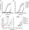
(a) Pore formation in HEK293T cells stably transfected with equine CCR2, CCR5, C5aR, CXCRA, CXCR2 and the Duffy antigen receptor (DARC) upon treatment with LukPQ. Mean percentages of permeable cells ± SD are shown (n = 3). (b) HEK293T cells stably transfected with equine CXCRA, CXCR2 and CCR5 were incubated with LukED and analysed for pore formation. Mean percentages of permeable cells ± SD are shown (n = 3). (c) Relative calcium mobilization by CXCRA and CXCR2 transfected HEK293T cells preincubated with buffer or 10 μg/ml LukP upon stimulation with CXCL5, CXCL6 and CXCL8. Bars indicate SD with n = 3. Statistically significant effects of preincubation with LukP are indicated. **P < 0.01 and *P < 0.05. Pre-incubation with LukP resulted in a significant decrease in calcium mobilization in both CXCRA and CXCR2 cells stimulated with CXCL6 (p < 0.01 and p < 0.05 respectively), and in CXCR2 cells stimulated with CXCL5 (p < 0.05). A trend was seen in CXCRA cells stimulated with CXCL8 (p = 0.06).
Figure 4
Figure 4. LukPQ and LukED exhibit distinct species specificities in an F-component-dependent manner.
Pore formation in equine (a), bovine (b) and human (c) neutrophils upon incubation with LukPQ, LukED, LukEQ or LukPD. Mean percentages of permeable cells ± SD are shown (n = 3–5).
Figure 5
Figure 5. Unique residues in F-components may underlie functional specificity.
(a) Structure-guided alignment of the DR4 region (highlighted yellow) in the rim domain of LukE, LukP, HlgA and LukM. (b) Homology model of the LukPQ heterodimer with LukP as a cartoon and LukQ as a surface representation. Residues unique to LukQ, but identical between LukD and LukF’ are coloured yellow; residues that differ in all three toxins are coloured cyan. The position of isoleucine 285 in the rim domain is annotated.

References

    1. Viana D. et al. Adaptation of Staphylococcus aureus to ruminant and equine hosts involves SaPI-carried variants of von Willebrand factor-binding protein. Mol. Microbiol. 77, 1583–1594 (2010). - PubMed
    1. Guinane C. M. et al. Evolutionary genomics of Staphylococcus aureus reveals insights into the origin and molecular basis of ruminant host adaptation. Genome Biol. Evol. 2, 454–466 (2010). - PMC - PubMed
    1. Viana D. et al. A single natural nucleotide mutation alters bacterial pathogen host tropism. Nat. Genet. 47, 361–366 (2015). - PMC - PubMed
    1. Alonzo F. III. & Torres V. J. The bicomponent pore-forming leucocidins of Staphylococcus aureus. Microbiol. Mol. Biol. Rev. 78, 199–230 (2014). - PMC - PubMed
    1. McCarthy A. J. & Lindsay J. A. Staphylococcus aureus innate immune evasion is lineage-specific: A bioinfomatics study. Infect. Genet. Evol. 19, 7–14 (2013). - PubMed

Publication types