

Innate immune defense in the inner ear - mucines are expressed by the human endolymphatic sac
- PMID: 28106268
- PMCID: PMC5244465
- DOI: 10.1111/joa.12559
Innate immune defense in the inner ear - mucines are expressed by the human endolymphatic sac
Abstract
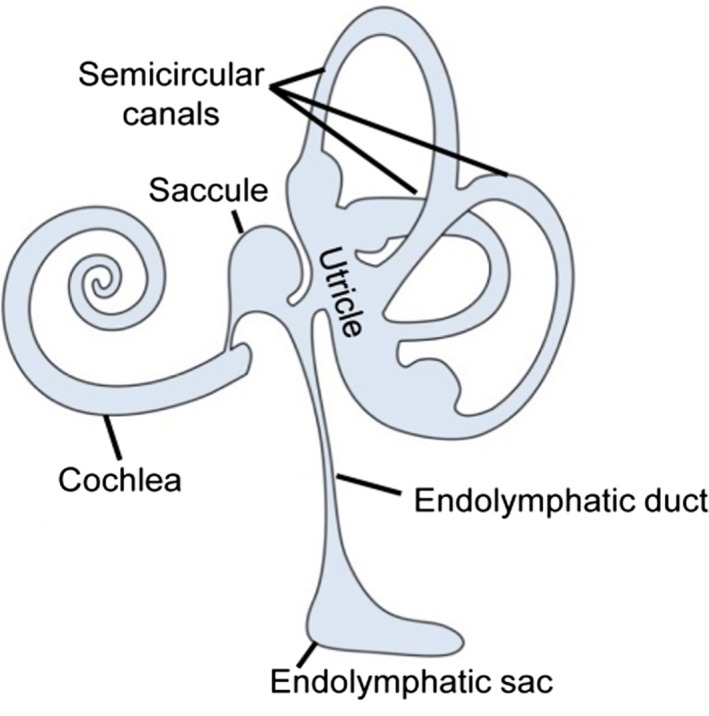
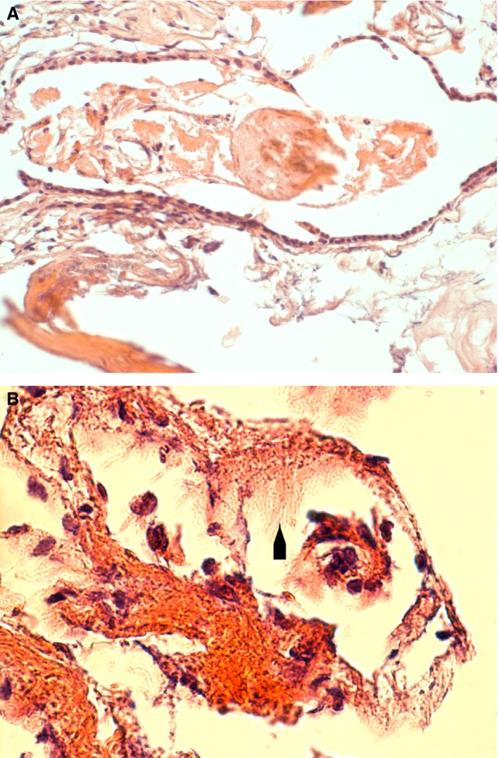
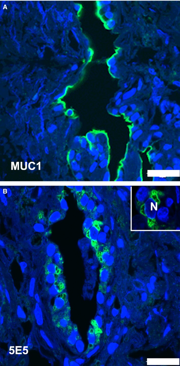
The human endolymphatic sac has been shown recently to have immunological capacities and has thus been proposed as the main entity protecting the inner ear from pathogen invasion, equivalent to mucosa-associated lymphoid tissue (MALT). Although the sac expresses molecules of the innate immune system, the potential expression of members of the important mucin family has not been detailed. Thus, this paper explores endolymphatic sac expression of a number of mucins and mucin precursors. Twelve fresh tissue samples from the human endolymphatic sac were obtained during translabyrinthine surgery. The expression of Mucin 1, 2, 5B/AC and 16, as well as the core structure elements (mucin precursors) T-antigen, Tn-antigen and Sialyl-Tn-antigen was investigated by immunohistochemistry. The endolymphatic sac epithelium expressed MUC1 (both apically towards the endolymphatic sac (ES) lumen and basally towards the capillary network), MUC 16 and Tn-antigen. There was no labeling after incubation with antibodies against T-antigen, sialyl-Tn-antigen, MUC2 and MUC5B/AC. We conclude that the human endolymphatic sac epithelium expresses a number of mucin molecules, which supports the hypothesis of the sac as the primary immunological tissue structure of the inner ear, equivalent to MALT in other organs. The mucins may also play a role in the formation and continuous homeostasis of the inner ear fluids, as well as the pathogenesis of Meniere's disease.
Keywords: endolymphatic sac; immunohistochemistry; mucins.
© 2016 Anatomical Society.
Figures

Similar articles
-
Gene expression demonstrates an immunological capacity of the human endolymphatic sac.Laryngoscope. 2015 Aug;125(8):E269-75. doi: 10.1002/lary.25242. Epub 2015 Mar 16. Laryngoscope. 2015. PMID: 25779626
-
The human endolymphatic sac expresses natriuretic peptides.Laryngoscope. 2017 Jun;127(6):E201-E208. doi: 10.1002/lary.26074. Epub 2017 Mar 14. Laryngoscope. 2017. PMID: 28295370
-
The endolymphatic sac: its importance in inner ear immune responses.Laryngoscope. 1986 Jun;96(6):685-91. doi: 10.1288/00005537-198606000-00018. Laryngoscope. 1986. PMID: 3713414
-
[The endolymphatic sac: its roles in the inner ear].Med Sci (Paris). 2004 Mar;20(3):304-10. doi: 10.1051/medsci/2004203304. Med Sci (Paris). 2004. PMID: 15067575 Review. French.
-
Functional morphology of the human endolymphatic sac. A review.ORL J Otorhinolaryngol Relat Spec. 1992;54(4):173-8. doi: 10.1159/000276294. ORL J Otorhinolaryngol Relat Spec. 1992. PMID: 1484698 Review.
Cited by
-
A Micro-CT and Synchrotron Imaging Study of the Human Endolymphatic Duct with Special Reference to Endolymph Outflow and Meniere's Disease.Sci Rep. 2020 May 19;10(1):8295. doi: 10.1038/s41598-020-65110-0. Sci Rep. 2020. PMID: 32427861 Free PMC article.
-
RNA-sequencing study of peripheral blood mononuclear cells in sporadic Ménière's disease patients: possible contribution of immunologic dysfunction to the development of this disorder.Clin Exp Immunol. 2018 Apr;192(1):33-45. doi: 10.1111/cei.13083. Epub 2017 Dec 11. Clin Exp Immunol. 2018. PMID: 29164594 Free PMC article.
-
A Nationwide, Population-based Cohort Study on Potential Autoimmune Association of Ménière Disease to Atopy and Vitiligo.Sci Rep. 2019 Mar 13;9(1):4406. doi: 10.1038/s41598-019-40658-8. Sci Rep. 2019. PMID: 30867466 Free PMC article.
-
Up-Regulated Expression of Interferon-Gamma, Interleukin-6 and Tumor Necrosis Factor-Alpha in the Endolymphatic Sac of Meniere's Disease Suggesting the Local Inflammatory Response Underlies the Mechanism of This Disease.Front Neurol. 2022 Feb 23;13:781031. doi: 10.3389/fneur.2022.781031. eCollection 2022. Front Neurol. 2022. PMID: 35280304 Free PMC article.
-
Bidirectional Transport of IgE by CD23 in the Inner Ear of Patients with Meniere's Disease.J Immunol. 2022 Feb 15;208(4):827-838. doi: 10.4049/jimmunol.2100745. Epub 2022 Jan 19. J Immunol. 2022. PMID: 35046106 Free PMC article.
References
-
- Altermatt HJ, Gebbers JO, Arnold W, et al. (1990) The epithelium of the human endolymphatic sac: immunohistochemical characterization. ORL J Otorhinolaryngol Relat Spec 52, 113–120. - PubMed
-
- Andrulis M, Ellert E, Mandel U, et al. (2014) Expression of MUC‐1 in multiple myeloma and its precursors: correlation with glycosylation and subcellular localization. Histopathology 64, 799–806. - PubMed
-
- Bryne M, Gravdahl C, Koppang HS, et al. (1995) Is the carbohydrate sialosyl‐Tn a marker for altered malignant activity in squamous epithelium the head and neck region? J Pathol 175, 237–242. - PubMed
-
- Clausen H, Stroud MR, Parker J, et al. (1988) Monoclonal antibodies directed to the blood group A associated structure, galactosyl‐A: specificity and relation to the Thomson‐Friedenreich antigen. Mol Immunol 25, 199–204. - PubMed
MeSH terms
Substances
LinkOut - more resources
Full Text Sources
Other Literature Sources
Research Materials
Miscellaneous
