. 2017 Feb;96(2):225-232.
doi: 10.1177/0022034516677529. Epub 2016 Nov 16.

Insulin-Like Growth Factor-1-Mediated DNA Repair in Irradiated Salivary Glands Is Sirtuin-1 Dependent

Affiliations

Insulin-Like Growth Factor-1-Mediated DNA Repair in Irradiated Salivary Glands Is Sirtuin-1 Dependent

S Meyer et al. J Dent Res. 2017 Feb.

Abstract

Ionizing radiation is one of the most common cancer treatments; however, the treatment leads to a wide range of debilitating side effects. In patients with head and neck cancer (HNC), the surrounding normal salivary gland is extremely sensitive to therapeutic radiation, and damage to this tissue results in various oral complications and decreased quality of life (QOL). In the current study, mice treated with targeted head and neck radiation showed a significant increase in double-stranded breaks (DSB) in the DNA of parotid salivary gland cells immediately after treatment, and this remained elevated 3 h posttreatment. In contrast, mice pretreated with insulin-like growth factor-1 (IGF-1) showed resolution of the same amount of initial DNA damage by 3 h posttreatment. At acute time points (30 min to 2 h), irradiated parotid glands had significantly decreased levels of the histone deactylase Sirtuin-1 (SirT-1) which has been previously shown to function in DNA repair. Pretreatment with IGF-1 increased SirT-1 protein levels and increased deacetylation of SirT-1 targets involved in DNA repair. Pharmacological inhibition of SirT-1 activity decreased the IGF-1-mediated resolution of DSB. These data suggest that IGF-1 promotes DNA repair in irradiated parotid glands through the maintenance and activation of SirT-1.

Keywords: DNA repair; IGF-1; Sirtuin-1; radiation; salivary glands; xerostomia.


Conflict of interest statement

The authors declare no potential conflicts of interest with respect to the authorship and/or publication of this article.

Figures
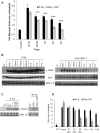
Figure 1.
Figure 1.
Pretreatment with insulin-like growth factor-1 (IGF-1) before radiation leads to resolution of double-stranded DNA breaks (DSB) at earlier time points as compared with radiation-only controls. The head and neck region of FVB mice was exposed to 5 Gy radiation, and the parotid salivary glands were removed at predetermined time points following irradiation. (A) Tissue samples were analyzed via a neutral comet assay. Average tail moment scores were determined from each parotid gland (n = 8 separate glands) and the data are graphed as fold over untreated controls. Asterisks denote significant differences (P ≤ 0.05) between 5 Gy and 5 Gy plus IGF-1 at each time point. (B) Protein samples from FVB mice were collected following exposure to 5 Gy radiation (left side) or 5 Gy radiation plus IGF-1 (right side) and immunoblotted for histone H2A variant phosphorylated on Ser139 (γ-H2AX). Membranes were stripped and reimmunoblotted for total histone 2A and total extracellular signal-regulated kinase (ERK) as loading controls. (C) Protein samples from FVB mice treated with 5 Gy or 5 Gy plus IGF-1 were collected at 5 min (left) or 30 min (right) post-treatment and immunoblotted for γ-H2AX. Membranes were stripped and reimmunoblotted for total ERK as a loading control. (D) Blots were analyzed by densitometry, with γ-H2AX normalized to H2A, and the data are graphed as mean intensity over untreated controls. A minimum of 4 mice per treatment group were analyzed.
Figure 2.
Figure 2.
Proteins involved in homologous recombination and non-homologous end joining repair appear to be similar between mice treated with radiation with or without insulin-like growth factor-1 (IGF-1). Mice were treated with targeted head and neck radiation, as described in Figure 1. (A) Protein samples were collected following exposure to 5 Gy (left) or 5 Gy plus IGF-1 (right) and immunoblotted for replication protein A (RPA) phosphorylated on serine (Ser)4/8. Membranes were stripped and re-immunoblotted for total extracellular signal-regulated kinase (ERK) as a loading control. (B) Blots were analyzed by densitometry, with phosphorylated RPA normalized to total ERK, and the data are graphed as mean intensity over untreated controls. A minimum of 4 mice per treatment group were analyzed. (C) Rad51 was immunoprecipitated from parotid tissue following exposure to 5 Gy or 5 Gy plus IGF-1. Membranes were immunoblotted for BRCA1 to determine binding to Rad51 (representative blot) and stripped and reimmunoblotted for total Rad51 as a loading control. (D) Paraffin-embedded tissue sections from parotid glands were prepared from mice untreated or treated for 30 min with 5 Gy. Slides were immunostained for Rad51 and representative images from untreated mice (left) and mice treated with 5 Gy (right) are shown. Scale bars represents 25 µm. DAPI, 4’,6-diamidino-2-phenylindole; UT, untreated.
Figure 3.
Figure 3.
Sirtuin-1 (SirT-1) and SirT-3 are increased with insulin-like growth factor-1 (IGF-1) pretreatment as compared with radiation-only controls. Mice were treated with targeted head and neck radiation as described in Figure 1. (A) Protein samples were collected from mice treated with 5 Gy or 5 Gy plus IGF-1 at predetermined time points and immunoblotted for SirT-1, -2 or -3. Membranes were stripped and reimmunoblotted for total extracellular signal-regulated kinase (ERK) as a loading control (representative blot). (B) Blots were analyzed by densitometry, with SirT-1 normalized to total ERK, and data are graphed as mean intensity over untreated controls. A minimum of 4 mice per treatment group were analyzed. (C) The mRNA expression level for SirT-1 was determined by quantitative RT-PCR. (D) Mice were injected with IGF-1 alone and parotid salivary glands were collected at predetermined time points. Protein samples were immunoblotted for SirT-1. Membranes were stripped and reimmunoblotted for total ERK as a loading control. (E) Parotid tissue sections were collected 30 min after radiation treatment and immunostained for SirT-1. Representative images from untreated mice (left) and mice treated with 5 Gy (right) are shown. Scale bars represents 25 µm. UT, untreated.
Figure 4.
Figure 4.
Pretreatment with insulin-like growth factor-1 (IGF-1) increases NSB1 serine (Ser)343 phosphorylation and decreases p53 acetylation. Mice were treated with targeted head and neck radiation as described in Figure 1. (A) Protein samples were collected 30 min after treatment and immunoblotted for NBS1 phosphorylation on Ser343. Membranes were stripped and reimmunoblotted for acetylated p53 and total extracellular signal-regulated kinase (ERK) as a loading control. (B) Blots were analyzed by densitometry, with phosphorylated NBS1 or acetylated p53 normalized to total ERK, and data are graphed as mean intensity over untreated controls. A minimum of 4 mice per treatment group were analyzed. (C) The mRNA expression level for E2F1 was determined by quantitative RT-PCR. UT, untreated.
Figure 5.
Figure 5.
Maintenance of sirtuin-1 (SirT-1) activity is critical for efficient DNA repair in primary salivary acinar cells. Primary acinar cells were isolated from the parotid glands of FVB mice as described in the Materials and Methods. (A) After 4 d in culture, cells were treated with 5 Gy or pretreated with insulin-like growth factor-1 (IGF-1) for 5 min before radiation. Cell lysates were collected 30 min after radiation and immunoblotted for SirT-1. Membranes were stripped and re-immunoblotted for total Akt as a loading control. (B) Primary cells were treated with 5 Gy, treated with IGF-1 before radiation (IR + IGF), pretreated with a sirtuin inhibitor (EX 527, 98 nM), or treated with IGF-1 and 98 nM of EX527 before radiation (EX + IR + IGF). Single-cell suspensions were collected 30 min after radiation and analyzed by neutral comet assay. Average tail moment scores were determined from each plate (n = 6) and are graphed as fold over untreated controls. Treatment groups with the same letter are not statistically different from each other. UT, untreated; IR, irradiated.

Similar articles

Cited by

References

    1. Al-Ejeh F, Kumar R, Wiegmans A, Lakhani SR, Brown MP, Khanna KK. 2010. Harnessing the complexity of DNA-damage response pathways to improve cancer treatment outcomes. Oncogene. 29(46):6085–6098. - PubMed
    1. Arany S, Benoit DS, Dewhurst S, Ovitt CE. 2013. Nanoparticle-mediated gene silencing confers radioprotection to salivary glands in vivo. Mol Ther. 21(6):1182–1194. - PMC - PubMed
    1. Avila JL, Grundmann O, Burd R, Limesand KH. 2009. Radiation-induced salivary gland dysfunction results from p53-dependent apoptosis. Int J Radiat Oncol Biol Phys. 73(2):523–529. - PMC - PubMed
    1. Ceccaldi R, Rondinelli B, D’Andrea AD. 2016. Repair pathway choices and consequences at the double-strand break. Trends Cell Biol. 26(1):52–64. - PMC - PubMed
    1. Cerqueira A, Santamaria D, Martinez-Pastor B, Cuadrado M, Fernández-Capetillo O, Barbacid M. 2009. Overall Cdk activity modulates the DNA damage response in mammalian cells. J Cell Biol. 187(6):773–780. - PMC - PubMed

Publication types

LinkOut - more resources