. 2017 May;158(5):856-867.
doi: 10.1097/j.pain.0000000000000846.

Primidone inhibits TRPM3 and attenuates thermal nociception in vivo

Affiliations

Primidone inhibits TRPM3 and attenuates thermal nociception in vivo

Ute Krügel et al. Pain. 2017 May.

Abstract

The melastatin-related transient receptor potential (TRP) channel TRPM3 is a nonselective cation channel expressed in nociceptive neurons and activated by heat. Because TRPM3-deficient mice show inflammatory thermal hyperalgesia, pharmacological inhibition of TRPM3 may exert antinociceptive properties. Fluorometric Ca influx assays and a compound library containing approved or clinically tested drugs were used to identify TRPM3 inhibitors. Biophysical properties of channel inhibition were assessed using electrophysiological methods. The nonsteroidal anti-inflammatory drug diclofenac, the tetracyclic antidepressant maprotiline, and the anticonvulsant primidone were identified as highly efficient TRPM3 blockers with half-maximal inhibition at 0.6 to 6 μM and marked specificity for TRPM3. Most prominently, primidone was biologically active to suppress TRPM3 activation by pregnenolone sulfate (PregS) and heat at concentrations markedly lower than plasma concentrations commonly used in antiepileptic therapy. Primidone blocked PregS-induced Cai influx through TRPM3 by allosteric modulation and reversibly inhibited atypical inwardly rectifying TRPM3 currents induced by coapplication of PregS and clotrimazole. In vivo, analgesic effects of low doses of primidone were demonstrated in mice, applying PregS- and heat-induced pain models, including inflammatory hyperalgesia. Thus, applying the approved drug at concentrations that are lower than those needed to induce anticonvulsive effects offers a shortcut for studying physiological and pathophysiological roles of TRPM3 in vivo.


Conflict of interest statement

Sponsorships or competing interests that may be relevant to content are disclosed at the end of this article.

Figures

Figure 1.
Figure 1.
Identification of diclofenac (A), maprotiline (B), and primidone (C) as inhibitors of TRPM3. Fluo-4-loaded HEKmTRPM3 cells were incubated with 20 μM of single compounds (black circles) or with 0.2% DMSO concentration (control, white circles), and fluorescence intensities were measured during injection of 35 μM pregnenolone sulfate as indicated by the bars. Fluo-4 fluorescence intensities F were normalised to the respective initial intensities F0 and depicted as time course. Traces extracted from the original screening data set, performed in a 384-well plate format, are shown along with the chemical structures of the respective drugs.
Figure 2.
Figure 2.
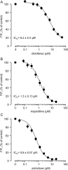
Concentration-dependent inhibition of pregnenolone sulfate–induced Ca2+ entry through TRPM3. Concentration–response curves for diclofenac (n = 9) (A), maprotiline (n = 8) (B), and primidone (n = 8) (C) were obtained by incubating HEKmTRPM3 cells with various concentrations of the respective drug and measuring the pregnenolone sulfate–induced activation of TRPM3. Activation without an inhibitor (DMSO control) was set as 100%, and fluorescence intensities evoked by solutions containing inhibitors were normalized to this value. IC50 values were obtained by fitting a 4-parameter Hill equation to each experiment, and mean values and SEM were calculated as shown.
Figure 3.
Figure 3.
Electrophysiological characterisation of the maprotiline-induced TRPM3 inhibition. (A) Representative whole-cell currents from a HEKmTRPM3 cell after alternating application of 35 μM of pregnenolone sulfate (PregS) and different concentrations of maprotiline (Map). Data are extracted from voltage ramps and depict currents at 87 mV (upper trace) and −113 mV (lower trace). The zero current level is indicated as dotted line. (B) Current density–voltage relation for PregS-induced TRPM3 currents at time points as indicated in (A) before (1), during (2), and after (3, 4) application of different concentrations of maprotiline (μM). (C) Statistical analysis of peak current densities (n = 12 for control and pregnenolone sulfate; n = 13 for PregS plus maprotiline) performed as in (A). Background conductivity is shown as dotted line. (D–F) Similar measurement as performed in (A–C) but activation and inhibition of PregS-induced currents were performed in the presence of 2.5 μM of maprotiline in the pipette solution (n = 4 each). Data are presented as mean values ± SEM. *P < 0.05 vs PregS alone.
Figure 4.
Figure 4.
Pregnenolone sulfate (PregS)-induced currents are potently and reversibly inhibited by primidone in HEKmTRPM3 cells. (A) Whole-cell currents were stimulated with 35 μM PregS, and different concentrations of primidone (Prim) were added as illustrated by the horizontal bars. Data show currents at 87 mV (upper trace) and −113 mV (lower trace). Zero current level is indicated as dotted line. (B) Current density–voltage relation for basal (1), PregS-induced (2), and primidone-blocked current (3) at time points indicated in (A). (C) Statistical analysis of peak current densities performed as in (A) (n = 5 for PregS plus primidone 5 μM, all others n = 7). Dotted line: level of background conductivity. (D) Whole-cell currents were stimulated with 40 μM PregS and 10 μM clotrimazole (Clt). Primidone (10 μM) was added as indicated by the horizontal bars. Data show currents at 74 mV (upper trace) and −126 mV (lower trace). (E) Current density–voltage relation for basal (1), PregS-induced (2), clotrimazole plus PregS-induced (3), and primidone-blocked current (4) at time points indicated in (D). (F) Statistical analysis of peak current densities determined as shown in (D) (n = 6). Data are presented as mean values ± SEM. *P < 0.05 vs PregS alone or PregS+Clt.
Figure 5.
Figure 5.
Selectivity of TRPM3 inhibition by diclofenac, maprotiline, and primidone in comparison with other nociceptive TRP channels. [Ca2+]i signals in stably TRPV1-, TRPM8-, and TRPA1-expressing HEK293 cell lines were evoked by capsaicin (2 μM), menthol (300 μM), and AITC (30 μM), respectively. The TRPM3-inhibiting drugs diclofenac (A), maprotiline (B), or primidone (C) were applied at different concentrations up to 50 μM. Data are presented as mean values ± SEM of 4 independent measurements performed in duplicates each. For comparison of selectivity of drugs for TRPM3, the respective nonlinear fits from Figure 2 are replotted as gray lines.
Figure 6.
Figure 6.
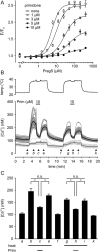
Primidone acts as an inhibitory allosteric modulator and prevents heat-induced activation of TRPM3. (A) Representative experiment of concentration-dependent pregnenolone sulfate (PregS)-induced Ca2+ entry in stably transfected murine HEKTRPM3 cells in the absence of primidone (open circles) and after incubation with different concentrations of primidone (1-10 μM, filled circles) for 60 seconds (n = 3 independent experiments in total). The left-most point per condition depicts controls without pregnenolone sulfate but primidone treatment. Data show mean values of quadruplets ± SEM. EC50 values (in μM) were obtained by fitting a 4-parameter Hill equation to experimental data. (B) Primidone inhibits heat activation of TRPM3 channels. Representative single-cell [Ca2+]i imaging in HEKmTRPM3. Parental HEK293 cells served as control. The upper graph depicts the temperature profile throughout the experiment. The lower graph shows changes in [Ca2+]i in individual HEKmTRPM3 cells (dark gray lines, n = 37) or in parental HEK293 cells (light gray, n = 15). The corresponding averages are given as black lines. Horizontal bars indicate the application of primidone (10 μM). Time points taken for analysis are marked (a-k). (C) Statistical analysis of heat-induced [Ca2+]i signals in HEKmTRPM3 cells determined as illustrated in (B). Bars indicate mean values ± SEM of 6 experiments. *P < 0.05 heat vs heat plus primidone.
Figure 7.
Figure 7.
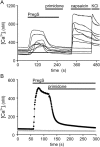
Primidone abrogates the pregnenolone sulfate (PregS)-induced increase in [Ca2+]i in freshly isolated rat DRG neurones and in transiently transfected HEK293 cells expressing human TRPM3. (A) Example traces of intracelluar calcium concentrations of single, freshly isolated rat DRG neurons, during application of PregS (35 μM) and primidone (25 μM) as well as of the TRPV1 activator capsaicin (2 μM) and KCl (10 mM) to select neuronal cells that express voltage-gated potassium channels. (B) Fluorescent measurements in HEK293 cells expressing human TRPM3α2 (black circles) or in nontransfected control cells (open circles). Pregnenolone sulfate (35 μM) and primidone (25 μM) were added as indicated by the horizontal bars.
Figure 8.
Figure 8.
Nociceptive response to chemical pain induced by pregnenolone sulfate (PregS) in mice is inhibited by primidone. Latency (A) and duration of nociceptive responses (B) to PregS (5 nmol) (licking, lifting, and shaking) injected into the right hind paw either as a mixture with primidone (10 nmol) or 5 minutes after pretreatment with primidone. Data are presented as mean values ± SEM (n = 12 each). *P < 0.05 vs vehicle group. Latency (C), number of responses (D), and total duration of nociceptive responses (E) to PregS (5 nmol, i.pl.) were registered after intraperitoneal pretreatment with indicated doses of primidone. Data are presented as mean values ± SEM (n = 8 each). *P < 0.05 vs vehicle group.
Figure 9.
Figure 9.
Systemically administered primidone attenuates nociceptive responses to noxious heat and thermal hyperalgesia in mice. (A) Effect of intraperitoneally administered primidone at indicated doses on the response latency of the hind paws on the hot plate (52°C). (B) Travelled distance of the same animals in an open field 120 minutes following drug administration (n = 10 each). (C) Effect of primidone (1 mg·kg−1, i.p.) on the response in the tail immersion test (48°C, n = 10 each). (D) Change in hot plate latency by treatment with primidone (1 mg·kg−1, i.p.) 24 hours after induction of thermal hyperalgesia by CFA injection into mice hind paws. Data are presented as mean values ± SEM (n = 8 each). *P < 0.05 vs vehicle (A–C) or vs CFA alone (D), +P < 0.05 vs vehicle group.

References

    1. Altman R, Bosch B, Brune K, Patrignani P, Young C. Advances in NSAID development: evolution of diclofenac products using pharmaceutical technology. Drugs 2015;75:859–77. - PMC - PubMed
    1. Alvin J, Goh E, Bush MT. Study of the hepatic metabolism of primidone by improved methodology. J Pharmacol Exp Ther 1975;194:117–25. - PubMed
    1. Bootman MD, Rietdorf K, Collins T, Walker S, Sanderson M. Ca2+-Sensitive fluorescent dyes and intracellular Ca2+ imaging. In: Parys JB, Bootman M, Yule DI, Bultynck G, editors. Calcium techniques. Cold Spring Habor: A Laboratory Manual Cold Spring Habor Laboratory Press, 2014. p. 25–41.
    1. Budakova L, Brozmanova H, Grundmann M, Fischer J. Simultaneous determination of antiepileptic drugs and their two active metabolites by HPLC. J Sep Sci 2008;31:1–8. - PubMed
    1. Carmody J, Knodler L, Murray S. Paradoxical modulation of nociception in mice by barbiturate agonism and antagonism: is a GABA site involved in nociception? Eur J Neurosci 1991;3:833–8. - PubMed

Publication types

MeSH terms