. 2017 Jan 19;22(1):163.
doi: 10.3390/molecules22010163.

Combination of Morroniside and Diosgenin Prevents High Glucose-Induced Cardiomyocytes Apoptosis

Affiliations

Combination of Morroniside and Diosgenin Prevents High Glucose-Induced Cardiomyocytes Apoptosis

Wen-Xia Pi et al. Molecules. .

Abstract

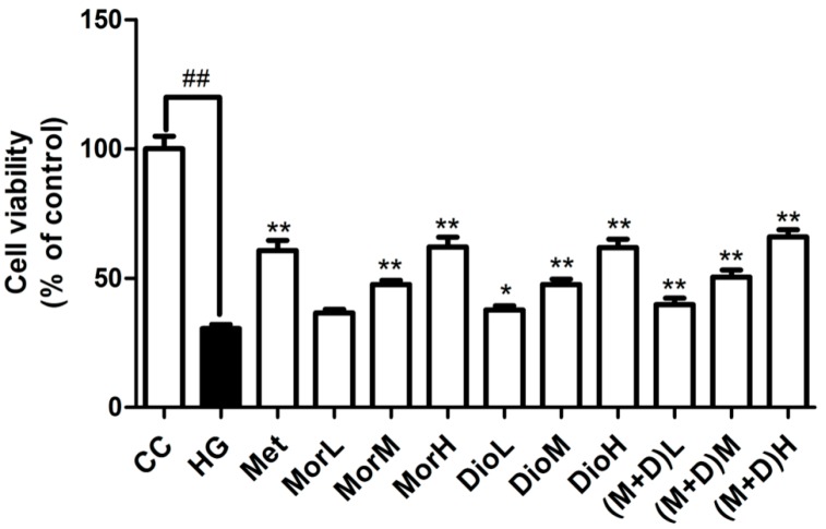
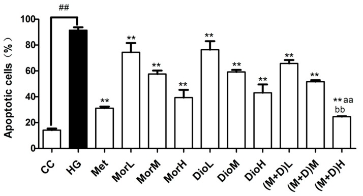
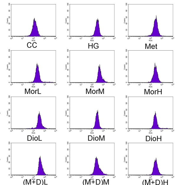
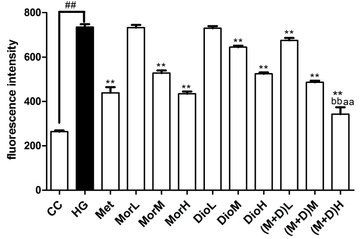
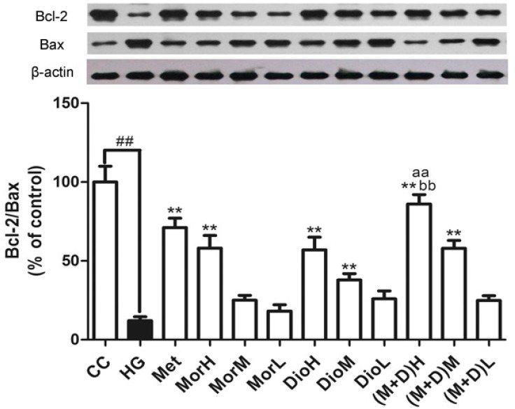
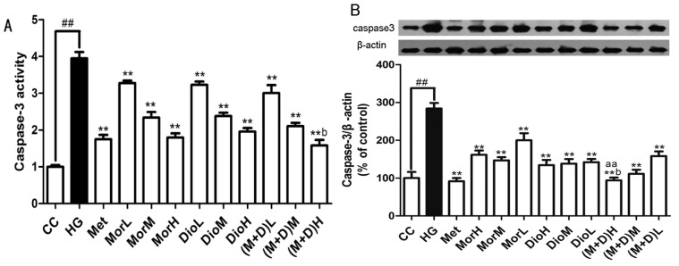
Cornus officinalis and Dioscorea opposita are two traditional Chinese medicines widely used in China for treating diabetes mellitus and its complications, such as diabetic cardiomyopathy. Morroniside (Mor) of Cornus officinalis and diosgenin (Dio) of Dioscorea opposita formed an innovative formula named M + D. The aims of the present study were to investigate myocardial protective effect of M + D on diabetic cardiomyopathy (DCM) through the inhibition of expression levels of caspase-3 protein, and identify the advantage of M + D compared with Mor, Dio, and the positive drug metformin (Met). We detected cell viability, cell apoptosis, intracellular reactive oxygen species (ROS) levels, and the expression levels of Bcl-2, Bax, and caspase-3 protein in rat cardiomyocytes. In result, Mor, Dio, and M + D increased cell viability, inhibited cell apoptosis and decreased ROS levels. Additionally, the expression of Bax and Bcl-2 protein was modulated and the expression levels of caspase-3 protein were markedly decreased. Among the treatment groups, M + D produced the most prominent effects. In conclusion, our data showed for the first time that Mor, Dio, and M + D prevented high glucose (HG)-induced myocardial injury by reducing oxidative stress and apoptosis in rat cardiomyocytes. Among all the groups, M + D produced the strongest effect, while Mor and Dio produced weaker effects.

Keywords: Bcl-2/Bax/caspase-3 signaling pathway; cardiomyocytes; diabetic cardiomyopathy; diosgenin; morroniside.


Conflict of interest statement

The authors declare no conflict of interest.

Figures

Figure 1
Figure 1
Effects of Mor (25, 50, 100 μg/mL), Dio (25, 50, 100 μg/mL), and M + D (25, 50, 100 μg/mL) on rat cardiomyocyte viability under HG-induced cell injury after pretreated 72 h. Values are expressed as the mean ± SD. ## p < 0.01 vs. CC; ** p < 0.01, * p < 0.05 vs. HG.
Figure 2
Figure 2
Effect of Mor (25, 50, 100 μg/mL), Dio (25, 50, 100 μg/mL), and M + D (25, 50, 100 μg/mL) on HG-induced cardiomyocyte apoptosis. (M + D)H significantly prevents from cardiomyocytes apoptosis as determined with TUNEL staining. Data are expressed as mean ± SD. ## p < 0.01 vs. CC; ** p < 0.01, vs. HG; aa p < 0.01, vs. MorH; bb p < 0.01, vs. DioH.
Figure 2
Figure 2
Effect of Mor (25, 50, 100 μg/mL), Dio (25, 50, 100 μg/mL), and M + D (25, 50, 100 μg/mL) on HG-induced cardiomyocyte apoptosis. (M + D)H significantly prevents from cardiomyocytes apoptosis as determined with TUNEL staining. Data are expressed as mean ± SD. ## p < 0.01 vs. CC; ** p < 0.01, vs. HG; aa p < 0.01, vs. MorH; bb p < 0.01, vs. DioH.
Figure 3
Figure 3
(M + D)H protects cardiomyocytes from mitochondrial production ROS induced with HG as determined with DCFH staining. Data are expressed as mean ± SD. ## p < 0.01 vs. CC; ** p < 0.01, vs. HG; aa p < 0.01, vs. MorH; bb p < 0.01, vs. DioH.
Figure 3
Figure 3
(M + D)H protects cardiomyocytes from mitochondrial production ROS induced with HG as determined with DCFH staining. Data are expressed as mean ± SD. ## p < 0.01 vs. CC; ** p < 0.01, vs. HG; aa p < 0.01, vs. MorH; bb p < 0.01, vs. DioH.
Figure 4
Figure 4
Regulation of proteins Bcl-2 and Bax associated with apoptosis by MorH, DioH, and (M + D)H. Data are expressed as mean ± SD. ## p < 0.01 vs. CC; ** p < 0.01, vs. HG; aa p < 0.01, vs. MorH; bb p < 0.01, vs. DioH.
Figure 5
Figure 5
(A) Effect of (M + D)H on caspase-3 activity by using a colorimetric activity assay kit. Data are expressed as mean ± SD. ## p < 0.01 vs. CC; ** p < 0.01 vs. HG; aa p < 0.01, vs. MorH; b p < 0.05 vs. DioH; and (B) the effect of (M + D)H on the expression levels of caspase-3 protein of cardiomyocytes after HG injury. Data are expressed as mean ± SD. ## p < 0.01 vs. CC; ** p < 0.01 vs. HG; aa p < 0.01, vs. MorH; b p < 0.05 vs. DioH.

References

    1. Rubler S., Dlugash J., Yuceoglu Y.Z., Kumral T., Branwood A.W., Grishman A. New type of cardiomyopathy associated with diabetic glomerulosclerosis. Am. J. Cardiol. 1972;30:595–602. doi: 10.1016/0002-9149(72)90595-4. - DOI - PubMed
    1. Cai L., Li W., Wang G., Guo L., Jiang Y., Kang Y.J. Hyperglycemia-induced apoptosis in mouse myocardium: Mitochondrial cytochrome C-mediated caspase-3 activation pathway. Diabetes. 2002;51:1938–1948. doi: 10.2337/diabetes.51.6.1938. - DOI - PubMed
    1. Zou M.H., Xie Z. Regulation of interplay between autophagy and apoptosis in the diabetic heart: New role of AMPK. Autophagy. 2013;9:624–625. doi: 10.4161/auto.23577. - DOI - PMC - PubMed
    1. Boudina S., Abel E.D. Diabetic cardiomyopathy revisited. Circulation. 2007;115:3213–3223. doi: 10.1161/CIRCULATIONAHA.106.679597. - DOI - PubMed
    1. Chen Y., Wu Y., Gan X., Liu K., Xing L.V., Shen H., Dai G., Xu H. Iridoid glycoside from Cornus officinalis ameliorated diabetes mellitus-induced testicular damage in male rats: Involvement of suppression of the AGEs/RAGE/p38 MAPK signaling pathway. J. Ethnopharmacol. 2016;194:850–860. doi: 10.1016/j.jep.2016.10.079. - DOI - PubMed

LinkOut - more resources