

Whole-brain vessel wall MRI: A parameter tune-up solution to improve the scan efficiency of three-dimensional variable flip-angle turbo spin-echo
- PMID: 28106936
- PMCID: PMC5519453
- DOI: 10.1002/jmri.25611
Whole-brain vessel wall MRI: A parameter tune-up solution to improve the scan efficiency of three-dimensional variable flip-angle turbo spin-echo
Abstract
Purpose: To propose and evaluate a parameter tune-up solution to expedite a three-dimensional (3D) variable-flip-angle turbo spin-echo (TSE) sequence for whole-brain intracranial vessel wall (IVW) imaging.
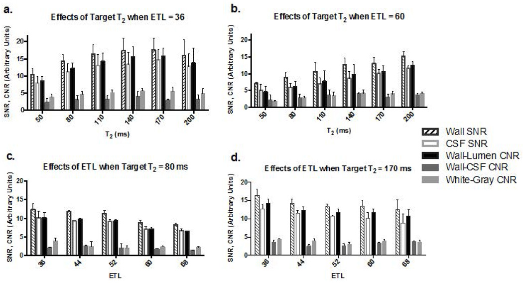
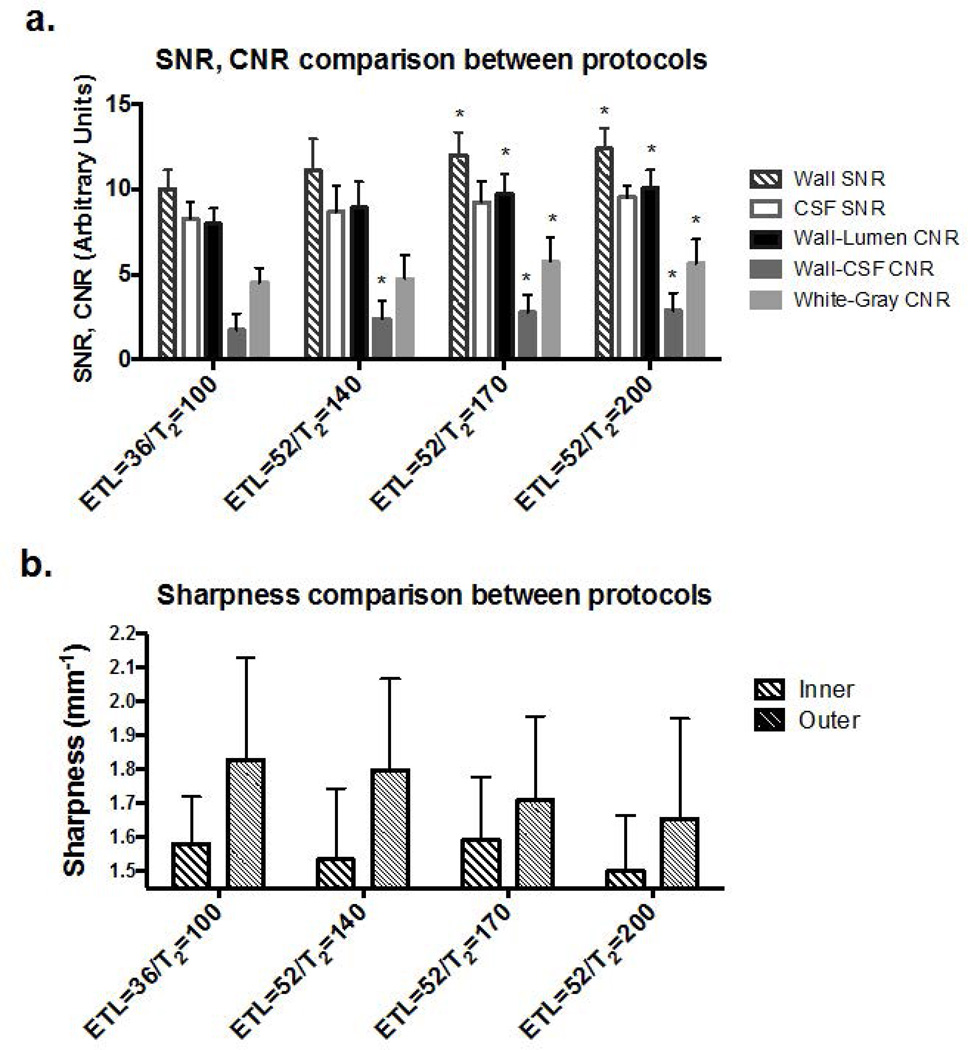
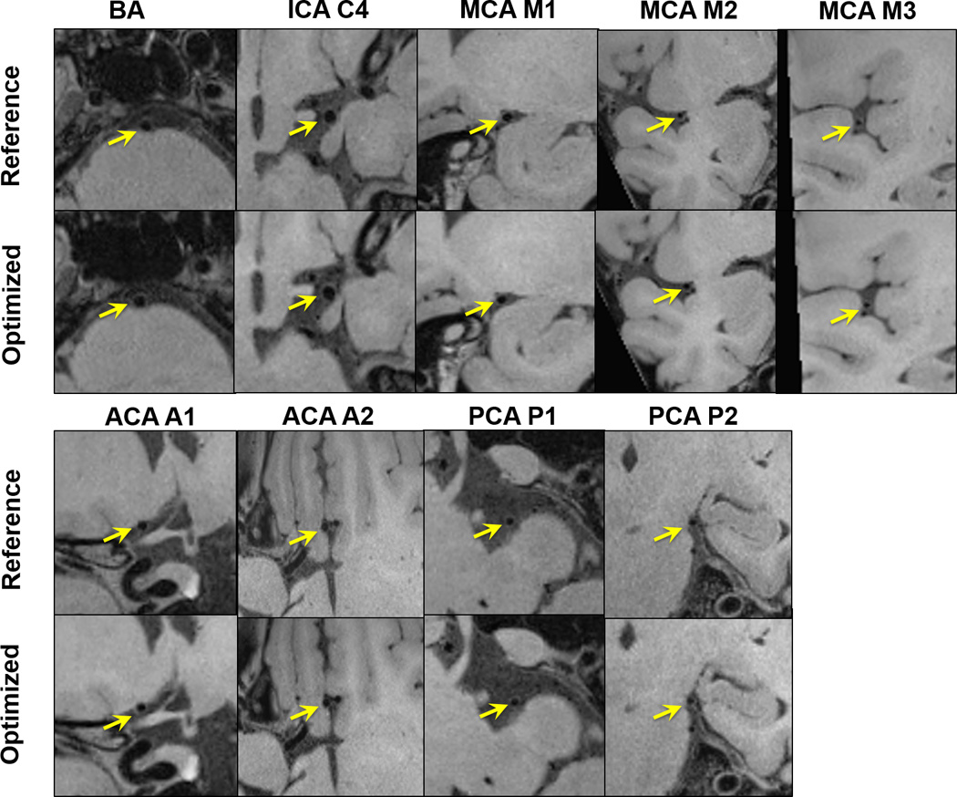
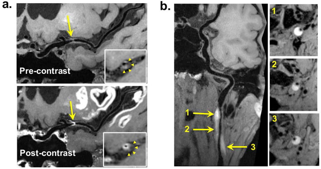
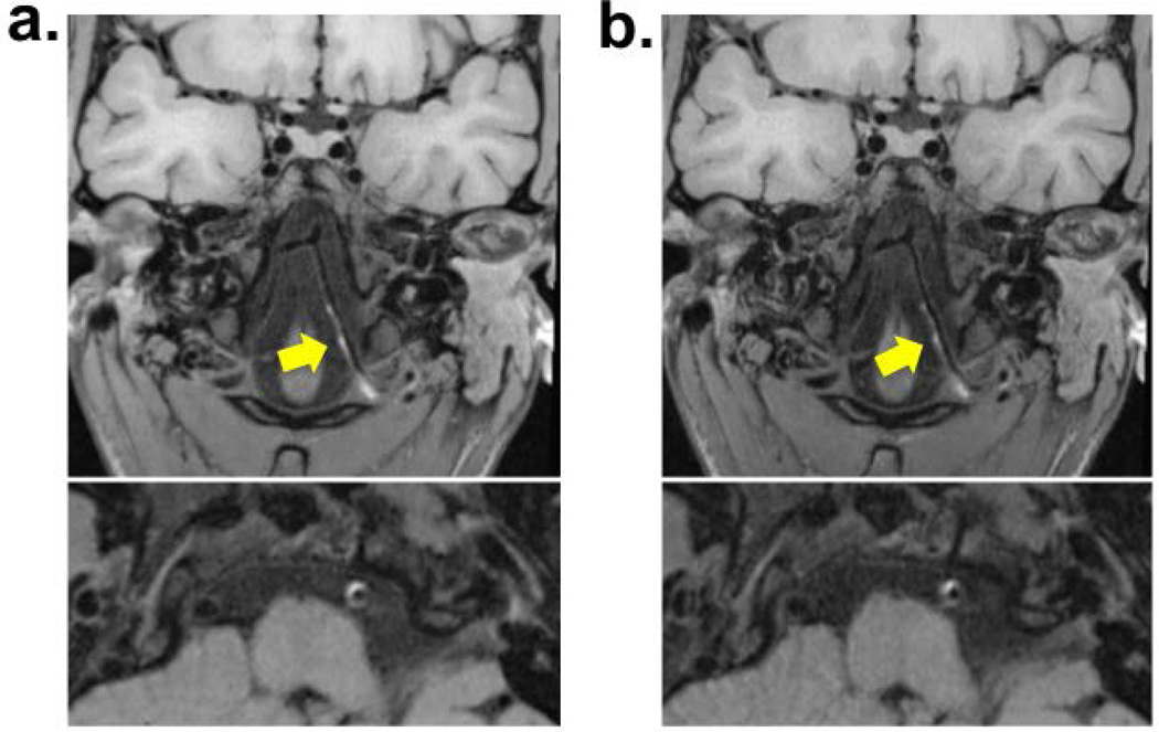
Materials and methods: Elliptical k-space sampling and prolonged echo train length (ETL), were used to expedite a 3D variable-flip-angle TSE-based sequence. To compensate for the potential loss in vessel wall signal, optimal combination of prescribed T2 and ETL was experimentally investigated on 22 healthy volunteers at 3 Tesla. The optimized protocol (7-8 min) was then compared with a previous protocol (reference protocol, 11-12 min) in terms of signal-to-noise ratio (SNR), contrast-to-noise ratio (CNR), vessel wall sharpness, and wall delineation quality on a 4-point scale (0:poor; 3:excellent) in 10 healthy volunteers. A pilot study of five patients was performed and lesion delineation score was used to demonstrate the diagnostic quality.
Results: A protocol with ETL = 52 and prescribed T2 = 170 ms was deemed an optimized one, which, compared with the reference protocol, provided significantly improved wall SNR (12.0 ± 1.3 versus 10.0 ± 1.1; P = 0.002), wall-lumen CNR (9.7 ± 1.2 versus 8.0 ± 0.9; P = 0.002), wall-CSF CNR (2.8 ± 1.0 versus 1.7 ± 1.0; P = 0.026), similar vessel wall sharpness at both inner (1.59 ± 0.18 versus 1.58 ± 0.14, P = 0.87) and outer (1.71 ± 0.25 versus 1.83 ± 0.30; P = 0.18) boundaries, and comparable vessel wall delineation score for individual segments (1.95-3; P > 0.06). In all patients, atherosclerotic plaques (10) or wall dissection (5) were identified with a delineation score of 3 or 2.
Conclusion: A parameter tune-up solution can accelerate 3D variable-flip-angle TSE acquisitions, particularly allowed for expedited whole-brain IVW imaging with preserved wall delineation quality.
Level of evidence: 2. Technical Efficacy: Stage 1 J. MAGN. RESON. IMAGING 2017;46:751-757.
Keywords: 3D TSE; intracranial vessel wall; magnetic resonance imaging; vessel wall imaging; whole brain.
© 2017 International Society for Magnetic Resonance in Medicine.
Figures
References
-
- Writing Group M. Mozaffarian D, Benjamin EJ, et al. Heart Disease and Stroke Statistics-2016 Update: A Report From the American Heart Association. Circulation. 2016;133(4):e38–e360. - PubMed
-
- Klein IF, Lavallee PC, Touboul PJ, Schouman-Claeys E, Amarenco P. In vivo middle cerebral artery plaque imaging by high-resolution MRI. Neurology. 2006;67(2) - PubMed
-
- Niizuma K, Shimizu H, Takada S, Tominaga T. Middle cerebral artery plaque imaging using 3-Tesla high-resolution MRI. Journal of Clinical Neuroscience. 2008;15(10):1137–1141. - PubMed
Publication types
MeSH terms
Grants and funding
LinkOut - more resources
Full Text Sources
Other Literature Sources
Molecular Biology Databases
Research Materials
