Review
. 2017 Apr 26;117(8):5619-5674.
doi: 10.1021/acs.chemrev.6b00571. Epub 2017 Jan 20.

Enzymatic Halogenation and Dehalogenation Reactions: Pervasive and Mechanistically Diverse

Affiliations
Review

Enzymatic Halogenation and Dehalogenation Reactions: Pervasive and Mechanistically Diverse

Vinayak Agarwal et al. Chem Rev. .

Abstract

Naturally produced halogenated compounds are ubiquitous across all domains of life where they perform a multitude of biological functions and adopt a diversity of chemical structures. Accordingly, a diverse collection of enzyme catalysts to install and remove halogens from organic scaffolds has evolved in nature. Accounting for the different chemical properties of the four halogen atoms (fluorine, chlorine, bromine, and iodine) and the diversity and chemical reactivity of their organic substrates, enzymes performing biosynthetic and degradative halogenation chemistry utilize numerous mechanistic strategies involving oxidation, reduction, and substitution. Biosynthetic halogenation reactions range from simple aromatic substitutions to stereoselective C-H functionalizations on remote carbon centers and can initiate the formation of simple to complex ring structures. Dehalogenating enzymes, on the other hand, are best known for removing halogen atoms from man-made organohalogens, yet also function naturally, albeit rarely, in metabolic pathways. This review details the scope and mechanism of nature's halogenation and dehalogenation enzymatic strategies, highlights gaps in our understanding, and posits where new advances in the field might arise in the near future.


Conflict of interest statement

COMPETING FINANCIAL INTERESTS

The authors declare no competing financial interests.

Figures

Figure 1
Figure 1
Examples of halogenated natural products relevant to human health and disease.
Figure 2
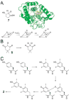
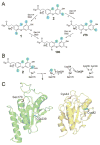
Figure 2. Heme-dependent haloperoxidases in biological halogenation reactions
(A) Structure (PDB: 2CPO) and mechanism of CPO that participates in the biosynthesis of 7. For the sake of simplicity, throughout the review, crystal structures of only the monomeric units of multimeric enzymes are shown. (B) Halogenation of 8 leads to loss in absorption at 277 nm. The MCD-assay has been widely used to monitor formation of freely diffusible hypohalite by halogenases. (C) Diiodination of two tyrosyl side chains of the thyroglobulin protein, followed by oxidative coupling, β-elimination, rearomatization, and proteolytic cleavage leads to the production of 2. Note that halogenation and bi-radical coupling is postulated to be affected within the same enzyme active site.
Figure 3
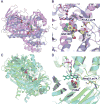
Figure 3. Structural similarity of FDHs with flavin-dependent oxygenases
(A) Overlay of crystal structures of FDH PrnA (PDB: 2AQJ) (in blue) and kynurenine-3-monooxygenase (PDB: 5FN0) (in magenta). The structures were overlaid so as to align the cofactor FAD isoalloxazine rings. FAD is shown in stick-ball representation. Chloride ions bound to PrnA is shown as an orange sphere. L-Trp (bound to 2AQJ) and kynurenine-3-monooxygenasae inhibitor (GSK180, bound to 5FN0) are shown as spheres with carbon atoms colored yellow. (B) Zoomed in view of the active sites, with the PrnA catalytic lysine (vide infra) shown in stick-ball representation with carbon atoms colored yellow. (C) Crystal structure of FDH Bmp2 (PDB: 5BVA) (in green) overlaid with 4-hydroxybenzoate hydroxylase (PDB: 1YKJ) (in cyan). 4-hydroxybenzoate (PHBA, bound to 1YKJ) is shown as spheres with carbon atoms colored yellow. (D) Zoomed in view of the active sites, with the Bmp2 catalytic lysine (vide infra) shown in stick-ball representation with carbon atoms colored yellow.
Figure 4
Figure 4. Reaction mechanism for FDHs
(A) The reaction scheme for FDHs and flavin-dependent oxygenases is identical until the formation of Fl-C4a-OOH. FDHs resolve this intermediate by the displacement of the distal oxygen atom by the halide anion, from where the hypohalite is captured by the catalytic lysine side chain by the formation of a haloamine intermediate, finally affecting an electrophilic aromatic substitution on an electron-rich substrate. (B) The relative positioning of the flavin cofactor isoalloxazine ring and the FDH catalytic lysine side chain as identified in the recently described crystal structure of the FDH Bmp2 (PDB: 5BVA). In the Bmp2 crystal structure, an ethylene glycol (EG) molecule was found in the vicinity of the cofactor proximal to the two tryptophan amino acids that are conserved in all FDH sequences. The dashed line represents the distance of 2.7 Å between the isoalloxazine N5 and one of the EG oxygen atoms. The EG binding site likely represents the region where Flred engages molecular oxygen, finally leading to the formation of the hypohalite that is then transferred to the lysine side chain.
Figure 5
Figure 5. Resemblance of bromination activity of single-component halogenase Bmp5 to single-component monooxygenases
(A) The two-step conversion of 4-hydroxybenzoate to 9 catalyzed by Bmp5. (B) Hydroxylation and (C) decarboxylative-hydroxylation of 4-hydroxybenzoate catalyzed by single-component monooxygenases. Aryl-rings bearing the halogen adducts are highlighted in blue in this section.
Figure 6
Figure 6. Regiospecific tryptophan halogenases and their participation in natural product biosynthesis
The halogenated indole rings are shaded blue. Note that the chlorination on the pyrrole ring of 10 is catalyzed not by PrnA but by PrnC, a second FDH encoded with the pyrrolnitrin biosynthetic gene cluster.
Figure 7
Figure 7
Structures of eukaryotic halogenated natural products biosynthesized by the action of FDHs.
Figure 8
Figure 8. Halogenation of acyl-S-CP substrates
(A) Post-translational acylation of apo-CP with CoA-derived phosphopantetheine generates holo-ACP that then undergoes thioesterification with L-proline and oxidation of the prolyl heterocycle to generate pyrrolyl-S-CP. The action of six different pyrrolyl-S-CP FDHs with corresponding natural product structures is shown. Note that the fourth bromine atom in 24 is also installed by Bmp2, while the dibromophenol moiety of 29 is generated by aforementioned single-component FDH Bmp5. Note that 30 is not derived from the action of Bmp2 and Bmp5. (B) Representative L-tyrosine derived halogenated natural products. Chlorination of β-hydroxytyrosine by SgcC3 is shown. The timing of halogenation for other natural products shown here has not been experimentally determined.
Figure 9
Figure 9. Proposed CP binding site in acyl-S-CP halogenases
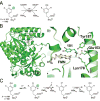
Active site of L-tryptophan chlorinase PrnA and pyrrolyl-S-CP halogenases Mpy16 and Bmp2 demonstrating conserved positioning of the FAD cofactor and the postulated haloamine bearing Lys side chain. However, the catalytic base in PrnA- Glu346, is replaced with Phe323 and Phe300 in Mpy16 and Bmp2, respectively. The product, 7-chlorotryptophan is also shown in the PrnA structure to identify the substrate recruitment site.
Figure 10
Figure 10. Proposed CP binding site in acyl-S-CP halogenases
Semi-transparent electrostatic surface representation of pyrrolyl-S-CP halogenases PltA (PDB: 5DBJ), Bmp2 (PDB: 5BVA) and Mpy16 (PDB: 5BUK) showing conserved positioning of a positively charged (in blue) concave cavity in the vicinity of the FAD cofactor (in stick-ball representation, carbon atoms colored yellow). In Mpy16, a chain of water molecules (in red) starting at the positively charged cavity traverses the FAD binding site terminating at the halogenation active site defined by the catalytic lysine side chain.
Figure 11
Figure 11. Incorporation of halogenated tryptophan in NRPS-derived peptide natural products
(A) 6-chlorotryptophan, generated by FDH Tar14 is converted to 4-chlorokyunerine. Both amino acids are incorporated in 36. (B) FDHs KtzQ and KtzR generate 6,7-dichlorotryptophan that is incorporated as a pyrroloindoline in the kutznerides family of natural products, such as 38. (C) The natural product 39 also bears the chlorinated pyrroloindoline motif that is generated via an as yet unidentified enzymatic route. (D) The FDH CmdE encoded in the biosynthetic gene cluster for the production of 45 bears homology to acyl-S-CP utilizing FDHs rather than Tar14, KtzQ, and KtzR (vide infra). It is thus likely that the mechanism for incorporation of the 2-chlorotryptophan moiety in 45, and in closely related jasplakindolide (46), might differ from that for 36 and 38.
Figure 12
Figure 12. Halogenated NRPS derived glycopeptide antibiotics
A CP loaded hexa-peptide was used as a substrate for the in vitro reconstitution of the activity for the FDH VhaA. The VhaA reaction product would require further NRPS elongation by the 3,5-dihydroxyphenylglycine amino acid, oxidative coupling of the aryl rings, glycosylation, and offloading from the NRPS assembly line for maturation into 3. The order or amino acid addition during the NRPS assembly is denoted by numerals for 3 and halogenated aryl rings are shaded blue.
Figure 13
Figure 13. Chemical structure of chlorinated natural products 47–50
The halogenated rings are shaded in blue.
Figure 14
Figure 14. Chemical structure of the lanthipeptide antibiotic 51
The halogenated indole ring is shaded in blue.
Figure 15
Figure 15. Neighbor-joining tree showing the relatedness of FDHs
Phylogenetic analysis was performed using Mega. The scale bar indicates 0.2 changes per amino acid. Full length primary sequences of select FDHs that have been biochemically characterized, or identified within genetic context of natural product biosynthetic gene clusters are included as discussed in the text. The FDH Amm3 is implicated in the biosynthesis of the ammosamide alkaloids.
Figure 16
Figure 16. Overall mechanism and vanadate coordination in V-HPOs
(A) Proposed catalytic scheme for generating the halogenating agent X+ in V-HPOs (adapted from Winter and Moore, 2009). In the first step of catalysis, hydrogen peroxide is coordinated to the vanadium center in a side-on manner. The peroxide bond is broken through a nucleophilic attack on the partially positive oxygen by a halide resulting in the release of the hypohalite. If the appropriate substrate (R) is present, a halogenated compound will be formed with the loss of water and regeneration of vanadate. If the appropriate substrate is not present, the hypohalite can react with hydrogen peroxide to regenerate the halide and form dioxygen in the singlet state. (B) Active site of native V-CPO from Curvularia inaequalis (PDB accession number 1IDQ). Active site residues are shown as yellow ball and sticks and vanadate is shown coordinated to the conserved His496 residue.
Figure 17
Figure 17
V-BPO-catalyzed cyclization of 52 to form the brominated sesquiterpene marine natural products. Co-VBPO: C. officinalis V-BPO.
Figure 18
Figure 18. V-BPO-catalyzed chemoenzymatic synthesis of brominated acetogenins
(A) Bromolactonization of 61 to 62 using the L. nipponica V-BPO (Ln-VBPO). (B) Bromolactonization of 63 using partially purified Ln-VBPO. (C) Conversion of 65 to 59 and 66 using a partially purified Ln-VBPO.
Figure 19
Figure 19. V-BPO-catalyzed chemoenzymatic synthesis of brominated furanones
(A) Bromination of acylhomoserine lactone 67 to 68 was demonstrated using whole pieces of D. pulchra. (B) Bromolactonization of 69 to 70. (C) Bromofuranones isolated from D. pulchra.
Figure 20
Figure 20
Chloronium-mediated cyclization of the napyradiomycin intermediate 71 to 72 catalyzed by the bacterial V-CPO NapH1. Tetrahydroxynaphthalene is the PKS-derived building block and is assembled by a type III PKS encoded within the biosynthetic gene cluster.
Figure 21
Figure 21. Chloronium-mediated cyclization of the merochlorin meroterpenoids
Mcl40 and Mcl24 are characterized V-CPOs from the merochlorin biosynthetic cluster, whereas Mcl23 is a characterized prenyltransferase that attaches the isosesquilavandulyl terpenoid moiety onto the tetrahydroxynapthalene-derived polyketide building block.
Figure 22
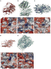
Figure 22. Comparison of V-HPO structures
(A) V-CPO from C. inaequalis (PDB: 1IDQ); (B) V-CPO from S. sp. CNQ-525 (PDB: 3W36); (C) V-BPO from A. nodosum (PDB: 1QI9); (D) V-IPO from Z. galactanivorans (PDB: 4USZ); and (E) V-BPO from Corallina officinalis (PDB: 1QHB). (Top panel): Ribbon structures with active site residues highlighted in yellow. (Bottom panel): Electrostatic surface representation showing a cross section of the cavity leading to the active site with bound vanadate or phosphate. Active site side chains, vanadate and phosphate are shown in ball and stick representation.
Figure 23
Figure 23. Neighbor-joining tree showing the relatedness of V-BPOs, V-CPOs, V-IPOs and acid phosphatases identified from fungi, algae and bacteria
Phylogenetic analysis was performed using Mega. The scale bar indicates 0.2 changes per amino acid. Sequences in which crystal structures are available are in bold. Sequence identification codes include Ci_VCPO from Curvularia inaequalis (GenBank accession number CAA59686); Rb_VCPO from Rhodopirellula baltica SH1 (CAD72609); Ed_VCPO from Embellisia didymospora (CAA72622); NapH1 VCPO from Streptomyces sp. CNQ-525 (ABS50486); NapH3 VCPO from Streptomyces sp. CNQ-525 (ABS50491); NapH4 VCPO from Streptomyces sp. CNQ-525 (ABS50492); Mcl40 VCPO from S. sp. CNH-189 (469658154); Mcl24 V-CPO from S. sp. CNH-189 (469658138); Cs_VCPO from Cellulophaga sp. MED134 (ZP_01050453); Ss1_VCPO1 from Streptomyces sp. CNQ-766 (WP_018841016); Ss2 VCPO from Streptomyces sp. CNQ-766 (WP_018836294); Ss3 VCPO from Streptomyces sp. CNQ-329(WP_027774460); Ss4_VCPO from Streptomyces sp. CNT-371 (WP_027745869); Ss5_VCPO from Streptomyces sp. CNQ-865 (WP_027769314); Ss6_VCPO from Streptomyces sp. CNT-371 (WP_027744001); Ss7_VCPO from Streptomyces sp. CNQ-509 (WP_027745869); Ss8_VCPO from Streptomyces sp. SBT349 (WP_049575625); Ss9_VCPO from Streptomyces sp. CNQ-509 (WP_047020372); Ss10_VCPO from Streptomyces sp. CNS-335 (WP_018842950); Ss11_VCPO from Streptomyces sp. MMG1121 (WP_053666624); Sac_VCPO from Saccharomonospora saliphila (WP_019819575); Am_VCPO from Actinopolyspora mortivallis (WP_019852972); An_VBPO from Ascophyllum nodosum (P81701); Co_VBPO from Corallina officinalis (AAM46061); Cp1_VBPO from Corallina pilulifera (BAA31261); Cp2_VBPO from Corallina pilulifera (BAA31262); Sys_VBPO from Synechococcus sp. CC9311 (YP_731869); Fd_VBPO from Fucus distichus (AAC35279); Ld1_VBPO from Laminaria digitata (CAD37191); Ld2_VBPO from Laminaria digitata (CAD37192); Ld1_VIPO from Laminaria digitata (CAF04025); Ld3_VIPO from Laminaria digitata (CAQ51446); Am1_VIPO from Algoriphagus marincola HL-49 (KPQ20079); Am2_VIPO from Algoriphagus marincola HL-49 (KPQ13775); Zg1_VIPO from Zobellia galactanivorans (4USZ); Zg2_VIPO from Zobellia galactanivorans (CAZ96246); Pi_ACP acid phosphatase from Prevotella intermedia, phoC (AB017537); Kp_ACP acid phosphatase from Klebsiella pneumoniae, phoC (AJ250377); St_ACP acid phosphatase from Salmonella typhimurium, phoN (X63599); Ps_ACP acid phosphatase from Providencia stuartii¸ phoN (X64820); and Eb_ACP acid phosphatase from Escherichia blattae (AB020481).
Figure 24
Figure 24. Natural products bearing halogen atoms on non-activated aliphatic carbon centers
Representative natural products shown here are biosynthesized via the action of NHFe halogenases as described in the text.
Figure 25
Figure 25. Halogenation assisted biosynthesis of cyclopropane rings and vinyl chlorides in natural products
(A) Structures of 86 and 87. (B) The biosynthetic origin of both the vinyl chloride (Jam, 88 and 89) and cyclopropane group (Cur, 87) arises from the initial action of an NHFe halogenase (Hal). A conserved dehydratase domain (ECH1, 94% sequence identity between Cur/Jam) is responsible for an identical dehydration reaction, however, the pathways then deviate through differing activities of the respective decarboxylase domains (ECH2). In jamaicamide biosynthesis, this results in the vinyl chloride moiety present in both 89 and 90. In the biosynthesis of 88, the product of the ECH2 domain is further modified by a novel NADPH-dependent enoyl reductase (ER) forming the cyclopropyl moiety.
Figure 26
Figure 26
Alkaloids with chlorine atoms installed via the action of NHFe halogenases.
Figure 27
Figure 27. Structural conservation between NHFe halogenases and hydroxylases
Crystal structures of NHFe halogenases (A) SyrB2 (PBD: 2FCT), (B) CytC3 (PBD: 3GJB), (C) Cur Hal (PBD: 3NNF), (D) WelO5 (PBD: 5IQT), and (E) the NHFe hydroxylase TauD (PDB: 1GY9) are shown in cartoon representation with Fe(II) as orange spheres and α-ketoglutarate in stick-ball representation with carbon atoms colored blue. Note the conservation of the anti-parallel β-sandwich motif (in grey) that binds the Fe(II) ion and α-ketoglutarate. For the sake of simplicity, active site comparisons with bound substrates and halide ions are shown separately in Figure 28B–D.
Figure 28
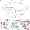
Figure 28. Reaction mechanism and active site comparisons of NHFe-dependent enzymes
(A) Scheme of the general reaction mechanism for both NHFe halogenases and hydroxylases. Comparison of the active sites of (B) TauD, (C) Cur Hal, and (D) WelO5, with substrate-bound, where available. The conserved 2 His, 1 carboxylate coordination of Fe (orange sphere) observed in the NHFe hydroxylase TauD is analogous to a 2His, 1 halide (green sphere) arrangement, wherein the halide replaces the carboxylate in the same coordination position. The substrate is bound in an orientation that is perpendicular to the plane in which the halide resides. The side chains of the active site amino acids are shown in stick-ball representation. Taurine (panel B), 12-epi-fischerindole U (panel D), and α-ketoglutarate (panel B-D) are shown in stick-ball representation with the carbon atoms colored yellow.
Figure 29
Figure 29. Reaction catalyzed by nucleophilic, SAM-dependent halogenases of the 5′-halo-5′-deoxyadenosine synthase class
Fluorinase FlA catalyzes the first committed step in the biosynthesis of 94 and 95 in the soil bacterium Streptomyces cattleya. Analogously, chlorinase SalL catalyzes the first committed step towards chloroethylmalonyl-CoA, a precursor to 96.
Figure 30
Figure 30. Crystal structures of 5′-halo-5′-deoxyadenosine synthases
(A) Overview structure of fluorinase FlA (PDB: 1RQR) displaying monomers colored in pink, green and cyan (top view). Note that the active site lies in the interface between monomers, with three active sites per homotrimer (ligands 5′-FDA and L-met shown as spheres and colored by element). The fluorinase has been shown to be a hexamer in solution (dimer of trimers), according to gel filtration, whereas the chlorinase is a trimer in solution according to ultracentrifugation studies. (B) Fluorinase active site with bound SAM (PDB: 1RQP); chains are colored the same way as in panel A, with the ligand colored by element and hydrogen bonds with key enzyme residues shown as yellow dashes. (C) Chlorinase SalL Y70T mutant (PDB: 2Q6O) active site with bound substrates SAM (colored by element) and chloride (green sphere). (D) Chlorinase SalL Y70T mutant active site showing a view through the chloride ion. Note that the chloride makes a hydrogen bond with the amide backbone of Gly131. The water molecule shown would be displaced by the Tyr70 residue in the wild-type enzyme. (E) Overview of one monomer each of fluorinase FlA (PDB: 1RQR, pink) and chlorinase SalL G131S Y70T double mutant (PDB: 2Q6L, wheat) highlighting a 23-residue loop in FlA (magenta, position 93 to 115) which is absent in SalL (yellow, position 87 to 90) and sits just above the active site. (F) Close up view of the active sites of panel E. Overlay of the active sites of fluorinase FlA (PDB: 1RQR, pink) and chlorinase SalL G131S Y70T double mutant (PDB: 2Q6L, wheat) with bound products 5′-FDA and 5′-ClDA, respectively, highlighting the displacement of the chlorinase loop carrying Ser131 away from the product when compared to fluorinase.
Figure 31
Figure 31. SAM hydroxide adenosyltransferases
(A) Overview structure of DUF-62 PH0463 from archaeon Pyrococcus horikoshii (PDB: 1WU8) showing the same fold as 5′-halo-5′-deoxyadenosine synthases. The three monomers of the homotrimer are colored magenta, green and cyan. (B) Previously proposed mechanism for water activation involving a conserved Asp-Arg-His triad not found in 5′-halo-5′-deoxyadenosine synthases.
Figure 32
Figure 32. SAM-dependent halide methyltransferase
(A) Reaction catalyzed by SAM-dependent halide methyltransferases. (B) Overview structure of SAM-dependent halide methyltransferase from A. thaliana (PDB: 3LCC) with SAH ligand shown as spheres. (C) Halide methyltransferase active site. Waters coordinating to Tyr172 are shown as red spheres; SAH is shown with carbon atoms colored yellow.
Figure 33
Figure 33. Selenoprotein iodothyronine deiodinase structure-mechanism
(A) Transformations catalyzed by Dio1–3. (B) Peroxiredoxin-like mechanism proposed for dehalogenation of 2 with residues corresponding to mouse Dio3 in panel ‘C’. Direct attack of the leaving iodine by the selenolate of Sec170 leads to formation of a selenyl iodide intermediate. The selenyl-iodine bond is broken by the addition of the thiolate from the Cys239 side chain to form an endogenous selenyl-sulfide bond in line with the mechanism of 2-Cys peroxiredoxins. The selenyl-sulfide bond is subsequently reduced by attack of the thiolate side chain of a third cysteine residue (Cys168) leading to formation of an endogenous disulfide bridge that is then reduced by a small protein thioredoxin to complete the catalytic cycle. (C) Crystal structure of the mouse Dio3cat (PDB: 4TR3) in green, and 2-Cys peroxiredoxin PtGPx5 (PDB: 2P5Q) in yellow showing the side chains of the catalytic selenocysteine and cysteine residues. Note that the selenocysteine (Sec170) in Dio3cat is mutated to a cysteine in the crystal structure.
Figure 34
Figure 34. Structure-function of IYD
(A) Net reaction catalyzed by IYD. (B) mIYD (PDB: 3GFD) co-crystal structure with the substrate 101 (left), and close-up of active site (right) showing FMN cofactor (yellow) and 101 (blue). The substrate 101 bridges active site residues of mIYD with the isoalloxazine ring of the FMN cofactor. (C) Single electron transfer mechanism proposed for deiodination of iodotyrosines beginning with keto-enol tautomerization followed by a single electron transfer from FMN hydroquinone (hq) to release iodide and form phenoxyl radical. The phenoxyl radical is then reduced by a second single electron transfer from the FMN semiquinone (sq) to afford the deiodinated tyrosine.
Figure 35
Figure 35. Dehalogenation in natural products biosynthesis
(A) The oxidative coupling reaction in the final step in the biosynthesis of 29 necessitates the dehalogenation of the highly brominated intermediate 24 to 103 catalyzed by the dehalogenase Bmp8. (B) Scheme depicting the role of the active site CXXC Cys residues in the Bmp8 reaction mechanism. The attacking Cys residue is denoted ‘CysA’, while the resolving Cys residue denoted ‘CysR’. (C) Proposed role for a Bmp8-like dehalogenase in the biosynthesis of 104.
Figure 36
Figure 36. Structure-mechanism B12-dependent RDHs
(A) Dehalogenation reactions catalyzed by PceA (top) and NpRdhA (bottom). (B) Overall crystal structures of PceA (PDB: 4UR0) in magenta and NpRdhA (PDB: 4RAS) in green. (C) Active sites of PceA and NpRdhA showing the B12 cofactor in yellow and the Fe-S clusters. In the PceA structure, two alternate conformations of 106 are modeled above the Co, while the similar site in NpRdhA is occupied by a chloride ion shown as a grey sphere. (D) Scheme showing hypothesized mechanistic routes for dehalogenation by B12-dependent RDHs illustrated with NpRdhA substrate 107.
Figure 37
Figure 37. Bacterial degradation of 109
(A) Bacterial pathway for degradation of 109. (B) Mechanism for dehalogenation of 109 by GST-like PcpC. Keto-enol tautomerization is followed by conjugation to glutathione (GS). Attack by an active site cysteine thiolate forms a protein-glutathione thioester and the dehalogenated product. The catalytic cycle is completed by the resolution of the protein-glutathione thioester by an exogenous glutathione thiolate.
Figure 38
Figure 38. Structure-mechanism for HDHs with a catalytic triad
(A) Proposed reaction mechanism for haloalkane/haloacid HDHs. (B) Overall structure (left) and active site (right) for a recently reported structure of a haloalkane HDH from a marine Rhodobacteraceae sp (PDB: 4C6H). The catalytic Asp residue is shown with the carbon atoms colored cyan. The His and Glu residue comprising of the catalytic triad are shown with carbon atoms colored green. The in situ generated product from the substrate 1-bromohexane used in the crystallization trials, 1-hexanol, is shown bound to the active site in two alternate conformations in yellow. A chloride ion bound in a hydrophobic pocket in the vicinity of the product is shown as a grey sphere and likely denotes the binding site for the substrate halogen atom. (C) Proposed reaction mechanism for 4-chlorobenzoyl-CoA HDH.
Figure 39
Figure 39. Structure-mechanism for SDR-like HDHs
(A) Proposed reaction mechanism for halohydrin dehalogenases and recruitment of a cyanide anion to resolve the dehalogenated epoxide product to generate β-hydroxynitriles. (B) Structural similarity of between halohydrin HDHs (in green, PDB: 1PWZ) and lanthipeptide oxidoreductase SDR ElxO (in brown, PDB: 4QEC). NAD+ bound to ElxO is shown in yellow sticks. (C) Active site of the halohydrin HDH HheC with an epoxide product bound to the active site (in yellow), and the side chains of the catalytic Tyr (in cyan), and the Ser and Arg (in green) residues. A chloride ion bound in the vicinity of the product is shown as a grey sphere.

References

    1. Gribble GW. Naturally occurring organohalogen compounds–a comprehensive survey. Fortschr Chem Org Naturst. 1996;68:1–423. - PubMed
    1. Gribble GW. Naturally Occurring Organohalogen Compounds – A Comprehensive Update. Springer; Vienna: 2010.
    1. Gribble Gw. The natural production of organobromine compounds. Environ Sci Pollut Res Int. 2000;7:37–47. - PubMed
    1. Emerson S, Hedges J. Chemical Oceanography and the Marine Carbon Cycle. 2008
    1. Lohman DC, Edwards DR, Wolfenden R. Catalysis by desolvation: the catalytic prowess of SAM-dependent halide-alkylating enzymes. J Am Chem Soc. 2013;135:14473–14475. - PubMed

LinkOut - more resources