. 2017 Jan 20;12(1):e0163614.
doi: 10.1371/journal.pone.0163614. eCollection 2017.

Syk Regulates Neutrophilic Airway Hyper-Responsiveness in a Chronic Mouse Model of Allergic Airways Inflammation

Affiliations

Syk Regulates Neutrophilic Airway Hyper-Responsiveness in a Chronic Mouse Model of Allergic Airways Inflammation

Sepehr Salehi et al. PLoS One. .

Abstract

Background: Asthma is a chronic inflammatory disease characterized by airways hyper-responsiveness (AHR), reversible airway obstruction, and airway inflammation and remodeling. We previously showed that Syk modulates methacholine-induced airways contractility in naïve mice and in mice with allergic airways inflammation. We hypothesize that Syk plays a role in the pathogenesis of AHR; this was evaluated in a chronic 8-week mouse model of house dust mite (HDM)-induced allergic airways inflammation.

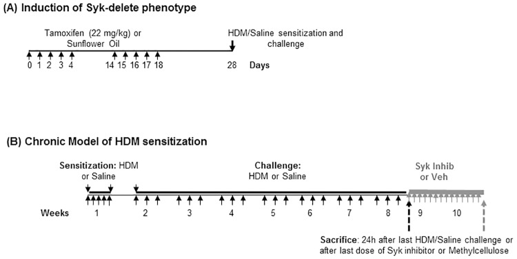
Methods: We used the Sykflox/flox//rosa26CreERT2 conditional Syk knock-out mice to assess the role of Syk prior to HDM exposure, and treated HDM-sensitized mice with the Syk inhibitor, GSK143, to evaluate its role in established allergic airways inflammation. Respiratory mechanics and methacholine (MCh)-responsiveness were assessed using the flexiVent® system. Lungs underwent bronchoalveolar lavage to isolate inflammatory cells or were frozen for determination of gene expression in tissues.

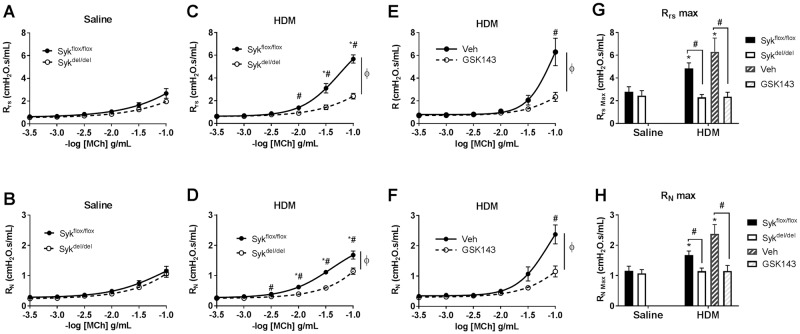
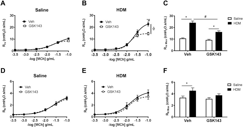
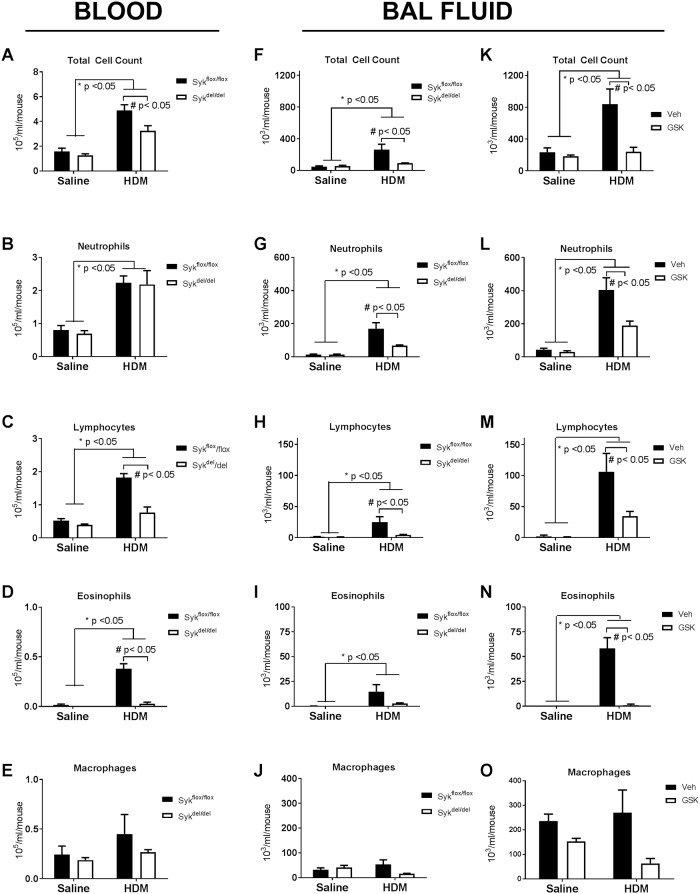
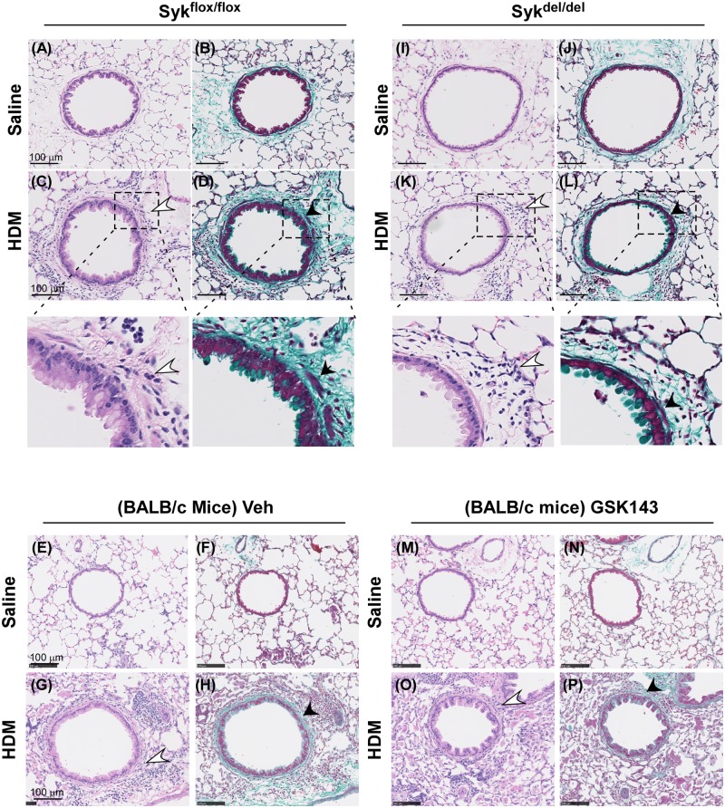
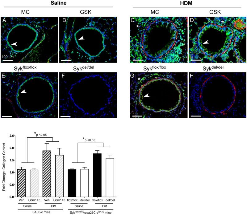
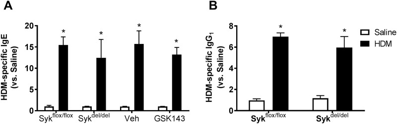
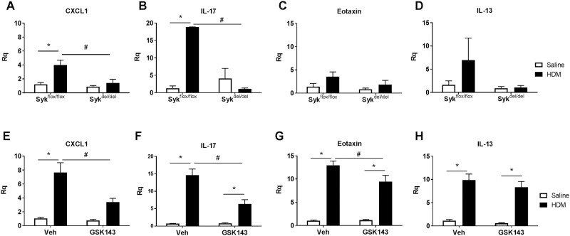
Results: MCh-induced AHR was observed following HDM sensitization in the Syk-intact (Sykflox/flox) and vehicle-treated BALB/c mice. MCh responsiveness was reduced to control levels in HDM-sensitized Sykdel/del mice and in BALB/c and Sykflox/flox mice treated with GSK143. Both Sykdel/del and GSK143-treated mice mounted appropriate immune responses to HDM, with HDM-specific IgE levels that were comparable to Sykflox/flox and vehicle-treated BALB/c mice. HDM-induced increases in bronchoalveolar lavage cell counts were attenuated in both Sykdel/del and GSK143-treated mice, due primarily to decreased neutrophil recruitment. Gene expression analysis of lung tissues revealed that HDM-induced expression of IL-17 and CXCL-1 was significantly attenuated in both Sykdel/del and GSK143-treated mice.

Conclusion: Syk inhibitors may play a role in the management of neutrophilic asthma.


Conflict of interest statement

The authors have declared that no competing interests exist.

Figures

Fig 1
Fig 1. Time-lines of tamoxifen-induced knock-out of Syk, chronic HDM exposure models of allergic airways inflammation and treatment with Syk inhibitor.
(A) Induction of the Syk knock-out in the Sykflox/flox//rosa26CreERT2 mice. Tamoxifen (Tam; 22 mg/kg) was administered by oral gavage. Control mice were gavaged with sunflower oil (SFo) as a vehicle control. (B) Timelines of the chronic HDM-exposure models of allergic airways inflammation and treatment with Syk inhibitor. In the Sykflox/flox//rosa26CreERT2 mice, HDM sensitization and challenge was initiated 10 days after the last treatment with tamoxifen or sunflower oil. Assessment of physiological endpoints occurred 24 h after the last dose of tamoxifen or sunflower oil. BALB/c and Sykflox/flox mice were exposed to HDM for 8 weeks, and then treated daily for 2 weeks with GSK143 (30 mg/kg) by oral gavage or 1% methylcellulose (Veh) control, prior to assessment of the physiological responses 24 h later.
Fig 2
Fig 2. Deletion of Syk prior to HDM and inhibition of Syk activity after development of chronic allergic airways inflammation abrogates airways hyperresponsiveness to methacholine in Sykflox/flox//rosa26CreERT2 mice.
In the Syk-expressing (Sykflox/flox) and Sykflox/flox mice treated with methylcellulose control (MC: panels A-F), the chronic (8-week) HDM sensitization and challenge protocol increased MCh responsiveness of the total respiratory resistance (Rrs, A, C, E) and central airways Newtonian resistance (RN, B, D, F) in a dose-dependent manner compared with the Saline-exposed group (*, p<0.05, n = 8-10/group). However, HDM failed to augment MCh responsiveness in the Syk-deleted state (Sykdel/del; C,D; open circles) or in Sykflox/flox mice treated with GSK143 (E, F; open circles). These responses were most obvious at the maximum Rrs and RN (G,H, *p<0.05 HDM vs. Saline, same genotype; #p<0.05, Sykflox/flox vs. Sykdel/del and Veh vs. GSK143; same exposure group, n = 8-10/group).
Fig 3
Fig 3. Daily treatment with GSK143 for 2 weeks following chronic HDM sensitization abrogates airways hyperresponsiveness to methacholine in BALB/c mice.
BALB/c mice underwent an 8-week HDM sensitization and challenge protocol followed by 2 weeks of treatment with GSK143 or Veh control. (A-C) HDM exposure increased MCh responsiveness of the total respiratory resistance (Rrs) compared with the Saline-exposed mice treated with MC; this was most evident at the maximal Rrs. HDM mice subsequently treated with GSK143 exhibited increased MCh responsiveness when compared naïve Saline-exposed mice; however, this response was significantly attenuated compared with HDM mice treated with MC. ϕ, p<0.05 dose response curve; *p<0.05 HDM vs. Saline, same treatment group; #p<0.05 GSK143 vs. Veh; same exposure group, n = 11-14/group. (D-F) HDM increased RN in the Veh treated group, but no differences between the VEH and GSK143 mice groups were observed. *p<0.05 HDM vs. Saline, same treatment group, n = 11-14/group.
Fig 4
Fig 4. Effect of loss of Syk activity on peripheral blood and bronchial alveolar lavage fluid total and differential leukocyte counts under basal conditions and following 8 weeks of HDM sensitization and challenge.
A-E: At 8 weeks after treatment with tamoxifen, as described in Fig 1, no significant differences in the total or differential blood leukocyte counts were observed between Sykflox/flox and Sykdel/del mice in the Saline groups. Furthermore, 8 weeks of HDM sensitization and challenge resulted in significant peripheral leukocytosis in both groups of mice, with Syk deletion impairing increases in the peripheral lymphocyte and eosinophil counts (*p<0.05, Saline vs. HDM; #p<0.05, Sykflox/flox vs. Sykdel/del, n = 4/group). F-J: HDM significantly increased BAL fluid total cell, neutrophil, lymphocyte and eosinophil cell counts compared with Saline mice (*p<0.05, 2-way ANOVA, n = 5-6/group); within the HDM mice, deletion of Syk significantly impaired recruitment of neutrophils and lymphocytes (#p<0.05, Sykflox/flox vs. Sykdel/del, n = 5-6/group). K-O: BALB/c mice exhibited a more pronounced BAL fluid leukocytosis following HDM exposure when compared with Sykflox/flox//rosa26CreERT2 mice; however, the pattern of response was similar. HDM lead to increases in the total BAL fluid cell counts, with significant increases in neutrophils, lymphocytes and eosinophils in the Veh control group, a reponse that was attenuated by treatment with GSK143 (*p<0.05, Saline vs. HDM, 2-way ANOVA; # p<0.05, Veh vs. GSK143, n = 5-6/group).
Fig 5
Fig 5. Loss of Syk activity does not abolish airway recruitment of inflammatory cell in the chronic HDM-challenge model.
A-H: Hematoxylin and eosin staining of lung sections revealed inflammatory cell peribronchial infiltrates (open arrows; C and inset, G) while Trichome Masson staining showed increased epithelial and basement membrane thickening and smooth muscle cell hypertrophy (closed arrows, D and inset, H; closed arrows) in the airways of HDM compared with Saline-exposed mice in the Sykflox/flox group and BALB/c mice treated with Veh control. Within the HDM groups of mice, the Sykdel/del (K,L) and BALB/c mice treated with GSK143 (O,P) did not exhibit significant differences in the inflammatory infiltrate, basement membrane thickening nor smooth muscle cell hypertrophy when compared with their respective Sykflox/flox (C,D) and MC-treated controls. (G.H). No differences were noted amongst the 4 Saline-exposed groups (A,B, I,J, E,F, M,N). Images are representative of 4 mice/group. Bar represents 100 μm.
Fig 6
Fig 6. Loss of Syk activity does not attenuate airway inflammation and remodeling or total lung collagen content in the chronic HDM-challenge model.
Immunostaining for Syk (green) revealed expression in airway epithelia (arrow) and leukocytes (*) under basal Saline conditions and following HDM exposure in all groups of mice (A-E, G) except the Syk-deficient (Sykdel/del) mice (F, H). Following HDM exposure, increased peribronchial leukocyte recruitment (DAPI staining, blue) was also observed (C,D,G,H), as was an increase in α-smooth muscle actin (stained red); this pattern was observed in the control Veh and Sykflox/flox mice (C,G) as well as the GSK-treated and Syk-deficient Sykdel/del mice (D,H). Images are representative of 4 mice/group. Bar represents 100 μm. I: Quantitative analysis revealed a 2-fold increase in the total lung collagen content in the HDM mice when compared with Saline control in all groups of mice (*p<0.05, HDM vs. Saline, 2 way ANOVA, n = 4/group). No differences were observed between Sykflox/flox and Sykdel/del mice, nor between BALB/c mice treated with Veh or GSK143.
Fig 7
Fig 7. Loss of Syk activity attenuates but does not abolish production of HDM-specific IgE and IgG1 in response to HDM-sensization and challenge.
HMD sensitization and challenge resulted in significant increases in the serum concencrations of HDM-specific IgE (A) and IgG1 (B) compared with Saline control (*p< 0.05, n = 6-9/group). In the HDM-exposed mice, no differences were observed between the Sykdel/del and Sykflox/flox mice nor between the BALB/c mice treated with GSK143 and Veh controls.
Fig 8
Fig 8. Syk regulates expression of CXCL-1 and IL-17 expression in the chronic HDM- model of allergic airways inflammation.
HDM sensitization and challenge for 8 weeks increased expression of CXCL-1 and IL-17, a response that was significantly abrogated in Sykdel/del mice (A,B) and following treatment with GSK143 (E,F). HDM-exposure induced the expression of eotaxin (C,G). Furthermore, a role for Syk in modulating this response was observed in the BALB/c mice treated with GSK143 (G), although no differences were observed between the Sykflox/flox and Sykdel/del mice (C). Increased IL-13 expression following HDM exposure was observed (D,H); however, a role of Syk in regulating this response was not consistently observed in the BALB/c and Sykflox/flox//rosa26CreERT2 mice. *p< 0.05, HDM vs. Saline, n = 5-6/group; # p<0.05, Sykflox/flox vs. Sykdel/del, n = 5-6/group.
Fig 9
Fig 9. Treatment with GSK143 impairs airway contractility to a similar degree as known inhibitors of smooth muscle contraction.
Precision cut lung slices were prepared from BALB/c mice following HDM (solid bars) or Saline exposure (open bars), and the MCh-induced airway contraction was measured. Airways from HDM-exposed mice showed significantly increased airway contraction compared with Saline-mice in the Veh-treated group (*p< 0.05, n = 12 /group), which was abolished by treatment with GSK143 (# p<0.05, n = 12/group). In the HDM mice (solid bars), incubation of the lung slices for 30 mins with inhibitors of PI3 kinase (50 μM), RhoK (1 μM) and MLCK (30 μM) significantly impaired airway contraction compared with slices incubated with DMSO, the diluent control (λ, p<0.05, n = 9-19/group); impairment of airway contraction was similar to that observed in the GSK143-treated airways. Within the DMSO group, HDM-exposed airways showed significantly increased airway contractility compared with Saline-exposed airways (*p<0.05, n = 9-19/group). In the airways of Saline-mice (open bars), inhibitors of PI3 kinase and RhoK significantly impaired airway contraction compared with DMSO-treated airways (ϕ, p<0.05, Saline group, n = 9-19/group), suggesting that these mediators play a role in maintaining normal airway tone.

Similar articles

Cited by

References

    1. Kim HY, DeKruyff RH, Umetsu DT (2010) The many paths to asthma: phenotype shaped by innate and adaptive immunity. Nat Immunol 11: 577–584. 10.1038/ni.1892 - DOI - PMC - PubMed
    1. Murphy DM, O'Byrne PM (2010) Recent advances in the pathophysiology of asthma. Chest 137: 1417–1426. 10.1378/chest.09-1895 - DOI - PubMed
    1. Barnes PJ (2010) New therapies for asthma: is there any progress? Trends Pharmacol Sci 31: 335–343. 10.1016/j.tips.2010.04.009 - DOI - PubMed
    1. Barnes PJ (2006) Novel signal transduction modulators for the treatment of airway diseases. Pharmacol Ther 109: 238–245. 10.1016/j.pharmthera.2005.08.001 - DOI - PubMed
    1. Bousquet J, Jeffery PK, Busse WW, Johnson M, Vignola AM (2000) Asthma. From bronchoconstriction to airways inflammation and remodeling. Am J Respir Crit Care Med 161: 1720–1745. 10.1164/ajrccm.161.5.9903102 - DOI - PubMed

Publication types

LinkOut - more resources