Review
. 2017 May;177(4):509-525.
doi: 10.1111/bjh.14510. Epub 2017 Jan 20.

Recent advances in understanding clonal haematopoiesis in aplastic anaemia

Affiliations
Review

Recent advances in understanding clonal haematopoiesis in aplastic anaemia

Natasha Stanley et al. Br J Haematol. 2017 May.

Abstract

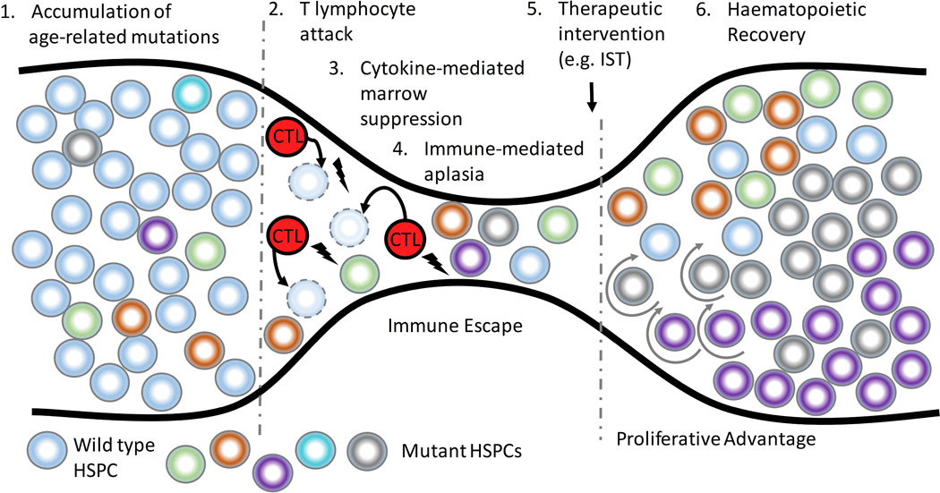
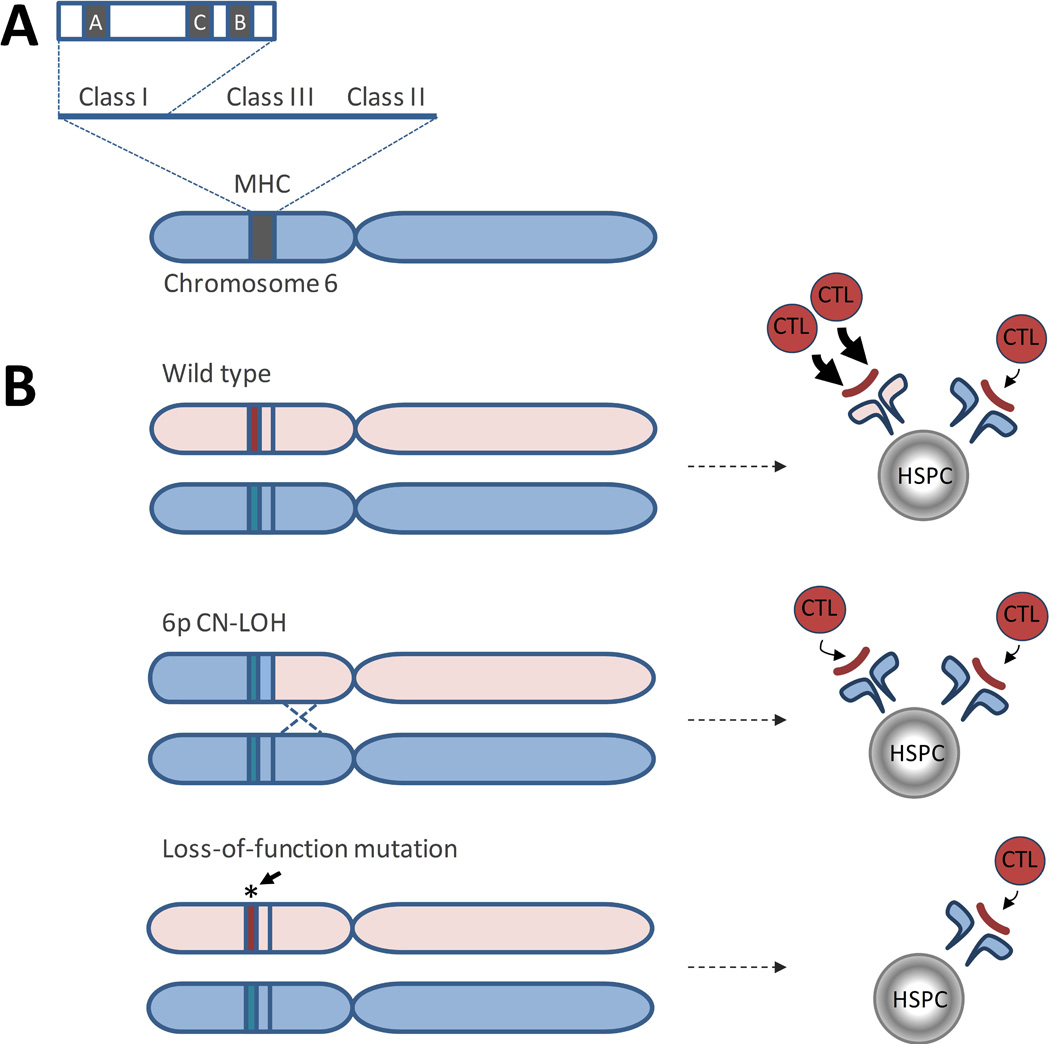
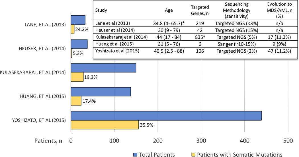
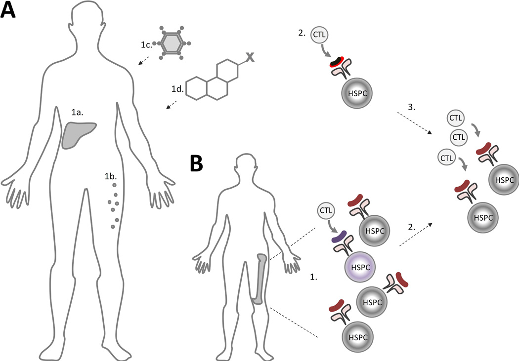
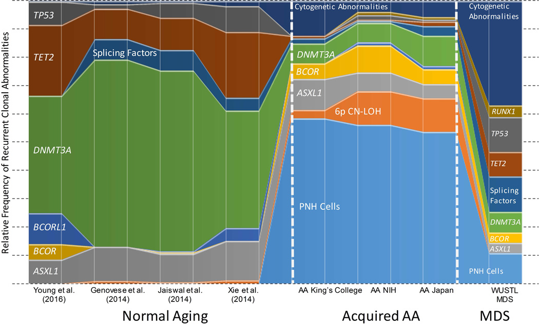
Acquired aplastic anaemia (AA) is an immune-mediated bone marrow failure disorder inextricably linked to clonal haematopoiesis. The majority of AA patients have somatic mutations and/or structural chromosomal abnormalities detected as early as at diagnosis. In contrast to other conditions linked to clonal haematopoiesis, the clonal signature of AA reflects its immune pathophysiology. The most common alterations are clonal expansions of cells lacking glycophosphotidylinositol-anchored proteins, loss of human leucocyte antigen alleles, and mutations in BCOR/BCORL1, ASXL1 and DNMT3A. Here, we present the current knowledge of clonal haematopoiesis in AA as it relates to aging, inherited bone marrow failure, and the grey-zone overlap of AA and myelodysplastic syndrome (MDS). We conclude by discussing the significance of clonal haematopoiesis both for improved diagnosis of AA, as well as for a more precise, personalized approach to prognostication of outcomes and therapy choices.

Keywords: CHIP; MDS; aplastic anaemia; bone marrow failure; clonal haematopoiesis.


Conflict of interest statement

Conflict of interest statement: The authors have no conflicts of interests to disclose.

Figures

Figure 1
Figure 1. Clonal Haematopoiesis in Aplastic anaemia
In Aplastic anaemia (AA), cytotoxic T lymphocyte (CTL)-mediated attack on the haematopoietic stem and progenitor cells (HSPCs) leads to an evolutionary “bottleneck”. Pre-existing age-related genetic mutations (1), depicted as circles of different colors, serve as a substrate for clonal selection. Cells that are either less immunogenic or more resistant to CTL-mediated apoptosis (2) or cytokine-mediated marrow suppression (3) have a relative growth advantage in the setting of autoimmunity, leading to immune escape of mutant HSPCs. During haematopoietic recovery (6), genetic events that increase HSPC replicative potential (depicted by circular arrows) lead to expansion of mutant clones. IST, immunosuppressive therapy.
Figure 2
Figure 2. Loss of Class I Human Leucocyte Antigen (HLA) Alleles in Aplastic anaemia
A) A diagram of the Major Histocompatibility Locus (MHC), located on the short arm of chromosome 6. B) A schematic showing two mechanisms of HLA class I allele loss in AA. In patients whose HLA alleles allow for enhanced cytotoxic T cell (CTL) immune recognition due to an immunogenic features of a particular HLA class I allele, HSPCs that undergo a somatic loss of that allele have a selective advantage compared to wild type cells. Top panel, haematopoietic stem and progenitor cell (HSPC) containing wild type HLA alleles, with differential immunogenicity (shown by arrows of different width) of an autoantigen (red line) presented in the context of two different HLA class I alleles. Middle panel, HLA allele loss due to an acquired 6p copy number-neutral loss of heterozygosity (CN-LOH) results in lessened immune recognition. Lower panel: HLA allele loss due to loss-of-function somatic mutations in HLA class I alleles can lead to decreased immune recognition by abolishing expression of the more immunogenic HLA allele.
Figure 3
Figure 3. Targeted Sequencing Studies of MDS-associated Genes in AA
A histogram summarizing targeted sequencing studies of patients with aplastic anaemia (AA). Individual studies are listed along the Y-axis, with the number of patients included in each study plotted along the X-axis. The blue bar represents the total number of patients in the study; the yellow bar shows the number of patients with identified somatic mutations, with corresponding percentage of patients with mutations stated to the right of each yellow bar. An embedded table depicts salient characteristics of each study. Age, median age (range) at diagnosis in years). Sensitivity, minimal % mutant allele frequency detected in each study. n/a: not available.*, the listed age corresponds to a larger patient cohort; ǂ, Cohort of 57 patients was screened for 835 genes; with a validation cohort of 93 patients screened for a mutations in a smaller subset of genes. AML, acute myeloid leukaemia; MDS, myelodysplastic syndrome; NGS, next generation sequencing.
Figure 4
Figure 4. Model of Autoimmune Pathogenesis of AA and of the AA/MDS Overlap Syndrome
A) In aplastic anaemia (AA), an immune trigger, such as type-negative hepatitis (1a), eosinophilic fasciitis (1b), or an exposure to an infectious agent (1c), drug or toxin (1d) leads to immune recognition of a neoantigen or an aberrantly expressed autoantigen (black and red line) presented in the context of a patient’s human leucocyte antigen (HLA) molecules (2). (3) Immune-mediated bone marrow aplasia is caused by subsequent clonal expansion of cytotoxic T lymphocytes (CTL) with autoreactivity against the normal haematopoietic stem and progenitor cells (HSPCs) due to epitope spread. B) In patients with coexisting AA and myelodysplastic syndrome (MDS), pre-existing MDS-related mutations may lead to presentation of an aberrantly-expressed or somatically modified neoantigen in the MDS HSPC (1) (shown in purple). Unlike the traditional model of AA, where a T-cell mediated immune response is directed at the healthy HSPCs, patients with the overlap AA/MDS have the immune response triggered by an underlying MDS clone with immune-mediated aplasia to the “bystander” wild type HSPCs (2).
Figure 5
Figure 5. Clonal Signature of AA Compared to MDS and Normal Aging
A schematic diagram comparing the relative frequencies of the most common clonal abnormalities found healthy individuals(Genovese, et al 2014, Jaiswal, et al 2014, Xie, et al 2014, Young, et al 2016), in three aplastic anaemia (AA)cohorts--King’s College (Kulasekararaj, et al 2014), National Institutes of Health (NIH) and Japan cohorts(Yoshizato, et al 2015), and in a myelodysplastic syndrome (MDS) cohort (Washington University in St. Louis, WUSTL) (Graubert, et al 2012, Walter, et al 2013). The prevalence of PNH cells in the healthy individuals and patients with MDS is depicted based on the published high-sensitivity PNH flow cytometry data in these populations (Kaiafa, et al 2008, Sutherland, et al 2012). The frequencies of cytogenetic abnormalities for healthy individuals are shown based on population-based studies of mosaic copy number abnormalities (Jacobs, et al 2012, Laurie, et al 2012). The prevalence of 6p copy number-neutral loss of heterozygosity (CN-LOH) in healthy individuals and in MDS is depicted based on a selection of studies (Jacobs, et al 2012, Laurie, et al 2012, Mohamedali, et al 2015).

References

    1. Abkowitz JL, Fialkow PJ, Niebrugge DJ, Raskind WH, Adamson JW. Pancytopenia as a clonal disorder of a multipotent hematopoietic stem cell. The Journal of clinical investigation. 1984;73:258–261. - PMC - PubMed
    1. Abkowitz JL, Catlin SN, McCallie MT, Guttorp P. Evidence that the number of hematopoietic stem cells per animal is conserved in mammals. Blood. 2002;100:2665–2667. - PubMed
    1. Afable MG, 2nd, Wlodarski M, Makishima H, Shaik M, Sekeres MA, Tiu RV, Kalaycio M, O'Keefe CL, Maciejewski JP. SNP array-based karyotyping: differences and similarities between aplastic anemia and hypocellular myelodysplastic syndromes. Blood. 2011;117:6876–6884. - PMC - PubMed
    1. Alter BP, Giri N, Savage SA, Peters JA, Loud JT, Leathwood L, Carr AG, Greene MH, Rosenberg PS. Malignancies and survival patterns in the National Cancer Institute inherited bone marrow failure syndromes cohort study. British journal of haematology. 2010;150:179–188. - PMC - PubMed
    1. Babushok DV, Xie HM, Roth JJ, Perdigones N, Olson TS, Cockroft JD, Gai X, Perin JC, Li Y, Paessler ME, Hakonarson H, Podsakoff GM, Mason PJ, Biegel JA, Bessler M. Single nucleotide polymorphism array analysis of bone marrow failure patients reveals characteristic patterns of genetic changes. British journal of haematology. 2014;164:73–82. - PMC - PubMed

MeSH terms

Substances