. 2017 Jan 19;65(2):260-271.
doi: 10.1016/j.molcel.2016.11.015.

PARP-1 Controls the Adipogenic Transcriptional Program by PARylating C/EBPβ and Modulating Its Transcriptional Activity

Affiliations

PARP-1 Controls the Adipogenic Transcriptional Program by PARylating C/EBPβ and Modulating Its Transcriptional Activity

Xin Luo et al. Mol Cell. .

Abstract

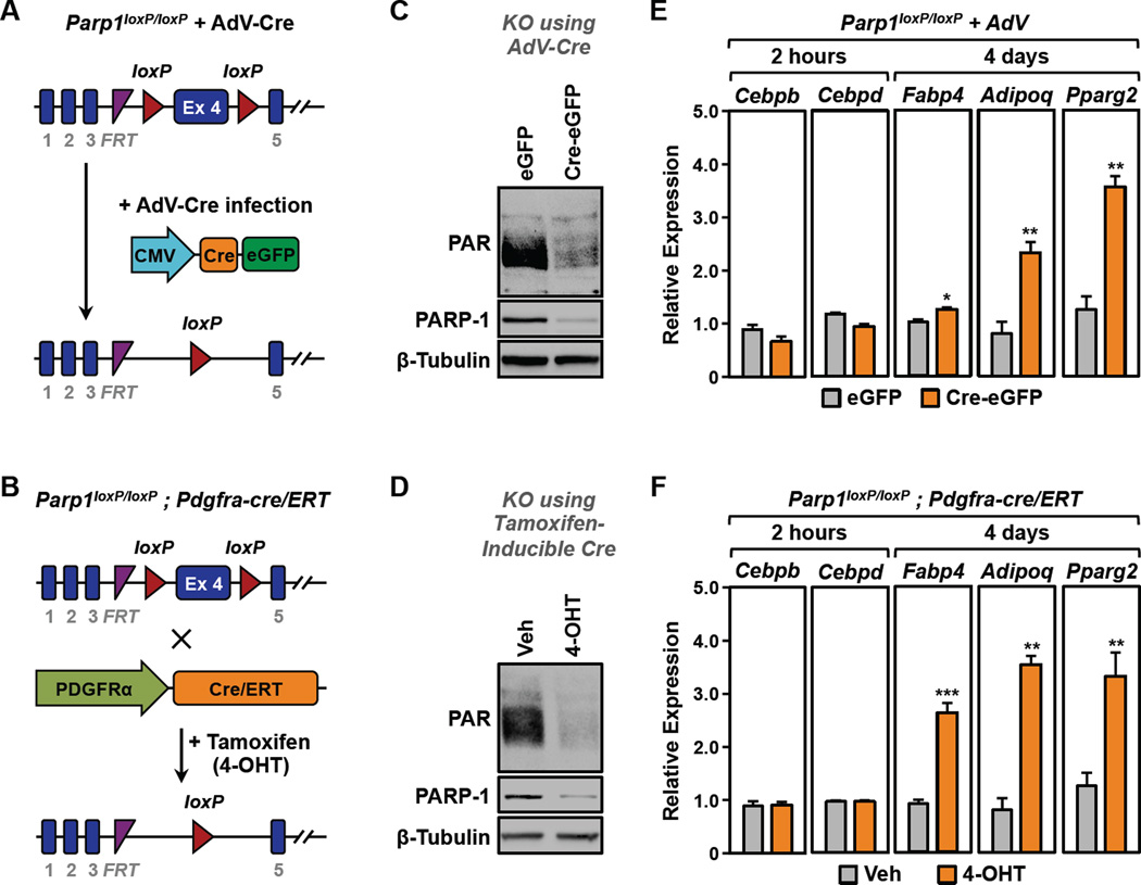
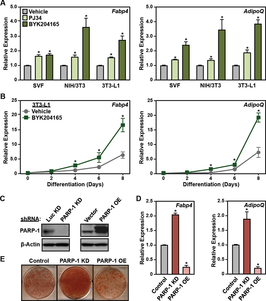
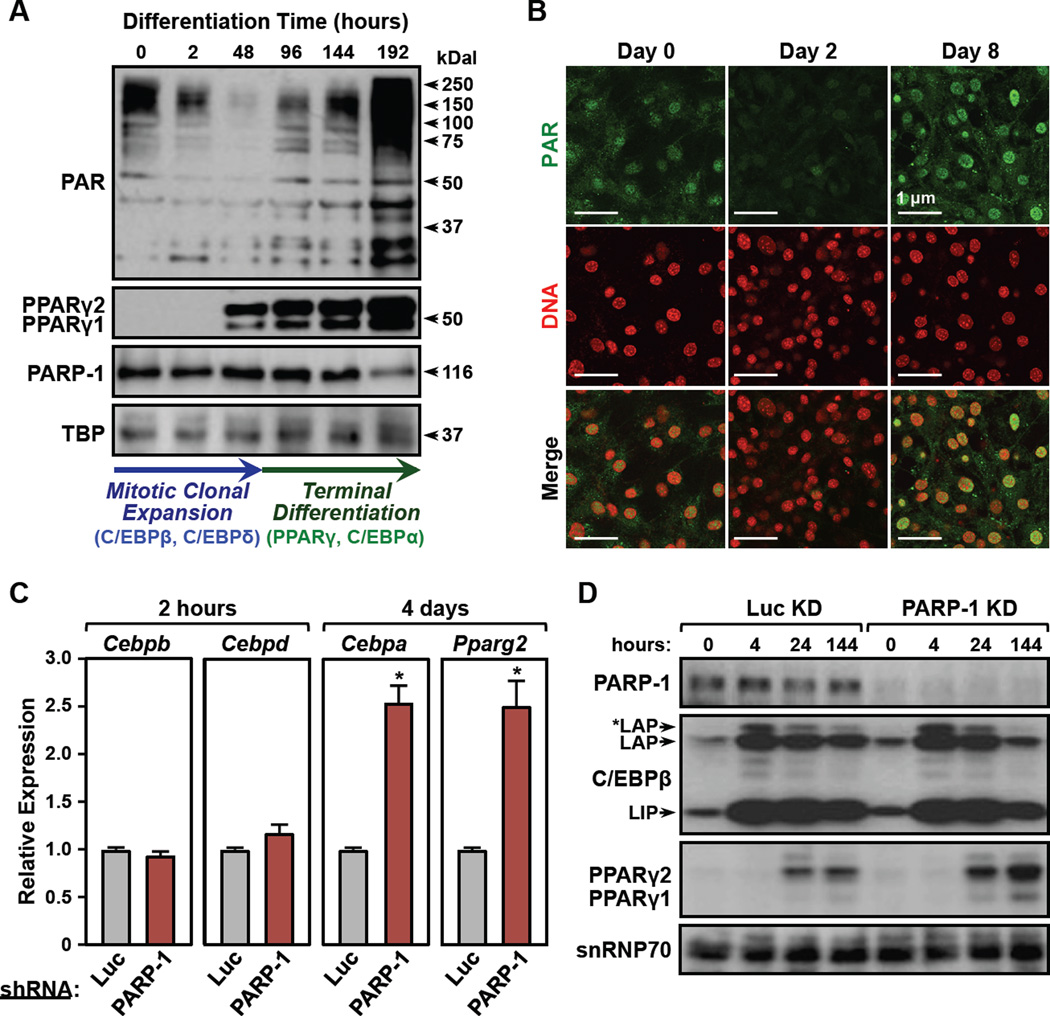
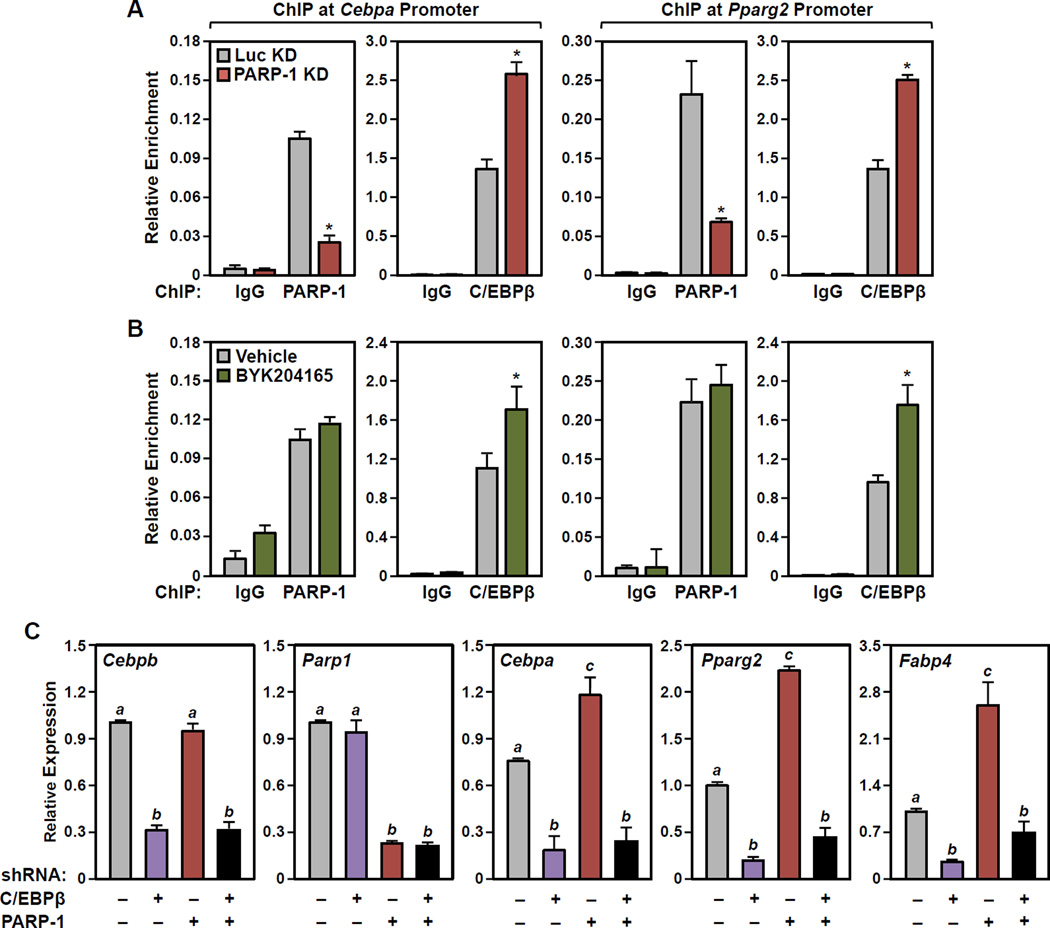
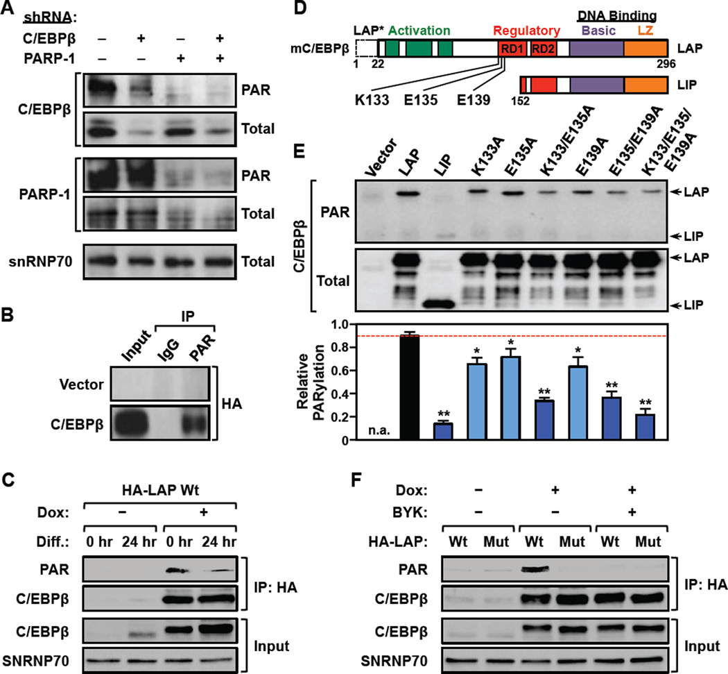
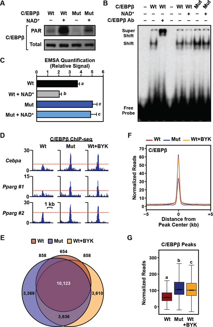
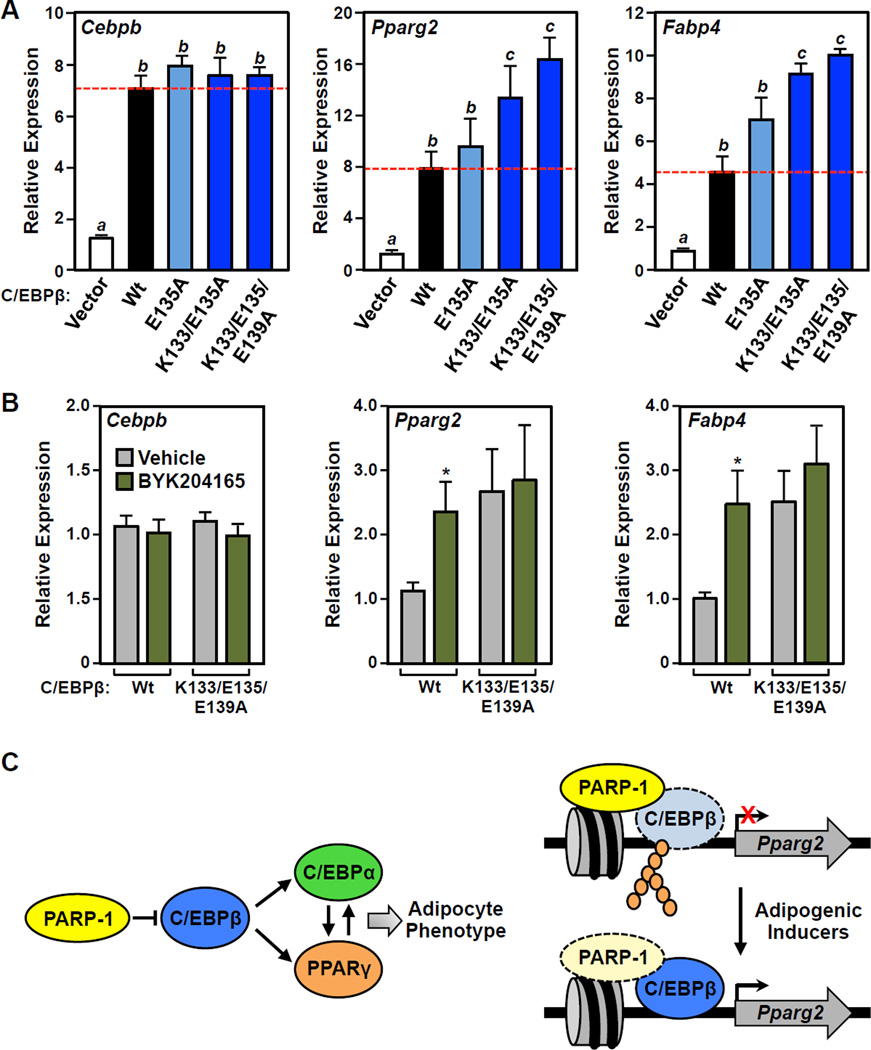
Poly(ADP-ribosyl)ation (PARylation) is a post-translational modification of proteins mediated by PARP family members, such as PARP-1. Although PARylation has been studied extensively, few examples of definitive biological roles for site-specific PARylation have been reported. Here we show that C/EBPβ, a key pro-adipogenic transcription factor, is PARylated by PARP-1 on three amino acids in a conserved regulatory domain. PARylation at these sites inhibits C/EBPβ's DNA binding and transcriptional activities and attenuates adipogenesis in various genetic and cell-based models. Interestingly, PARP-1 catalytic activity drops precipitously during the first 48 hr of differentiation, corresponding to a release of C/EBPβ from PARylation-mediated inhibition. This promotes the binding of C/EBPβ at enhancers controlling the expression of adipogenic target genes and continued differentiation. Depletion or chemical inhibition of PARP-1, or mutation of the PARylation sites on C/EBPβ, enhances these early adipogenic events. Collectively, our results provide a clear example of how site-specific PARylation drives biological outcomes.

Keywords: C/EBPβ; DNA binding; PARP inhibitor; PARP-1; PARylation; adipogenesis; gene expression; poly(ADP-ribose); transcription.


Figures

Figure 1
Figure 1. Genetic depletion of PARP-1 promotes the differentiation of primary preadipocytes from the stromal-vascular fraction (SVF)
(A and B) Primary SVF preadipocytes isolated from (A) Parp1loxP/loxP mice or (B) Parp1loxP/loxP;Pdgfra-cre/ERT mice were subjected to deletion of Parp1 in culture using adenovirus-Cre (AdV-Cre) or 4-OHT, respectively. (C and D) Western blots showing the relative levels of PARP-1 and PAR in primary SVF preadipocytes with or without deletion of Parp1. β-tubulin was used as a loading control. (E and F) SVF preadipocytes were subjected to Parp1 deletion using (E) AdV-Cre or (F) 4-OHT as shown in (A) and (B), respectively, prior to differentiation with MDI. Two hours or 4 days later, the relative expression of various adipocyte marker genes was assayed by RT-qPCR. Each bar represents the mean + SEM for three replicates. Bars marked with asterisks are statistically different from the control (Student’s t-test; *** p-value < 0.001 ** p-value < 0.01 or * p-value < 0.05). [See also Fig. S1]
Figure 2
Figure 2. Inhibition or depletion of PARP-1 promotes the differentiation of preadipocytes
(A) SVF, NIH/3T3, and 3T3-L1 cells were treated with 5 µM PJ34 or 20 µM BYK204165 prior to differentiation with MDI. Four days later, the relative expression of adipocyte marker genes Fabp4 and AdipoQ was assayed by RT-qPCR. Each bar represents the mean + SEM for three replicates. Bars marked with an asterisk are statistically different from the control (Student’s t-test; p-value < 0.05). (B) Time course of differentiation in 3T3-L1 cells in response to MDI ± 20 µM BYK204165. The relative expression of adipocyte marker genes Fabp4 and AdipoQ was assayed by RT-qPCR every two days. Each point represents the mean ± SEM for three replicates. Points marked with an asterisk are statistically different from the vehicle-treated control (Student’s t-test; p-value < 0.05). (C) Western blots showing the levels of PARP-1 after knockdown (KD) (left, using shRNAs targeting PARP-1 or luciferase, Luc,) or ectopic expression (right, Flag-tagged PARP-1) in 3T3-L1 cells. β-actin was used as a loading control. (D and E) 3T3-L1 cells, with or without stable knockdown or ectopic expression of PARP-1, as (C), were differentiated with MDI. (C) Oil Red-O staining at day 8. (D) RT-qPCR for adipocyte marker genes Fabp4 and AdipoQ at day 4. Each bar represents the mean + SEM for three replicates. Bars marked with an asterisk are statistically different from the control (Student’s t-test; p-value < 0.05). [See also Fig. S2]
Figure 3
Figure 3. Nuclear PAR levels fluctuate and demarcate the transition between two distinct waves of transcription during adipogenesis
(A) (Top) Western blots showing the levels of PAR, PPARγ1, PPARγ2, and PARP-1 in nuclear extracts from 3T3-L1 cells during a time course of differentiation with MDI. TBP was used as a loading control. (Bottom) The arrows indicate two distinct phases of adipogenesis: (1) mitotic clonal expansion (0 to 48 hours post MDI; elevated C/EBPβ and C/EBPδ; blue) and (2) terminal differentiation (>48 hours post MDI; elevated PPARγ and C/EBPα; green). (B) Immunofluorescent staining of 3T3-L1 cells for PAR (green) and DNA (red) during a time course of differentiation with MDI. (C) RT-qPCR analysis showing the expression of early transcription factors (2 hours post MDI; Cebpb, Cebpd), and late transcription factors (4 days post MDI; Cebpa, Pparg2) in 3T3-L1 cells ± knockdown using shRNAs targeting PARP-1 or luciferase (Luc, as a control). Each bar represents the mean + SEM for three replicates. Bars marked with an asterisk are statistically different from the control (Student’s t-test; p-value < 0.05). (D) Western blots showing the expression of PARP-1, C/EBPβ, and PPARγ in 3T3-L1 cells ± knockdown using shRNAs targeting PARP-1 or luciferase (Luc, as a control) during a time course of differentiation with MDI. snRNP70 was used as a loading control. [See also Fig. S3]
Figure 4
Figure 4. PARP-1 and PARylation modulate the binding of C/EBPβ to the promoters of C/EBPβ target genes encoding late transcription factors in 3T3-L1 cells
(A and B) Results from ChIP-qPCR assays for PARP-1 or C/EBPβ (as indicated) binding at the Cebpa (left) and Pparg2 (right) gene promoters performed in 3T3-L1 cells 24 hours after differentiation with MDI. The assays were performed (A) with knockdown using shRNAs targeting PARP-1 or luciferase (Luc, as a control), or (B) ± 20 µM BYK204165 (1 hour pretreatment, followed by 24 hours of treatment after adding MDI). Each bar represents the mean + SEM for three replicates. Bars marked with an asterisk are statistically different from the control (Student’s t-test; p-value < 0.05). (C) Results from RT-qPCR assays showing that the effects of PARP-1 knockdown on the expression of Cebpa, Pparg2, Fabp4 are abrogated by C/EBPβ knockdown in 3T3-L1 cells. Results for Parp1 and Cebpb are shown to confirm shRNA-mediated knockdown. Each bar represents the mean + SEM for three replicates. Bars marked with different letters are statistically different from each other (ANOVA; p-value < 0.05). [See also Fig. S3]
Figure 5
Figure 5. PARP-1 PARylates C/EBPβ at specific sites in 3T3-L1 cells
(A) PARP-1-dependent PARylation of C/EBPβ. Western blots showing the levels of PARylated and total C/EBPβ and PARP-1 in control, C/EBPβ-, or PARP-1-depleted 3T3-L1 cells. Knockdown was achieved by using shRNAs targeting C/EBPβ, PARP-1, or luciferase (Luc, as a control). snRNP70 was used as a loading control. The full blot of PARylated C/EBPβ is shown in Fig. S4A. (B) PARylated proteins were immunoprecipitated from 293T cells ectopically expressing HA-tagged C/EBPβ and analyzed by Western blotting as indicated. (C) Reduced PARylation of C/EBPβ in 3T3-L1 cells upon differentiation. The cells were differentiated with MDI for 24 hours and the expression of HA-tagged mouse C/EBPβ was induced from a transgene by treatment with Dox for 8 hours. The cells were then subjected to immunoprecipitation with an anti-HA antibody. The input and the immunoprecipitate (IP) were analyzed by Western blotting for C/EBPβ and PAR as indicated. snRNP70 was used as a loading control. (D) Schematic diagram of mouse C/EBPβ showing the ADP-ribosylation site determined by mass spectrometry (E135), as well as two adjacent sites determined by mutagenesis (K133, E139). C/EBPβ isoforms: LAP, common full-length, LAP*, 22 amino acid N-terminal extension; LIP, N-terminally truncated. (E)Top, Extracts from 293T cells ectopically expressing wild-type or mutant HA-tagged C/EBPβ proteins were analyzed by Western blotting for PARylated and total C/EBPβ. Bottom, Quantification of the relative PARylation of C/EBPβ proteins in Western blots like the one shown in the panel above. Each bar represents the mean + SEM for three independent experiments. Bars marked with asterisks are statistically different from the control (Student’s t-test; ** p-value < 0.01 or * p-value < 0.05). (F) A PARylation site mutant of C/EBPβ is not PARylated in 3T3-L1 cells. The expression of HA-tagged wild-type (Wt) or PARylation site mutant (K133A/E135A/E139A; Mut) mouse C/EBPβ was induced from a transgene by treatment with Dox. The cells were induced to differentiate with MDI for 24 hours ± 20 µM BYK204165 (BYK) and then subjected to immunoprecipitation (IP) with an anti-HA antibody. The input and the immunoprecipitated material were analyzed by Western blotting for C/EBPβ and PAR. snRNP70 was used as a loading control. [See also Fig. S4]
Figure 6
Figure 6. PARylation of C/EBPβ by PARP-1 at specific sites inhibits C/EBPβ binding to DNA in vitro and in cells
(A through C) Mutation of PARP-1-dependent sites of PARylation render C/EBPβ resistant to PARylation-induced decreases in DNA binding. Wild-type or PARylation site point mutant (K133A/E135A/E139A; Mut) C/EBPβ proteins were expressed in 293T cells. Nuclei were collected and then treated ± 100 µM NAD+ for 30 min. to stimulate PARylation prior to preparation of nuclear extracts. (A) Western blots showing the levels of PARylated (top) and total (bottom) C/EBPβ in nuclear extracts prepared from the transfected 293T cells. (B) Representative EMSA experiment performed using nuclear extract and a labeled double stranded oligonucleotide probe corresponding to the C/EBP regulatory element in the Cebpa promoter (probe) ± supershifting using a C/EBPβ antibody. (C) Quantification of the results from experiments like those shown in (B). Each bar represents the mean + SEM for three independent experiments. Bars marked with different letters are statistically different from each other (ANOVA; p-value < 0.05). (D through F) Mutation of PARP-1-dependent sites of PARylation render C/EBPβ resistant to PARylation-induced decreases in chromatin binding in 3T3-L1 cells. Expression of HA-tagged wild-type (Wt) or PARylation site mutant (K133A/E135A/E139A; Mut) mouse C/EBPβ was induced from a transgene by treatment with Dox for 8 hours ± 20 µM BYK204165 (BYK). The cells were crosslinked and subjected to ChIP-seq for C/EBPβ. (D) ChIP-seq browser tracks for C/EBPβ in the three conditions tested (Wt, Mut, and Wt+BYK) at genomic loci with C/EBPβ enhancers near the Cebpa and Pparg genes. (E) Venn diagram showing the overlap of statistically significant peaks of C/EBPβ in the three conditions tested. (F) Metaplot and (G) box plot analyses of C/EBPβ binding in 3T3-L1 cells by ChIP-seq in the three conditions tested. [See also Fig. S5]
Figure 7
Figure 7. Mutation of PARP-1-dependent of sites PARylation in C/EBPβ enhances adipogenesis and renders C/EBPβ resistant to the effects of PARP inhibitors
(A and B) Wild-type or mutant C/EBPβ were ectopically expressed in 3T3-L1 cells, as indicated, which induces spontaneous differentiation of the cells in the absence of MDI. The expression of Pparg and Fabp4 was determined by RT-qPCR performed 4 days post-transfection. Results for Cebpb are shown to confirm equal ectopic expression of the mutants. (A) Mutation of PARP-1-dependent sites of PARylation in C/EBPβ enhances adipogenesis. Bars marked with different letters are statistically different from each other (ANOVA; p-value < 0.05). (B) PARylation site C/EBPβ mutants are resistant to the effects of PARP inhibitor. The cells were treated ± 20 µM BYK204165 (1 hour pretreatment, followed by 4 days of treatment after adding MDI). Bars marked with an asterisk are statistically different from the corresponding control (Student’s t-test; p-value < 0.05). (C) Model for the role of PARP-1 in attenuating adipogenesis, as described in the text.

References

    1. Abdou HS, Atlas E, Hache RJ. A positive regulatory domain in CCAAT/enhancer binding protein beta (C/EBPBeta) is required for the glucocorticoid-mediated displacement of histone deacetylase 1 (HDAC1) from the C/ebpalpha promoter and maximum adipogenesis. Endocrinology. 2013;154:1454–1464. - PubMed
    1. Altmeyer M, Messner S, Hassa PO, Fey M, Hottiger MO. Molecular mechanism of poly(ADP-ribosyl)ation by PARP1 and identification of lysine residues as ADP-ribose acceptor sites. Nucleic Acids Res. 2009;37:3723–3738. - PMC - PubMed
    1. Asher G, Reinke H, Altmeyer M, Gutierrez-Arcelus M, Hottiger MO, Schibler U. Poly(ADP-ribose) polymerase 1 participates in the phase entrainment of circadian clocks to feeding. Cell. 2010;142:943–953. - PubMed
    1. Bai P, Canto C, Oudart H, Brunyanszki A, Cen Y, Thomas C, Yamamoto H, Huber A, Kiss B, Houtkooper RH, et al. PARP-1 inhibition increases mitochondrial metabolism through SIRT1 activation. Cell Metab. 2011;13:461–468. - PMC - PubMed
    1. Christy RJ, Kaestner KH, Geiman DE, Lane MD. CCAAT/enhancer binding protein gene promoter: binding of nuclear factors during differentiation of 3T3-L1 preadipocytes. Proc Natl Acad Sci U S A. 1991;88:2593–2597. - PMC - PubMed

MeSH terms

Substances