. 2017 May;23(9-10):415-425.
doi: 10.1089/ten.TEA.2016.0378. Epub 2017 Mar 31.

Hierarchically Ordered Porous and High-Volume Polycaprolactone Microchannel Scaffolds Enhanced Axon Growth in Transected Spinal Cords

Affiliations

Hierarchically Ordered Porous and High-Volume Polycaprolactone Microchannel Scaffolds Enhanced Axon Growth in Transected Spinal Cords

Dena Shahriari et al. Tissue Eng Part A. 2017 May.

Abstract

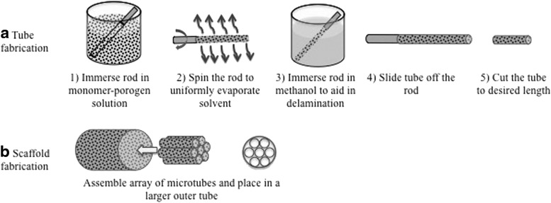
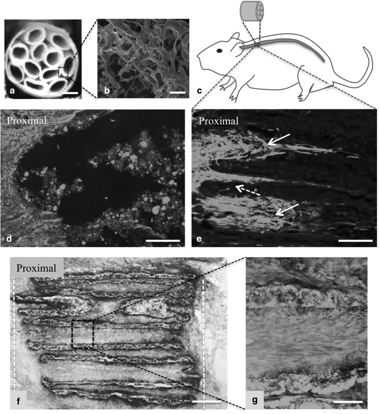
The goal of this work was to design nerve guidance scaffolds with a unique architecture to maximize the open volume available for nerve growth. Polycaprolactone (PCL) was selected as the scaffold material based on its biocompatibility and month-long degradation. Yet, dense PCL does not exhibit suitable properties such as porosity, stiffness, strength, and cell adhesion to function as an effective nerve guidance scaffold. To address these shortcomings, PCL was processed using a modified salt-leaching technique to create uniquely controlled interconnected porosity. By controlling porosity, we demonstrated that the elastic modulus could be controlled between 2.09 and 182.1 MPa. In addition, introducing porosity and/or coating with fibronectin enhanced the PCL cell attachment properties. To produce PCL scaffolds with maximized open volume, porous PCL microtubes were fabricated and translated into scaffolds with 60 volume percent open volume. The scaffolds were tested in transected rat spinal cords. Linear axon growth within both the microtubes as well as the interstitial space between the tubes was observed, demonstrating that the entire open volume of the scaffold was available for nerve growth. Overall, a novel scaffold architecture and fabrication technique are presented. The scaffolds exhibit significantly higher volume than state-of-the-art scaffolds for promising spinal cord nerve repair.

Keywords: controlled porosity; mechanical properties; nerve guidance scaffolds; nerve regeneration; polycaprolactone.


Conflict of interest statement

No competing financial interests exist.

Figures

<b>FIG. 1.</b>
FIG. 1.
Steps in fabricating PCL (a) tubes and (b) scaffolds. PCL, polycaprolactone.
<b>FIG. 2.</b>
FIG. 2.
Increasing the planetary ball-milling time reduces the particle size and size distribution. SEM images of NaCl (a) as-received, and NaCl ball-milled at 400 rpm for: (b) 1 min, (c) 5 min, (d) 30 min, (e) 60 min, and (f) 90 min. The inset scale bars are 25 μm; all other scale bars are 200 μm. rpm, Revolutions per minute; SEM, scanning electron microscopy.
<b>FIG. 3.</b>
FIG. 3.
Cross-sectional SEM analysis of PCL films. (a) One hundred percent PCL (no porosity). A 70 vol % porosity PCL film fabricated using 17 μm (average diameter) NaCl particles (b) before and (d) after salt-leaching. (c, e) 2× Magnification images of the dashed boxes shown in (b, d), respectively. The solid arrows in (c) point to NaCl particles and the dashed arrows point to intrinsic porosities created in addition to porosities created by the porogen. Scale bars: (a) 2.5 μm, (b, d) 10 μm, and (c, e) 5 μm. vol %, Volume percent.
<b>FIG. 4.</b>
FIG. 4.
The elastic modulus of PCL versus porosity percentage created by 17 μm NaCl particles.
<b>FIG. 5.</b>
FIG. 5.
Fibroblast and primary rat Schwann cell attachment on PCL films. SEM images of the top surface of (a) nonporous PCL film and (b) 70 vol % porosity PCL with corresponding magnified images demonstrate an increased surface roughness in porous PCL. NIH 3T3 fibroblasts were cultured on uncoated (c) tissue-culture treated flask as control, (d) nonporous PCL film and (e) 70 vol % porosity PCL film, and on fibronectin-coated (f) tissue culture-treated flask, (g) nonporous PCL film, and (h) 70 vol % porosity PCL film. Cells were fixed after 72 h and stained for actin and nuclei. Unlike nonporous PCL, 70 vol % porosity PCL films provided cell attachment comparable to the positive control. Both groups exhibited cell attachment after fibronectin coating. Similarly, primary rat Schwann cells were cultured on uncoated (i) PDL surfaces as control, (j) nonporous PCL, and (k) 70 vol % porosity PCL as well as fibronectin-coated (l) PDL surfaces, (m) nonporous PCL, and (n) 70 vol % porous PCL films. Cells were fixed and stained for actin in green and nuclei in blue 72 h postseeding. While Schwann cells did not attach to uncoated PCL films with or without porosity, cell attachment occurred on both fibronectin-coated PCL surfaces. Scale bars in the insets in (a, b) are 10 μm. All other scale bars are 100 μm.
<b>FIG. 6.</b>
FIG. 6.
PCL scaffold characterization and in vivo studies. (a) Cross-sectional image of a PCL scaffold with inner tubes of 260 μm in diameter, fabricated from 70 vol % porosity PCL. SEM image of an inner tube cross-section is shown in (b), which shows interconnected porosity. (c) A schematic of fibronectin-coated porous PCL scaffold is shown, which was inserted in the transected T3 section of a rat spinal cord. Four weeks post-implantation, animals were perfused and longitudinal sections were obtained and stained with neurofilament (NF200). (d) Transected spinal cord without an implant showed minimum axon penetration into the lesion. (e) The proximal site of the scaffold shows linear axon growth into PCL scaffolds. Solid white arrows in (e) demonstrate the axons grew linearly inside the microtubes, whereas the dashed arrow indicates some axons grew linearly in between the microtubes. (f) Toluidine Blue staining of PCL scaffold 4 weeks postimplantation shows that the scaffold microtubes were maintained and minimum scar tissue formation was observed around the scaffold. The white dashed lines show the boundary of the implanted scaffold in the transected spinal cord. The boxed region in (f) is magnified in (g), which highlights the intimate contact of cells with scaffold walls. Scale bars: (a) 300 μm, (b) 100 μm, (d, e) 200 μm, (f) 250 μm, and (g) 50 μm.

References

    1. Silva N.A., Sousa N., Reis R.L., and Salgado A.J. From basics to clinical: a comprehensive review on spinal cord injury. Prog Neurobiol 114, 25, 2014 - PubMed
    1. Varma A.K., Das A., Wallace G., IV, Barry J., Vertegel A.A., Ray S.K., and Banik N.L. 2013. Spinal cord injury: a review of current therapy, future treatments, and basic science frontiers. Neurochem Res 38, 895, 2013 - PMC - PubMed
    1. Mahoney M.J., and Anseth K.S. Three-dimensional growth and function of neural tissue in degradable polyethylene glycol hydrogels. Biomaterials 27, 2265, 2006 - PubMed
    1. Xu X.M., Guénard V., Kleitman N., and Bunge M.B. Axonal regeneration into Schwann cell‐seeded guidance channels grafted into transected adult rat spinal cord. J Comp Neurol 351, 145, 1995 - PubMed
    1. McDonald J.W., Liu X.Z., Qu Y., Liu S., Mickey S.K., Turetsky D., Gottlieb D.I., and Choi D.W. Transplanted embryonic stem cells survive, differentiate and promote recovery in injured rat spinal cord. Nat Med 5, 1410, 1999 - PubMed

Publication types