Review
. 2017 Aug 1;9(8):a028142.
doi: 10.1101/cshperspect.a028142.

Transition Zone Migration: A Mechanism for Cytoplasmic Ciliogenesis and Postaxonemal Centriole Elongation

Affiliations
Review

Transition Zone Migration: A Mechanism for Cytoplasmic Ciliogenesis and Postaxonemal Centriole Elongation

Tomer Avidor-Reiss et al. Cold Spring Harb Perspect Biol. .

Abstract

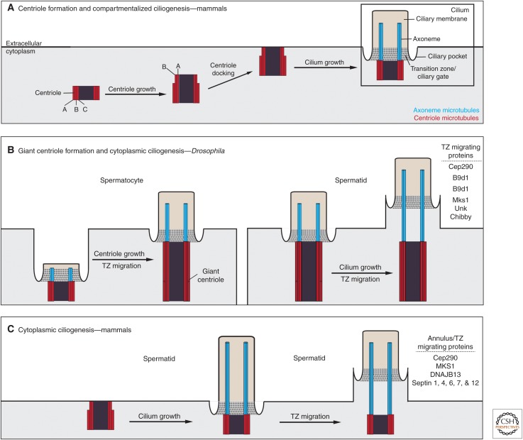
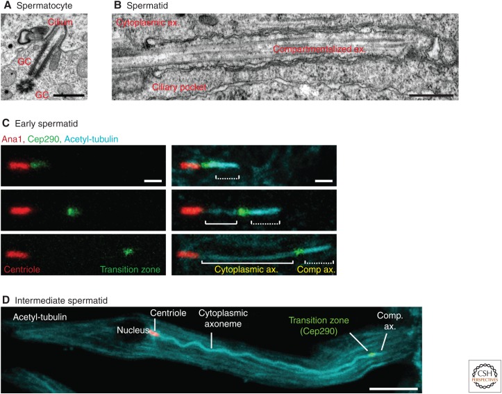
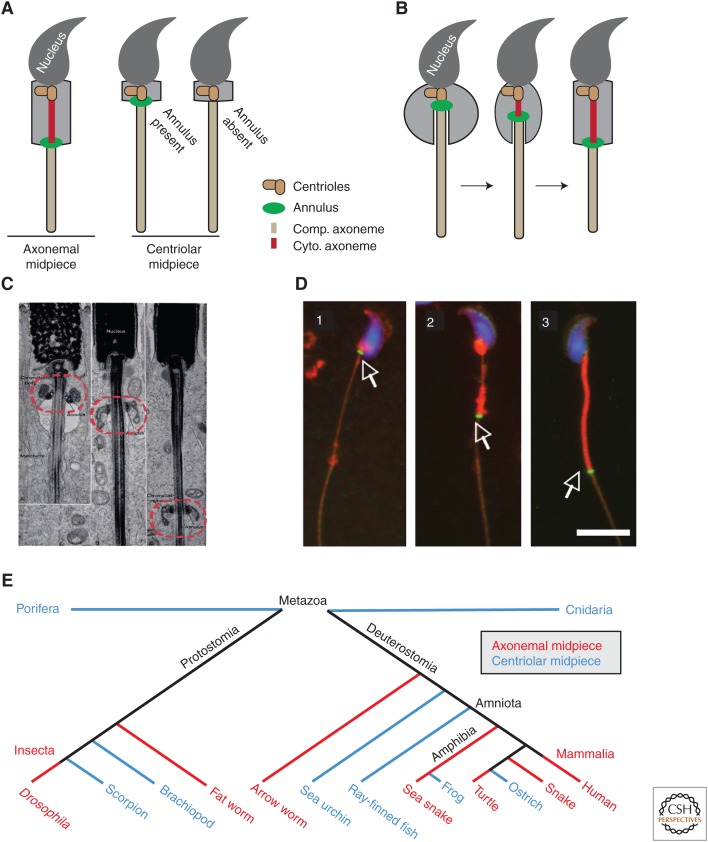
The cilium is an elongated and continuous structure that spans two major subcellular domains. The cytoplasmic domain contains a short centriole, which serves to nucleate the main projection of the cilium. This projection, known as the axoneme, remains separated from the cytoplasm by a specialized gatekeeping complex within a ciliary subdomain called the transition zone. In this way, the axoneme is compartmentalized. Intriguingly, however, this general principle of cilium biology is altered in the sperm cells of many animals, which instead contain a cytoplasmic axoneme domain. Here, we discuss the hypothesis that the formation of specialized sperm giant centrioles and cytoplasmic cilia is mediated by the migration of the transition zone from its typical location as part of a structure known as the annulus and examine the intrinsic properties of the transition zone that may facilitate its migratory behavior.


Figures

Figure 1.
Figure 1.
Models of transition zone migration in centriole formation and ciliogenesis. (A) In a typical cell, the centriole forms in the cytoplasm. At its base, the centriole is comprised of triplet microtubules known as the A, B, and C microtubules, with only the A and B microtubules extending to its tip. The centriole then either associates with a vesicle that fuses with the plasma membrane (not shown) or migrates directly to the membrane for docking. There, the microtubules at the distal end of the centriole elongate to form an axoneme, which is surrounded by a specialized membrane known as the ciliary membrane. At the base of the cilium, this membrane forms a pocket known as the ciliary pocket, and the axoneme is embedded in a network of proteins that serves as a ciliary gate known as the transition zone. (B) During Drosophila spermatogenesis, ciliogenesis begins in the premeiotic diploid spermatocytes and is completed in the postmeiotic haploid spermatids. In the spermatocyte, the centriole docks to the plasma membrane and forms a short cilium. After the axoneme is initiated, both the centriole and the cilium elongate. Presumably, the centriole grows as a result of transition zone migration along the axoneme. In the spermatid, the cilium continues to grow and the transition zone continues to migrate along the axoneme, exposing axonemal microtubules to the cytoplasm. (C) During mammalian spermatogenesis, ciliogenesis starts and is completed in spermatids. In the spermatid, the centriole docks to the plasma membrane and forms a full-length cilium. After the cilium is formed, the transition zone and annulus migrate along the axoneme. Proteins involved in transition zone migration and annulus formation and migration are indicated in B and C.
Figure 2.
Figure 2.
Transition zone migration in Drosophila melanogaster. (A) Electron micrograph showing two giant centrioles, one of which forms a short cilium. Scale bar, 500 nm. (B) Electron micrograph showing a spermatid cytoplasmic and compartmentalized axoneme segment. The ciliary pocket is observed at the base of the compartmentalized axoneme. Scale bar, 500 nm. (C) Light microscopy demonstrating transition zone migration in early spermatids. The centriole is labeled by Ana1-tdTomato (red), transition zone is labeled by Cep290-GFP (green), and axoneme (ax) is labeled with antiacetylated-tubulin (cyan). (D) A full-length intermediate spermatid with centriole, cytoplasmic axoneme, transition zone, and compartmentalized axoneme. (D, From Basiri et al. 2014; reprinted, with permission, from Elsevier © 2014.)
Figure 3.
Figure 3.
Annulus migration occurs in mammalian spermatids. (A) Sperm can be classified into three types based on the location and presence of the annulus: sperm with an annulus separating cytoplasmic and compartmentalized axoneme segments (axonemal midpiece, left), sperm with an annulus separating the centriole from a fully compartmentalized axoneme (centriolar midpiece with annulus), and sperm lacking an annulus but containing a centriolar midpiece and a fully compartmentalized axoneme within the principal piece (centriolar midpiece without annulus). (B) Illustration of annulus migration in mammalian spermatids. (C) Longitudinal sections of Macaca mulatta spermatids demonstrating annulus migration during spermiogenesis. The annulus is indicated by a red dotted circle. (C, Reprinted from data in Fawcett et al. 1970.) (D) Septin 4 localization during mouse spermiogenesis (white arrow). The annulus indicated by Septin 4 antibody (green) is found near the nucleus (blue) in early stage elongating spermatids (1). The annulus then begins to migrate toward the growing end of the axoneme to form the midpiece (red) (2). Finally, the annulus reaches the distal end of the midpiece to define the midpiece-principal piece junction in mature sperm (3). (D, From Guan et al. 2009; reprinted under the terms of the Creative Commons Attribution License.) Scale bar, 10 µm. (E) Distribution of spermatids containing either a centriolar midpiece or an axonemal midpiece across animal phylogeny. (Data based on Afzelius 1955; Silveira and Porter 1964; Sato et al. 1967; Reger and Cooper 1968; Mattei 1988; Hess et al. 1991; Mita and Nakamura 1992; Medina 1994; Dallai et al. 1995; Iomini and Justine 1997; Reunov and Klepal 2003; Scheltinga et al. 2003; Smita et al. 2004; Al-Dokhi et al. 2007, ; Vignoli et al. 2008; Lipke et al. 2009.)

References

    1. Afzelius BA. 1955. The fine structure of the sea urchin spermatozoa as revealed by the electron microscope. Z Zellforsch Mikrosk Anat 42: 134–148. - PubMed
    1. Al-Dokhi O, Al-Onazee Y, Mubarak M. 2007. Fine structure of the epididymal sperm of the snake Eryx jayakari (Squamata, Reptilia). Int J Zool Res 3: 1–13.
    1. Avidor-Reiss T, Leroux MR. 2015. Shared and distinct mechanisms of compartmentalized and cytoplasmic ciliogenesis. Curr Biol 25: R1143–R1150. - PMC - PubMed
    1. Avidor-Reiss T, Maer AM, Koundakjian E, Polyanovsky A, Keil T, Subramaniam S, Zuker CS. 2004. Decoding cilia function: Defining specialized genes required for compartmentalized cilia biogenesis. Cell 117: 527–539. - PubMed
    1. Baccetti B. 1982. The evolution of the sperm tail. Symp Soc Exp Biol 35: 521–532. - PubMed

LinkOut - more resources