. 2018 Mar 1;28(3):974-987.
doi: 10.1093/cercor/bhx005.

mGluR2 versus mGluR3 Metabotropic Glutamate Receptors in Primate Dorsolateral Prefrontal Cortex: Postsynaptic mGluR3 Strengthen Working Memory Networks

Affiliations

mGluR2 versus mGluR3 Metabotropic Glutamate Receptors in Primate Dorsolateral Prefrontal Cortex: Postsynaptic mGluR3 Strengthen Working Memory Networks

Lu E Jin et al. Cereb Cortex. .

Abstract

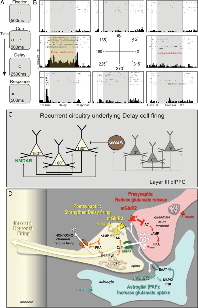
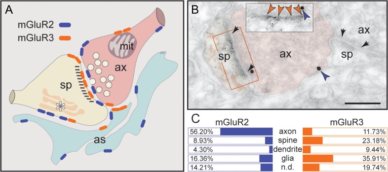
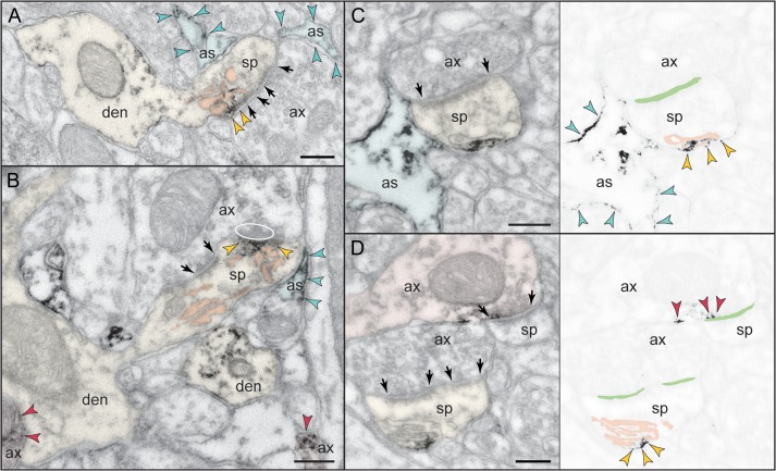
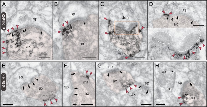
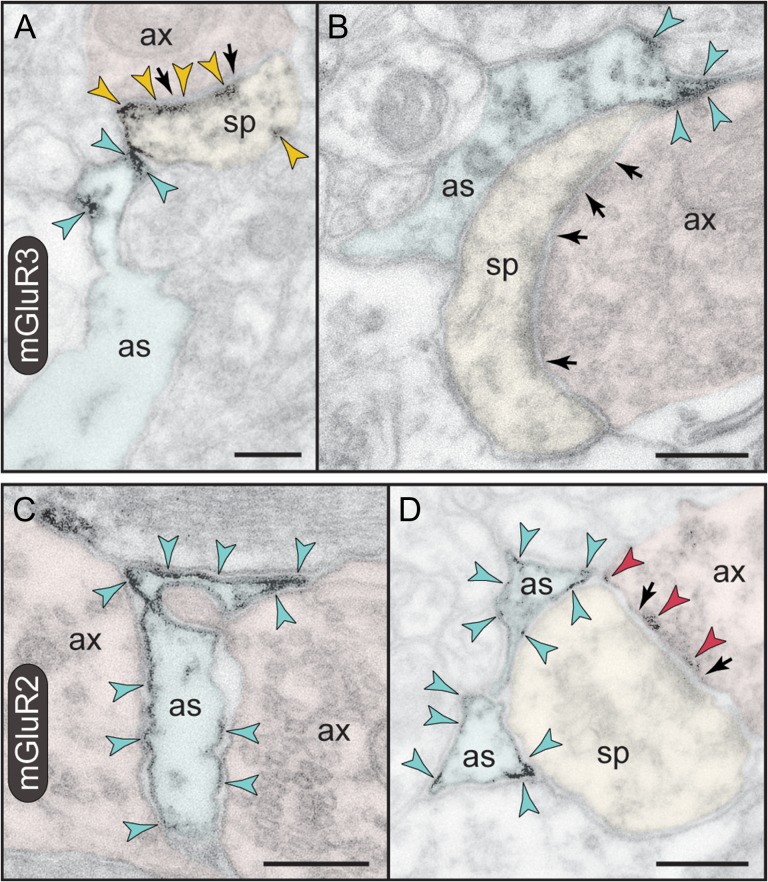
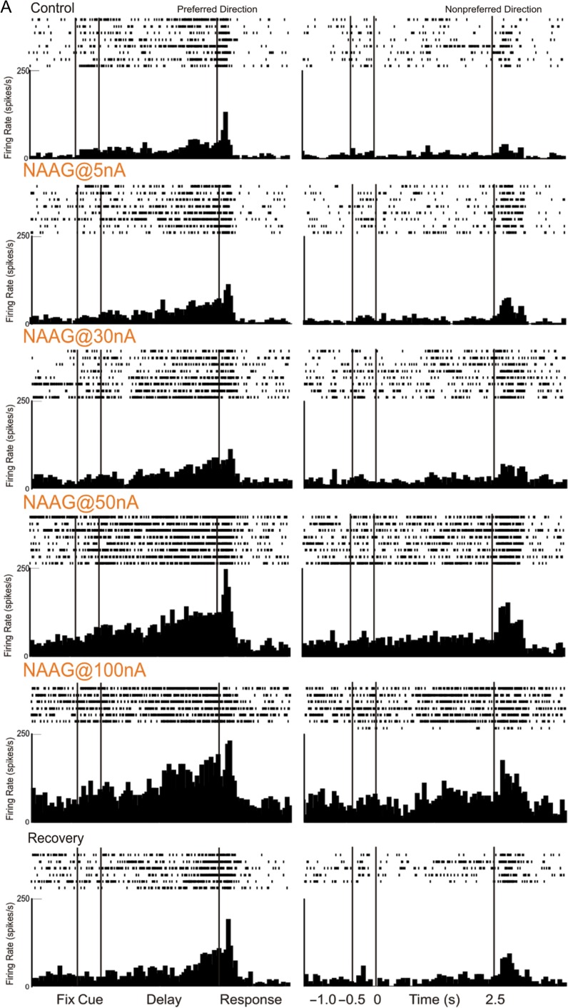
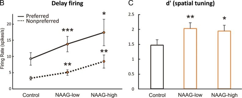
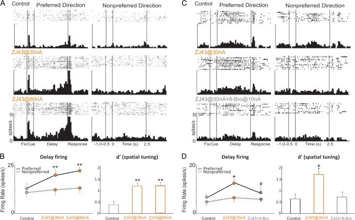
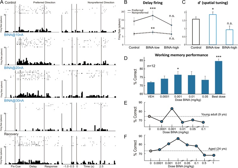
The newly evolved circuits in layer III of primate dorsolateral prefrontal cortex (dlPFC) generate the neural representations that subserve working memory. These circuits are weakened by increased cAMP-K+ channel signaling, and are a focus of pathology in schizophrenia, aging, and Alzheimer's disease. Cognitive deficits in these disorders are increasingly associated with insults to mGluR3 metabotropic glutamate receptors, while reductions in mGluR2 appear protective. This has been perplexing, as mGluR3 has been considered glial receptors, and mGluR2 and mGluR3 have been thought to have similar functions, reducing glutamate transmission. We have discovered that, in addition to their astrocytic expression, mGluR3 is concentrated postsynaptically in spine synapses of layer III dlPFC, positioned to strengthen connectivity by inhibiting postsynaptic cAMP-K+ channel actions. In contrast, mGluR2 is principally presynaptic as expected, with only a minor postsynaptic component. Functionally, increase in the endogenous mGluR3 agonist, N-acetylaspartylglutamate, markedly enhanced dlPFC Delay cell firing during a working memory task via inhibition of cAMP signaling, while the mGluR2 positive allosteric modulator, BINA, produced an inverted-U dose-response on dlPFC Delay cell firing and working memory performance. These data illuminate why insults to mGluR3 would erode cognitive abilities, and support mGluR3 as a novel therapeutic target for higher cognitive disorders.

Keywords: Alzheimer's disease; GRM2; GRM3; dendritic spine; schizophrenia.


Figures

Figure 1.
Figure 1.
Neuronal basis of visuospatial representation during a working memory task. (A) The ODR task. The monkey initiates a trial by fixating a central point; a visual cue appears at 1 of 8 locations; the monkey must remember the cued location over a delay period and make a saccade to the correct location to get reward. The cued location varies from trial to trial, requiring constant updating of information held in working memory. (B) A typical Delay cell, with spatially tuned persistent firing across the delay period in the neuron's preferred direction (180°) but not for nonpreferred directions (e.g., 0°). (C) Deep layer III dlPFC microcircuits that generate Delay cell firing. Pyramidal neurons with similar preferred directions excite each other through NMDAR synapses, for example, a cluster of 180° neurons maintain firing across the delay following a cue at 180°, but reduce firing at 0° (nonpreferred direction) via lateral inhibition from GABAergic interneurons. (D) Working model of mGluR2 and mGluR3 actions in a layer III dlPFC glutamatergic synapse. Postsynaptic mGluR3 reside near the synapse and mGluR2 near the Ca2+-storing spine apparatus (asterisk). They inhibit cAMP production, close HCN and KCNQ channels, strengthen synaptic efficacy, and enhance Delay cell firing. In contrast, presynaptic mGluR2 on axon terminals reduce glutamate release. mGluR3 on PAPs increase glial glutamate uptake by increasing EAAT expression via mitogen-activated protein kinase and phosphoinositide 3-kinase signaling (Aronica et al. 2003). The functions of presynaptic mGluR3 near mitochondria, and of mGluR2 on astrocytes at a distance from the synapse are unknown.
Figure 2.
Figure 2.
Summary of mGluR2 versus mGluR3 expression patterns in layer III dlPFC. (A) mGluR2 is principally a presynaptic receptor, targeted to synapses as well as preterminal axons. Postsynaptic expression is limited to extrasynaptic spine membranes, in association with the spine apparatus (asterisk), and rarely to perisynaptic membranes. Neuronal mGluR3 is primarily a postsynaptic receptor targeted to long, thin spine synapses, the spine type that is (1) most prevalent in layer III dlPFC, (2) most associated with cAMP-K+ channel modulation, and (3) most vulnerable to loss with advancing age. In contrast, there is little presynaptic expression of mGluR3, specifically along the axonal membrane facing mitochondria (mit) and rarely perisynaptically. Both mGluR2 and mGluR3 are localized in astrocytes in primate dlPFC. The localization of mGluR2 in glia is distinct from mGluR3, as it is not targeted to the glial leaflets ensheathing the synapse, which is the typical mGluR3 pattern. (B) Dual immunolabeling for mGluR2 (immunogold, blue arrowheads) and mGluR3 (DAB, orange arrowheads) reveals presynaptic mGluR2 and postsynaptic mGluR3 juxtapositioned at the same axospinous synapse (frame and inset). The labeled axon (ax) and spine (sp) are pseudocolored for clarity. A nonlabeled axospinous synapse is shown for comparison; synapses are between arrows. Scale bar, 200 nm. (C) The prevalence of mGluR2 versus mGluR3 in various cellular profiles in layer III of the dlPFC neuropil, expressed as percentage of an mGluR2 or mGluR3 profile (e.g., axon) per total mGluR2 or mGluR3 profiles, respectively (see quantitative assessment in the “Materials and Methods” section). Nondetermined (n.d.) are profiles that could not be unequivocally categorized.
Figure 3.
Figure 3.
Postsynaptic expression of mGluR3 in monkey dlPFC. mGluR3 is prominently expressed in dendritic spines, both within the synaptic active zone (A) and perisynaptically (B,C); label in C is found at the central perforation of a perforated synapse. (D,E) The framed images are edited to facilitate receptor visualization at the synapse (the synaptic cleft is marked in green). Note in D the typical mGluR3 localization in PAPs. (F,G) mGluR3 is additionally expressed at nonsynaptic spine membranes. In F, one section of a perforated synapse is additionally labeled. Extrasynaptic mGluR3 in G is found next to the spine apparatus (pink-pseudocolored) in the spine neck of a prototypical thin spine; a second spine, not shown in its entirety, emanates from the parent dendrite (curved arrow). The enlarged frame in G shows the common postsynaptic expression of mGluR3 at the synapse. Labeled spines (sp) and astrocytes (as) are pseudocolored for clarity; color-coded arrowheads point to mGluR3; synapses are between arrows. ax, axon; den, dendrite. Scale bars, 200 nm.
Figure 4.
Figure 4.
Postsynaptic expression of mGluR2 in monkey dlPFC. (A,B) mGluR2 is weakly expressed in dendritic spines perisynaptically. Its association with the glutamatergic-like synapse is selective to stubby (A) and mushroom-type (B) spines; in B, the obliquely sectioned synaptic disk is marked with oval. (C,D). However, mGluR2 in spines is primarily an extrasynaptic receptor. Remarkably, mGluR2 was distinctly associated with the plasma membrane facing the spine apparatus. This is best appreciated in the edited images C and D, where the synaptic cleft is marked in green and the apparatus is pink-pseudocolored. Note also the astrocytic as well as axonal labeling for mGluR2. Labeled axons (ax), dendrites (den), spines (sp), and astrocytes (as) are pseudocolored for clarity; color-coded arrowheads point to mGluR2; synapses are between arrows. Scale bars, 200 nm.
Figure 5.
Figure 5.
Presynaptic expression of mGluR2 versus mGluR3 in monkey dlPFC. (A–D) mGluR2 is expressed in glutamatergic-like axons establishing asymmetric synapses. Label is found within the synaptic active zone, and is typically over one section of a perforated synapse (A). Perisynaptic mGluR2 is visualized on membranes flanking the synapse (cross section in B and D) or in a halo surrounding the synaptic disk (ovals; oblique section in C and inset). Note that mGluR2 is also present extrasynaptically (A–C), and in preterminal axons (not captured here; see Supplementary Fig. 2). (E–H) Axons are weakly reactive against mGluR3. Unlike mGluR2, label is restricted to patches of the plasma membrane with a predilection for membranes apposing mitochondria (mit in E and F). As seen in E–G, axonal mGluR3 is for the most part extrasynaptic. However, there are rare examples when mGluR3 can be found perisynapticaly as in the synaptic triad in H. Labeled axons (ax) are pseudocolored for clarity; color-coded arrowheads point to mGluR2 or mGluR3; synapses are between arrows. den, dendrite; sp, spine. Scale bars, 200 nm.
Figure 6.
Figure 6.
Astrocytic expression of mGluR3 versus mGluR2 in monkey dlPFC. (A,B) PAPs are selectively labeled for mGluR3; note that the receptor is not distributed uniformly on the PAP plasma membrane but is placed immediately next to the synapse in the path of escaping glutamate. A spine in A is also reactive against mGluR3 at the synapse, perisynaptically, and extrasynaptically. (C,D) In contrast to mGluR3, mGluR2 is not targeted to the PAPs ensheathing the synapse, but presents a more uniform distribution on astrocytic membranes. In D, presynaptic mGluR2 is also captured at the synapse. Axons (ax), spines (sp), and astrocytes (as) are pseudocolored for clarity; color-coded arrowheads point to mGluR3 or mGluR2; synapses are between arrows. Scale bars, 200 nm.
Figure 7.
Figure 7.
mGluR3 stimulation by iontophoresis of NAAG enhances Delay cell firing. (A) Example of a Delay cell where NAAG had a linear, enhancing effect on neuronal firing. The increase was significant even when the dose was raised to 100 nA; firing recovered to control levels after washout. (B) The averaged neuronal firing rate during the delay epoch for the population of Delay cells (n = 22) under Control, NAAG low-dose condition (5–30 nA) and NAAG high-dose condition (50–100 nA), shown for the neurons’ preferred direction (solid line), and a representative nonpreferred direction (dashed line); mean ± SEM. Both low and high doses significantly increased delay firing, especially for the neuron's preferred direction. (C) The averaged d′ for neuronal firing during the delay epoch for the population of Delay cells under Control, NAAG low-dose, and NAAG high-dose conditions; mean + SEM. Both low and high doses of NAAG enhanced the neural representation of visual space as measured by d′. *P < 0.05, **P < 0.01, ***P < 0.001, n.s. nonsignificant, compared with Control condition.
Figure 7.
Figure 7.
mGluR3 stimulation by iontophoresis of NAAG enhances Delay cell firing. (A) Example of a Delay cell where NAAG had a linear, enhancing effect on neuronal firing. The increase was significant even when the dose was raised to 100 nA; firing recovered to control levels after washout. (B) The averaged neuronal firing rate during the delay epoch for the population of Delay cells (n = 22) under Control, NAAG low-dose condition (5–30 nA) and NAAG high-dose condition (50–100 nA), shown for the neurons’ preferred direction (solid line), and a representative nonpreferred direction (dashed line); mean ± SEM. Both low and high doses significantly increased delay firing, especially for the neuron's preferred direction. (C) The averaged d′ for neuronal firing during the delay epoch for the population of Delay cells under Control, NAAG low-dose, and NAAG high-dose conditions; mean + SEM. Both low and high doses of NAAG enhanced the neural representation of visual space as measured by d′. *P < 0.05, **P < 0.01, ***P < 0.001, n.s. nonsignificant, compared with Control condition.
Figure 8.
Figure 8.
Increasing endogenous NAAG stimulation of mGluR3 by iontophoresis of the glutamate carboxypeptidase II inhibitor, ZJ43, enhances Delay cell firing, and is reversed by 8-Bromo-cAMP. (A) Example of a Delay cell where ZJ43 had a linear, enhancing effect on neuronal firing. (B) The averaged neuronal firing rate (left) and d′ for neuronal firing (right) during the delay epoch for the population of Delay cells (n = 6) under Control, ZJ43@30 nA, and ZJ43@60 nA conditions, shown for the neurons’ preferred direction (solid line), and a representative nonpreferred direction (dashed line); mean ± SEM. Both doses significantly increased delay firing for the neuron's preferred direction and enhanced the neural representation of visual space as measured by d′. (C) Example of a Delay cell where 8-Bromo-cAMP reversed the enhancing effect of ZJ43 on neuronal firing. (D) The averaged neuronal firing rate (left) and d′ for neuronal firing (right) during the delay epoch for the population of Delay cells (n = 4) under Control, ZJ43@30 nA and ZJ43@30 nA + 8-Bromo-cAMP@10 nA conditions, shown for the neurons’ preferred direction (solid line), and a representative nonpreferred direction (dashed line); mean ± SEM. Co-iontophoresis of 8-Bromo-cAMP with ZJ43 reversed the enhancing effects of ZJ43 (30 nA) on Delay cell firing and spatial tuning. *P < 0.05, **P < 0.01 compared with Control condition; #P < 0.05 compared with ZJ43 alone.
Figure 9.
Figure 9.
mGluR2 stimulation with BINA has an inverted-U dose–response effect on Delay cell neuronal firing rates, d′ measures of Delay cell spatial tuning, and working memory performance in monkeys. (A) Example of a Delay cell where iontophoresis of low doses of BINA (10 and 20 nA) increased Delay cell firing, whereas a higher dose of 30 nA greatly reduced the firing. Neuronal firing recovered to control levels after drug washout. (B) The averaged neuronal firing rate during the delay epoch for the population of Delay cells under Control, BINA low-dose condition (5–40 nA), and BINA high-dose condition (50–100 nA). Averaged firing rate is shown for the neurons’ preferred direction (solid line) and a representative nonpreferred direction (dashed line); mean ± SEM. Low doses increased while higher doses reduced Delay cell firing. (C) The averaged d′ measure of spatial tuning for neuronal firing during the delay epoch for the population of Delay cells under Control, BINA low-dose condition (5–40 nA), and BINA high-dose condition (50–100 nA); mean + SEM. Low doses improved while higher doses reduced d′. (D) Average percent correct on the delayed response task following systemically administered BINA or vehicle control; mean + SEM, n = 12. BINA produced an inverted-U dose–response, with modest improvement at lower doses. (E,F) Individual examples of dose–response curves of a young adult (E) and an aged monkey (F), where BINA produced an inverted-U dose–response influence on working memory performance. *P < 0.05, **P < 0.01, ***P < 0.001, n.s. nonsignificant, compared with Control condition.

Similar articles

Cited by

References

    1. Arion D, Corradi JP, Tang S, Datta D, Boothe F, He A, Cacace AM, Zaczek R, Albright CF, Tseng GF, et al. . 2015. Distinctive transcriptome alterations of prefrontal pyramidal neurons in schizophrenia and schizoaffective disorder. Mol Psychiatry. 20:1397–1405. - PMC - PubMed
    1. Arnsten AFT, Wang M, Paspalas CD. 2012. Neuromodulation of thought: flexibilities and vulnerabilities in prefrontal cortical network synapses. Neuron. 76:223–239. - PMC - PubMed
    1. Aronica E, Gorter JA, Ijlst-Keizers H, Rozemuller AJ, Yankaya B, Leenstra S, Troost D. 2003. Expression and functional role of mGluR3 and mGluR5 in human astrocytes and glioma cells: opposite regulation of glutamate transporter proteins. Eur J Neurosci. 17:2106–2118. - PubMed
    1. Benneyworth MA, Xiang Z, Smith RL, Garcia EE, Conn PJ, Sanders-Bush E. 2007. A selective positive allosteric modulator of metabotropic glutamate receptor subtype 2 blocks a hallucinogenic drug model of psychosis. Mol Pharmacol. 72:477–484. - PubMed
    1. Bishop JR, Reilly JL, Harris MS, Patel SR, Kittles R, Badner JA, Prasad KM, Nimgaonkar VL, Keshavan MS, Sweeney JA. 2015. Pharmacogenetic associations of the type-3 metabotropic glutamate receptor (GRM3) gene with working memory and clinical symptom response to antipsychotics in first-episode schizophrenia. Psychopharmacology. 232:145–154. - PMC - PubMed

Publication types

MeSH terms

Substances