. 2017 Mar 15;77(6):1465-1475.
doi: 10.1158/0008-5472.CAN-16-1646. Epub 2017 Jan 20.

Fibrinolytic Enzyme Cotherapy Improves Tumor Perfusion and Therapeutic Efficacy of Anticancer Nanomedicine

Affiliations

Fibrinolytic Enzyme Cotherapy Improves Tumor Perfusion and Therapeutic Efficacy of Anticancer Nanomedicine

Ameya R Kirtane et al. Cancer Res. .

Abstract

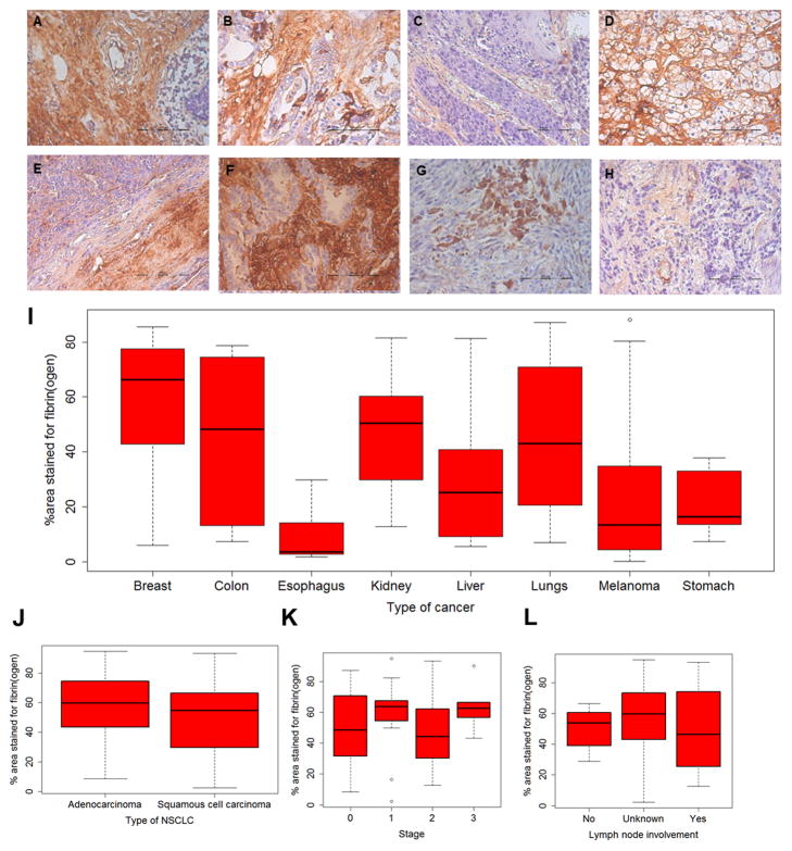
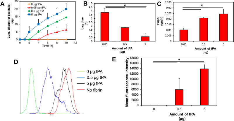
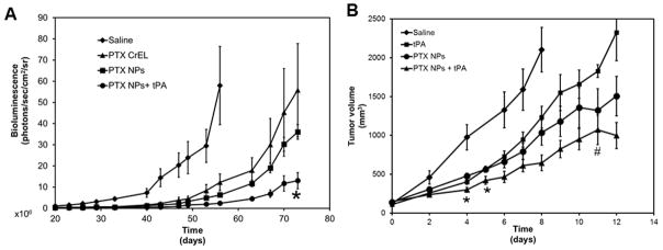
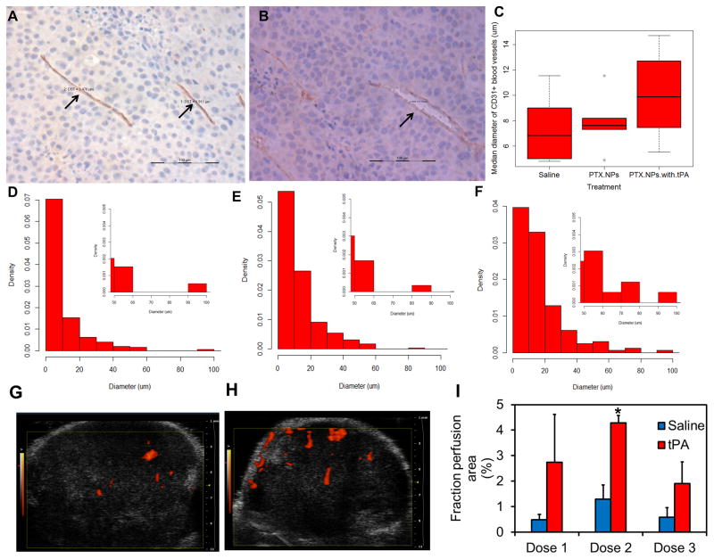
Elevated interstitial fluid pressure and solid stress within tumors contribute to poor intratumoral distribution of nanomedicine. In this study, we hypothesized that the presence of fibrin in tumor extracellular matrix contributes to hindered intratumoral distribution of nanocarriers and that this can be overcome through the use of a fibrinolytic enzyme such as tissue plasminogen activator (tPA). Analysis of fibrin expression in human tumor biopsies showed significant fibrin staining in nearly all tumor types evaluated. However, staining was heterogeneous across and within tumor types. We determined the effect of fibrin on the diffusion, intratumoral distribution, and therapeutic efficacy of nanocarriers. Diffusivity of nanocarriers in fibrin matrices was limited and could be improved significantly by coincubation with tPA. In vivo, coadministration of tPA improved the anticancer efficacy of nanoparticle-encapsulated paclitaxel in subcutaneous syngeneic mouse melanoma and orthotopic xenograft lung cancer models. Furthermore, treatment with tPA led to decompression of blood vessels and improved tumor perfusion. Cotreatment with tPA resulted in greater intratumoral penetration of a model nanocarrier (Doxil), leading to enhanced availability of the drug in the tumor core. Fibrinolytics such as tPA are already approved for other indications. Fibrinolytic cotherapy is therefore a rapidly translatable strategy for improving therapeutic effectiveness of anticancer nanomedicine. Cancer Res; 77(6); 1465-75. ©2017 AACR.


Conflict of interest statement

Conflict of interest: J.P., T.S. and A.R.K. have a US patent application regarding the use of fibrinolytics to improve chemotherapeutic efficacy of nanoparticles

Figures

Figure 1
Figure 1. Fibrin(ogen) staining of human tumor biopsies
Tissue microarrays of human tumors were stained for fibrin(ogen). Shown here are representative images of fibrin(ogen) staining for cancers of (A) breast, (B) colon, (C) esophagus, (D) kidney, (E) liver, (F) lungs, (G) melanoma, and (H) stomach. Scale bar in (A)–(H) is 100 μm. (I) shows fraction of area stained for fibrin(ogen). For lung tumors, 228 images of samples acquired from 59 patients were analyzed. For melanoma, 120 images of samples acquired from 30 patients were analyzed. For the other tumor types, 20 images of samples acquired from 5 patients were analyzed. (J–L) show fibrin(ogen) staining in non-small cell lung carcinomas as a function of type, stage and lymph node involvement.
Figure 2
Figure 2. Effect of tPA on nanoparticle mobility in fibrin gels
Transwell® assay was used to analyze the migration of nanoparticles (with or without tPA) across fibrin matrix. (A) Cumulative amount of paclitaxel accumulating in the bottom chamber as a function of time. (B) Lag time for the first appearance of paclitaxel in the bottom chamber with increasing amounts of tPA. Results are represented as mean ± S.D., n=3, *p<0.05, one-way ANOVA. (C) Apparent permeability of paclitaxel from the top to bottom chamber. Data represented as mean ± S.D., n=3, *p<0.05, one-way ANOVA (D) Cellular uptake of nanoparticles across fibrin gels was determined using Transwell® assay. Representative histograms indicating cellular uptake of fluorescently labeled nanoparticles. (E) Quantitation of cellular uptake of nanoparticles as function of tPA concentration. Results are represented as mean ± S.D., n=4, *p<0.05, one-way ANOVA.
Figure 3
Figure 3. Efficacy of paclitaxel nanoparticles-tPA combination therapy
(A) SCID-beige mice bearing orthotopic lung tumors were treated with paclitaxel. Tumor growth was monitored by measuring bioluminescence using IVIS in vivo imaging. Results shown as mean ± S.E.M, n=5–6, *p<0.05 compared to paclitaxel nanoparticles, Student’s t-test (B) C57Bl/6 mice bearing subcutaneous B16F10 melanoma tumors were treated with tPA, or paclitaxel nanoparticles or a combination of paclitaxel nanoparticles and tPA. Tumor volumes were measured over time using a digital caliper. Data represented as mean ± S.E.M., n=6–9 animals/group. *p<0.05 compared to paclitaxel nanoparticles, # indicates p<0.05 compared to tPA, Student’s t-test
Figure 4
Figure 4. Effect of tPA treatment on tumor blood vessel diameter and tumor perfusion
Representative pictures showing (A) compressed blood vessel (arrows) in saline-treated tumor and (B) decompressed blood vessel (arrow) in a tumor treated with tPA-paclitaxel nanoparticle combination therapy. Scale bar in A and B is 100 μm. (C) Median tumor blood vessel diameter in animals that received different treatments. Representative histograms showing tumor blood vessel diameter in animals treated with (D) saline, (E) paclitaxel nanoparticles, and (F) paclitaxel nanoparticles and tPA. B16F10 tumor bearing animals were treated with three doses of saline or tPA. Ultrasound images were taken after each dose. Representative sonograms of B16F10 tumors after receiving the second dose of (G) saline or (H) tPA. (I) Mean perfusion area in saline or tPA treated tumors. Data represented as mean ± S.E.M., n=3–5, *p<0.05, Student’s t-test.
Figure 5
Figure 5. Intratumoral and intraorgan distribution of Doxil® in B16F10 tumors
Tumor bearing mice were treated with either Doxil or a combination of Doxil and tPA. Animals were sacrificed 24 h later, tumors were collected and fluorescence of doxorubicin was imaged using confocal microscopy. Shown here are representative micrographs of tumors treated with (A) Doxil® or (B) combination of Doxil® and tPA. Arrows indicate fluorescence in tumor sections. Relative fluorescence intensity normalized to area in the entire tissue section and the central 40% and 10% of the tissue area in (C) tumor (D) liver (E) lung (F) spleen (G) brain. Data is represented as mean ± S.E.M., n=3 animals/group. Three tumor sections and one section of normal tissue from each animal were analyzed.

Similar articles

Cited by

References

    1. Minchinton AI, Tannock IF. Drug penetration in solid tumours. Nat Rev Cancer. 2006;6:583–92. - PubMed
    1. Moghimi SM, Hunter AC, Murray JC. Long-circulating and target-specific nanoparticles: theory to practice. Pharmacol Rev. 2001;53:283–318. - PubMed
    1. Torchilin VP. Polymer-coated long-circulating microparticulate pharmaceuticals. J Microencapsul. 1998;15:1–19. - PubMed
    1. Albanese A, Tang PS, Chan WC. The effect of nanoparticle size, shape, and surface chemistry on biological systems. Annu Rev Biomed Eng. 2012;14:1–16. - PubMed
    1. Chauhan VP, Stylianopoulos T, Boucher Y, Jain RK. Delivery of molecular and nanoscale medicine to tumors: transport barriers and strategies. Annu Rev Chem Biomol Eng. 2011;2:281–98. - PubMed

Publication types

MeSH terms