. 2017 Feb;216(2):367-377.
doi: 10.1083/jcb.201608128. Epub 2017 Jan 20.

VAPs and ACBD5 tether peroxisomes to the ER for peroxisome maintenance and lipid homeostasis

Affiliations

VAPs and ACBD5 tether peroxisomes to the ER for peroxisome maintenance and lipid homeostasis

Rong Hua et al. J Cell Biol. 2017 Feb.

Abstract

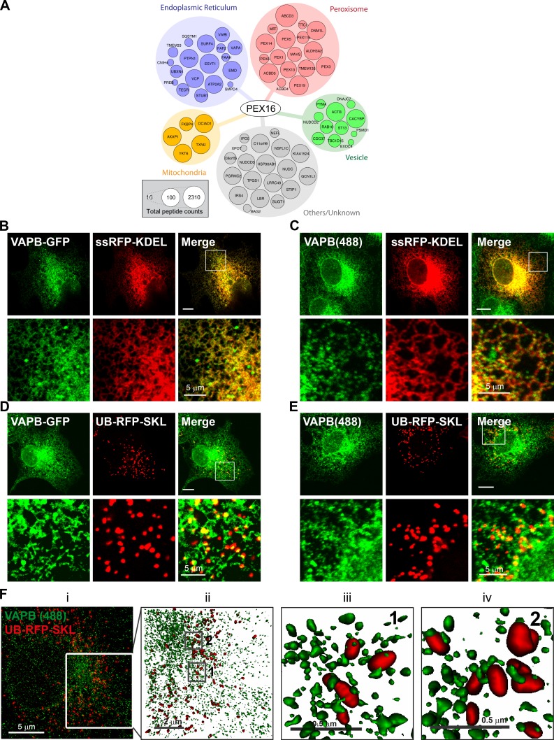
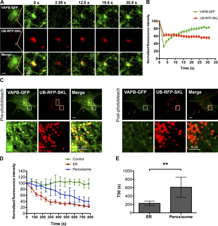
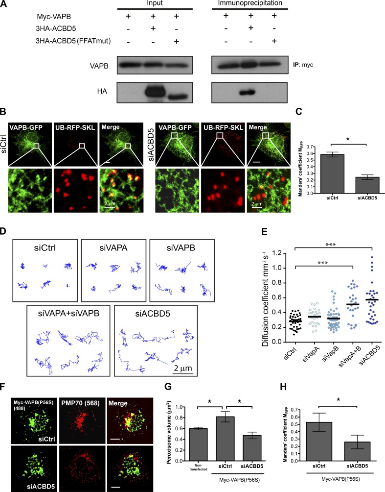
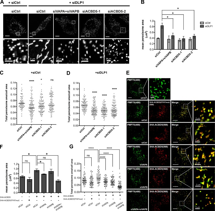
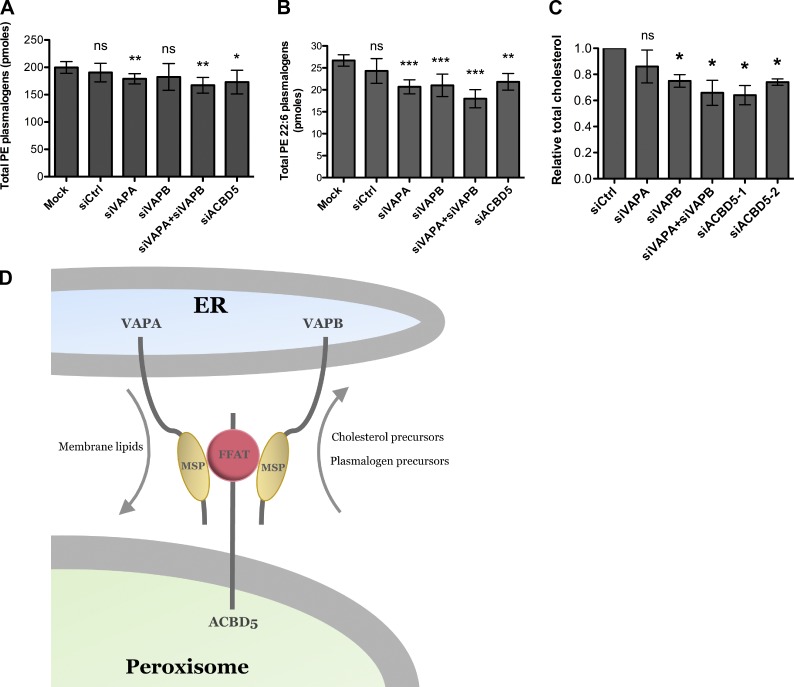
Lipid exchange between the endoplasmic reticulum (ER) and peroxisomes is necessary for the synthesis and catabolism of lipids, the trafficking of cholesterol, and peroxisome biogenesis in mammalian cells. However, how lipids are exchanged between these two organelles is not understood. In this study, we report that the ER-resident VAMP-associated proteins A and B (VAPA and VAPB) interact with the peroxisomal membrane protein acyl-CoA binding domain containing 5 (ACBD5) and that this interaction is required to tether the two organelles together, thereby facilitating the lipid exchange between them. Depletion of either ACBD5 or VAP expression results in increased peroxisome mobility, suggesting that VAP-ACBD5 complex acts as the primary ER-peroxisome tether. We also demonstrate that tethering of peroxisomes to the ER is necessary for peroxisome growth, the synthesis of plasmalogen phospholipids, and the maintenance of cellular cholesterol levels. Collectively, our data highlight the importance of VAP-ACBD5-mediated contact between the ER and peroxisomes for organelle maintenance and lipid homeostasis.


Figures

Figure 1.
Figure 1.
VAPB punctate structures colocalize with peroxisomes. (A) PEX16 interactome. Node size is proportional to peptide counts detected. COS7 cells transiently expressing ssRFP-KDEL (B and C) or UB-RFP-SKL (D and E) and either coexpressing VAPB-GFP (B and D) or immunostained for endogenous VAPB (C and E). The white boxes indicate the magnified area shown below each panel. Bars, 10 µm (or as indicated). (F, left to right) Maximum intensity projection of VAPB–Alexa 488 (green) and UB-RFP-SKL (red) acquired via SIM (i). (ii) Surface projection of the region denoted in panel i of F. Boxes denoted by 1 and 2 in panel ii are magnified in panels iii and iv, respectively. The surface projections demonstrate the apposition of the two organelles in 3D space.
Figure 2.
Figure 2.
VAPB localizes in juxtaposition to peroxisomes on the ER. (A) FRAP assay performed in a COS7 cell transiently coexpressing VAPB-GFP and UB-RFP-SKL. Yellow squares indicate the photobleached ROI. (B) FRAP curves. Shown is the normalized fluorescence intensity of VAPB-GFP and UB-RFP-SKL punctate structures within each ROI. Mean ± SD (n = 12). (C) FLIP assay performed in a COS7 cell transiently coexpressing VAPB-GFP and UB-RFP-SKL in a yellow rectangular ROI. Shown is the first frame before photobleaching (pre-photobleach) and the first image after repeated photobleaching (post-photobleach). The white boxes indicate the magnified area shown below each panel. Bars, 10 µm (or as indicated). (D) FLIP curves. Shown is the normalized fluorescence intensity of VAPB-GFP punctate structure juxtaposed to a peroxisome and that of ER-localized VAPB-GFP. The fluorescence intensity of VAPB-GFP in an adjacent cell from the same image serves as a control for imaging induced photobleaching. Mean ± SD (n = 6). (E) Bar graph illustrating the time taken by the ER localized VAPB-GFP and peroxisomal VAPB-GFP in D to drop to 50% of its original level (T50). Mean ± SD (n = 6). **, P < 0.01.
Figure 3.
Figure 3.
VAPB-ACBD5 tethers peroxisomes to the ER. (A) Coimmunoprecipitation performed in HEK293 cells transiently expressing Myc-VAPB (34 kD) with wild-type or FFAT-motif mutant (mut) ACBD5 (63 kD). (B) COS7 cells treated with indicated siRNAs, and coexpressing VAPB-GFP and UB-RFP-SKL. The white boxes indicate the magnified area shown below each panel. Bars, 10 µm (or as indicated). (C) Bar graph illustrating the Manders’ colocalization coefficient MRFP for UB-RFP-SKL and VAPB-GFP in B. Mean ± SD (n = 3; 20 cells/trial). (D) Representative trajectories of HeLa cells treated with indicated siRNAs and expressing UB-RFP-SKL. Z stacks of single cells were acquired at 40 frames/min, and the center of peroxisomes was tracked over 2 min. (E) The median diffusion coefficient of >27 cells from 3 experiments are graphed (dots) along with the mean (bars). Each video analyzed contained >30 trajectories and each condition >6,000 trajectories. (F) COS7 cells treated with indicated siRNAs, and expressing Myc-VAPB(P56S). Cells were immunostained for Myc tag and endogenous peroxisomal PMP70. Bars, 10 µm. (G and H) Quantification of mean peroxisome volume (G) and the Manders’ colocalization coefficient MRFP for PMP70 and Myc-VAPB(P56S) in F. Mean ± SD (n = 3; 20 cells/trial). *, P < 0.05; ***, P < 0.001. IP, immunoprecipitation.
Figure 4.
Figure 4.
Loss of VAP–ACBD5 tether prevents peroxisomal membrane expansion. (A) HeLa cells treated with indicated siRNAs and immunostained for PMP70. Bars, 10 µm (or as indicated). (B) Quantification of mean peroxisome area in HeLa cells treated with indicated siRNAs. Mean ± SD (n = 3; 30 cells/trial). Quantification of total peroxisome area in HeLa cells either in the absence (C) or presence (D) of DLP1 knockdown and treated with indicated siRNAs. The total peroxisome area of >90 cells from 3 experiments for each siRNA condition are graphed (dots) along with the medium (bars). One-way analysis of variance with Bonferroni correction. (E) COS7 cells treated with indicated siRNAs and expressing wild-type or the FFAT-motif mutant (mut) ACBD5. Cells were immunostained for HA tag and endogenous PMP70. The white boxes indicate the magnified area shown below each panel. Bars, 10 µm (or as indicated). (F) Quantification of mean peroxisome area in COS7 cells in E. Mean ± SD (n = 3; 30 cells/trial). (G) Quantification of total peroxisome area in COS7 cells in E. Quantification similar to C. *, P < 0.05; ****, P < 0.0001.
Figure 5.
Figure 5.
Loss of VAP–ACBD5 tether affects cellular plasmalogen and cholesterol levels. Bar graphs of total PE plasmalogens (A) and total PE 22:6 plasmalogens (B) in HeLa cells treated with indicated siRNAs. Mean ± SD (n = 4). *, P < 0.1; **, P < 0.05; ***, P < 0.01 as compared with mock cells. (C) Quantification of total cholesterol levels in HeLa cells treated with indicated siRNAs using the Amplex Red Cholesterol reagent. The total cholesterol level for each siRNA treatment (n = 3; mean ± SD) was normalized to that in siCtrl-treated cells. *, P < 0.05 as compared with siCtrl-treated cells. (D) Model for ER–peroxisome contact sites. The ER-anchored VAPs bind directly to the FFAT motif containing peroxisomal protein ACBD5 via their major sperm protein (MSP) domains to allow for peroxisome tethering and lipid exchange.

Comment in

References

    1. Agrawal G., and Subramani S.. 2016. De novo peroxisome biogenesis: Evolving concepts and conundrums. Biochim. Biophys. Acta. 1863:892–901. 10.1016/j.bbamcr.2015.09.014 - DOI - PMC - PubMed
    1. Agrawal G., Joshi S., and Subramani S.. 2011. Cell-free sorting of peroxisomal membrane proteins from the endoplasmic reticulum. Proc. Natl. Acad. Sci. USA. 108:9113–9118. 10.1073/pnas.1018749108 - DOI - PMC - PubMed
    1. Alpy F., Rousseau A., Schwab Y., Legueux F., Stoll I., Wendling C., Spiegelhalter C., Kessler P., Mathelin C., Rio M.C., et al. . 2013. STARD3 or STARD3NL and VAP form a novel molecular tether between late endosomes and the ER. J. Cell Sci. 126:5500–5512. 10.1242/jcs.139295 - DOI - PubMed
    1. Antonenkov V.D., and Hiltunen J.K.. 2012. Transfer of metabolites across the peroxisomal membrane. Biochim. Biophys. Acta. 1822:1374–1386. 10.1016/j.bbadis.2011.12.011 - DOI - PubMed
    1. Aranovich A., Hua R., Rutenberg A.D., and Kim P.K.. 2014. PEX16 contributes to peroxisome maintenance by constantly trafficking PEX3 via the ER. J. Cell Sci. 127:3675–3686. 10.1242/jcs.146282 - DOI - PMC - PubMed

MeSH terms

Grants and funding