Review
. 2018 Mar 1;8(3):a024091.
doi: 10.1101/cshperspect.a024091.

Cell Biology and Pathophysiology of α-Synuclein

Affiliations
Review

Cell Biology and Pathophysiology of α-Synuclein

Jacqueline Burré et al. Cold Spring Harb Perspect Med. .

Abstract

α-Synuclein is an abundant neuronal protein that is highly enriched in presynaptic nerve terminals. Genetics and neuropathology studies link α-synuclein to Parkinson's disease (PD) and other neurodegenerative disorders. Accumulation of misfolded oligomers and larger aggregates of α-synuclein defines multiple neurodegenerative diseases called synucleinopathies, but the mechanisms by which α-synuclein acts in neurodegeneration are unknown. Moreover, the normal cellular function of α-synuclein remains debated. In this perspective, we review the structural characteristics of α-synuclein, its developmental expression pattern, its cellular and subcellular localization, and its function in neurons. We also discuss recent progress on secretion of α-synuclein, which may contribute to its interneuronal spread in a prion-like fashion, and describe the neurotoxic effects of α-synuclein that are thought to be responsible for its role in neurodegeneration.


Figures

Figure 1.
Figure 1.
Historical timeline of α-synuclein-related findings. Marked are the major cell biological discoveries (top, blue) and pathobiological findings (bottom, red).
Figure 2.
Figure 2.
Schematic of α-synuclein conformations associated with its physiological function and pathological activities. Soluble α-synuclein is natively unstructured and monomeric. After binding to highly curved membranes, such as synaptic vesicles, α-synuclein undergoes a conformational change and folds into an amphipathic α-helix, which is associated with multimerization and mediates its SNARE-complex chaperoning function. Under pathological conditions, soluble α-synuclein forms β-sheet-like oligomers (protofibrils), which convert into amyloid fibrils and eventually deposit into Lewy bodies. Protofibrils and fibrils may propagate from neuron to neuron in Parkinson’s disease and Lewy body dementia and from glia to glia in multiple system atrophy. (From Burré et al. 2015; reprinted, with permission, from the authors.)
Figure 3.
Figure 3.
Soluble native α-synuclein is predominantly an unstructured monomer. (A) Sodium dodecyl sulfate polyacrylamide gel electrophoresis (SDS-PAGE) analysis of five stages of α-synuclein purification from mouse brain. Purified α-synuclein was analyzed by immunoblotting and mass spectrometry as shown. (B) Size-exclusion chromatography multi-angle light scattering (SEC-MALS) shows that purified α-synuclein from mouse brain tissue is largely monomeric (main peak with a mass of 17 ± 1 kDa), but includes a minor component (plateau along the left shoulder with a mass of 58 ± 5 kDa) that contains little detectable α-synuclein (see immunoblot in boxed region). Calculated masses were extracted from marked areas. (From Burré et al. 2013; reprinted, with permission, from the authors.) (C) Two-dimensional 1H–15N nuclear magnetic resonance (NMR) spectra of α-synuclein in A2780 and SK-N-SH cells (red, selected regions) and of isolated N-terminally acetylated α-synuclein in buffer (black). (D) N-terminal α-synuclein residues experiencing site-selective signal attenuations (boxed) are expanded, with in-cell NMR contours plotted at 2.5-fold lower levels (red). In-cell NMR cross-peaks are superimposed with reference NMR signals of N-terminally acetylated α-synuclein in buffer (black), confirming the presence of this modification in mammalian cells. IEX, Anion exchange chromatography; HIC, hydrophobic interaction chromatography; AcM1, acetylated Met1. (From Theillet et al. 2016; reprinted, with permission, from Nature Publishing Group © 2016.)
Figure 4.
Figure 4.
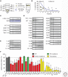
Binding of α-synuclein mutants to phospholipid membranes. (A,B) Phospholipid binding assay. Liposomes mixed with wild-type (WT) and mutant α-synuclein were floated by density gradient centrifugation (A). Based on liposome distribution in the gradient (B), the top two fractions 1 and 2 were defined as lipid-bound fractions. (C) Lack of flotation of bovine serum albumin (BSA) and α-synuclein in the absence of liposomes or with uncharged liposomes, analyzed by Coomassie staining or by immunoblotting with antibodies to the myc-epitope fused to α-synuclein. (DF) Quantitation of phospholipid binding by WT and mutant α-synuclein. Flotation of α-synuclein point mutants (D) and truncations (E) with liposomes was quantitated as the sum of the top two fractions, and was plotted as the percentage of total α-synuclein in the gradient (F). Data are means ± SEM (**P < 0.01, ***P < 0.001 by Student’s t-test; n = 6 independent experiments). RT, Room temperature. (From Burré et al. 2012; reprinted, with permission, from the authors.)
Figure 5.
Figure 5.
α-Synuclein directly binds to synaptobrevin-2/vesicle-associated membrane protein 2 (VAMP2) in SNARE complexes. (A) Coimmunoprecipitation of α-synuclein with SNARE complexes reconstituted in HEK293T cells. Cell lysates were immunoprecipitated with antibodies to (left) α-synuclein or (right) SNAP-25 and analyzed by means of immunoblotting. (B,C) The C terminus of α-synuclein directly binds to the N terminus of synaptobrevin-2. α-Synuclein was immunoprecipitated from HEK293T cells coexpressing full-length (α-Syn) or C-terminally truncated α-synuclein (α-Syn1-95) with full-length (Syb2) or N-terminally truncated synaptobrevin-2 (Syb229-116). Immunoprecipitates were analyzed by means of immunoblotting of α-synuclein and synaptobrevin-2. (D) Diagram of the α-synuclein/synaptobrevin-2 complex on synaptic vesicles (SVs). (From Burré et al. 2010; reprinted, with permission, from the authors.)
Figure 6.
Figure 6.
α-Synuclein boosts SNARE-complex assembly and clusters liposomes. (A) Linear relationship between α-synuclein levels and SNARE-complex assembly in α, β, γ-synuclein triple-knockout neurons infected at DIV4 with increasing amounts of lentivirus expressing α-synuclein and analyzed by means of immunoblotting of nonboiled samples at DIV17 (n = 3 to 6 cultures). (From Burré et al. 2010; reprinted, with permission, from the authors.) (B) Native α-synuclein promotes liposome vesicle clustering in a concentration-dependent manner by binding to both synaptobrevin-2 and anionic membranes. Bar graph: Quantitation of interacting vesicles. (Bottom panel) Representative fluorescence images of interacting vesicles on the imaging surface. Data are means ± SD (***P < 0.001 by Student’s t-test; n = 15 random imaging locations in the sample channel). (From Diao et al. 2013; reprinted, with permission, from the authors.)

Similar articles

Cited by

References

    1. Abeliovich A, Schmitz Y, Farinas I, Choi-Lundberg D, Ho WH, Castillo PE, Shinsky N, Verdugo JM, Armanini M, Ryan A, et al. 2000. Mice lacking α-synuclein display functional deficits in the nigrostriatal dopamine system. Neuron 25: 239–252. - PubMed
    1. Ahmad M, Attoub S, Singh MN, Martin FL, El-Agnaf OM. 2007. γ-Synuclein and the progression of cancer. FASEB J 21: 3419–3430. - PubMed
    1. Ahn BH, Rhim H, Kim SY, Sung YM, Lee MY, Choi JY, Wolozin B, Chang JS, Lee YH, Kwon TK, et al. 2002. α-Synuclein interacts with phospholipase D isozymes and inhibits pervanadate-induced phospholipase D activation in human embryonic kidney-293 cells. J Biol Chem 277: 12334–12342. - PubMed
    1. Ahn KJ, Paik SR, Chung KC, Kim J. 2006a. Amino acid sequence motifs and mechanistic features of the membrane translocation of α-synuclein. J Neurochem 97: 265–279. - PubMed
    1. Ahn M, Kim S, Kang M, Ryu Y, Kim TD. 2006b. Chaperone-like activities of α-synuclein: α-synuclein assists enzyme activities of esterases. Biochem Biophys Res Commun 346: 1142–1149. - PubMed

Publication types

Substances