. 2017 Mar;205(3):1101-1112.
doi: 10.1534/genetics.116.196089. Epub 2017 Jan 20.

Meiotic Crossing Over in Maize Knob Heterochromatin

Affiliations

Meiotic Crossing Over in Maize Knob Heterochromatin

Stephen M Stack et al. Genetics. 2017 Mar.

Abstract

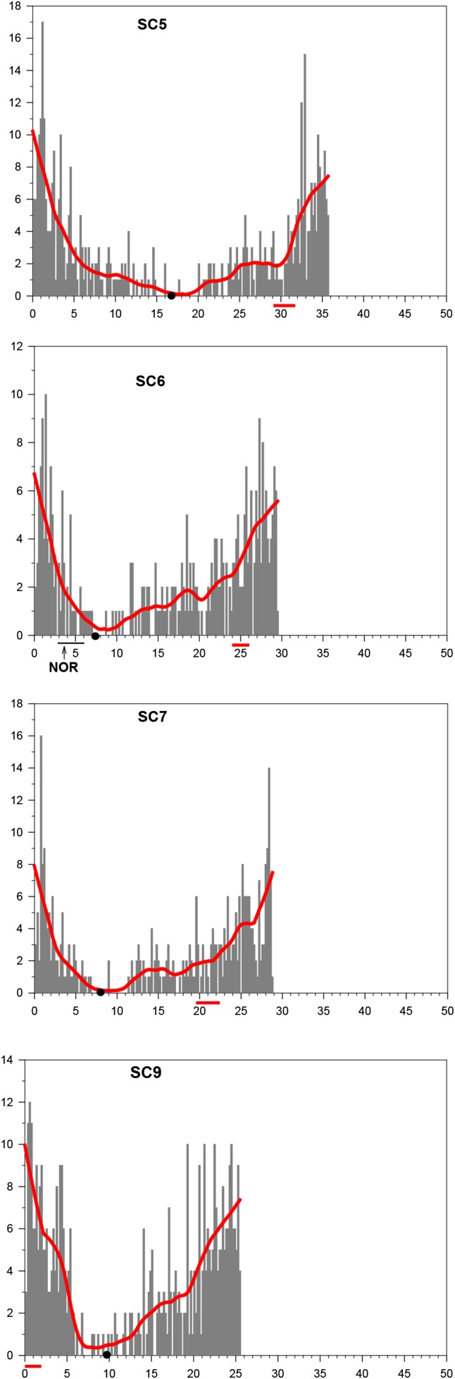
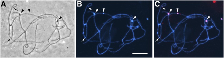
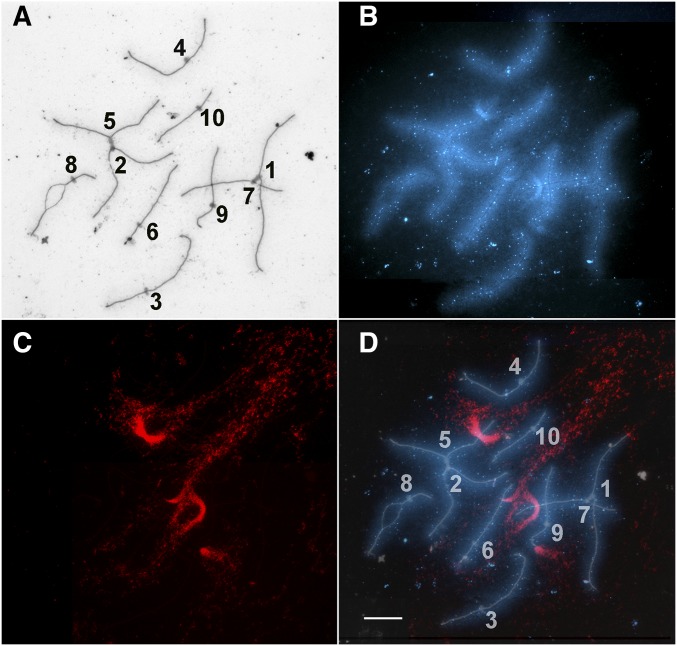
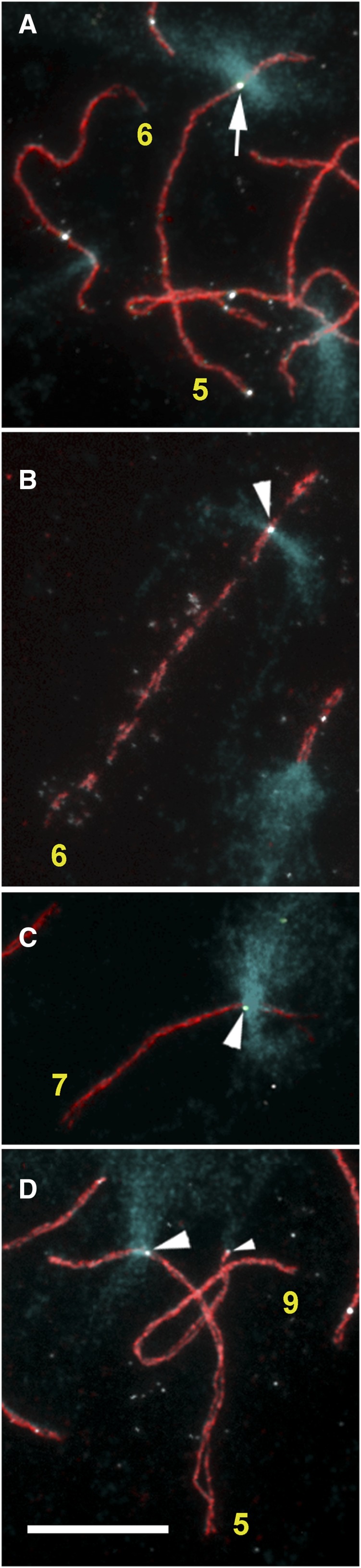
There is ample evidence that crossing over is suppressed in heterochromatin associated with centromeres and nucleolus organizers (NORs). This characteristic has been attributed to all heterochromatin, but the generalization may not be justified. To investigate the relationship of crossing over to heterochromatin that is not associated with centromeres or NORs, we used a combination of fluorescence in situ hybridization of the maize 180-bp knob repeat to show the locations of knob heterochromatin and fluorescent immunolocalization of MLH1 protein and AFD1 protein to show the locations of MLH1 foci on maize synaptonemal complexes (SCs, pachytene chromosomes). MLH1 foci correspond to the location of recombination nodules (RNs) that mark sites of crossing over. We found that MLH1 foci occur at similar frequencies per unit length of SC in interstitial knobs and in the 1 µm segments of SC in euchromatin immediately to either side of interstitial knobs. These results indicate not only that crossing over occurs within knob heterochromatin, but also that crossing over is not suppressed in the context of SC length in maize knobs. However, because there is more DNA per unit length of SC in knobs compared to euchromatin, crossing over is suppressed (but not eliminated) in knobs in the context of DNA length compared to adjacent euchromatin.

Keywords: crossing over; heterochromatin; knobs; maize; synaptonemal complex.


Figures

Figure 1
Figure 1
Histograms showing the distribution of RNs along the length of KYS maize SCs 5, 6, 7, and 9. Each SC is represented on the x-axis in micrometers with the short arm to the left and the position of the kinetochore marked by a black dot. Beneath the x-axis, the locations of knobs are marked by horizontal, thick red lines, and the position of the NOR on SC 6 is marked by a horizontal, thin black line. The histogram bars on the y-axis represent the total number of RNs observed in each 0.2-μm segment of SC length for 203 SC 5’s, 176 SC 6’s, 178 SC 7’s, and 234 SC 9’s. A red smoothing line is superimposed over each histogram to show the general trend of RN distribution. These histograms are used with permission from GENETICS (Anderson et al. 2003). RN, recombination nodule; SC, synaptonemal complex.
Figure 2
Figure 2
Model to explain how crossing over could be suppressed in maize knob heterochromatin while RN frequencies on SC in knobs are similar to RN frequencies on SC in nearby euchromatin. In this frontal view of an SC, short, paired sister loops of euchromatin (orange) extend laterally from their regularly spaced attachment sites on lateral elements of the SC. Here, homozygous knob chromatin (black) consists of long, paired sister loops in each homolog that are collapsed to cover a length of SC well beyond their small attachment sites on the lateral elements. Thus, most of the SC covered by a knob is actually in euchromatin. A RN in the central region of the SC is shown located in euchromatin near the attachment sites of the knob loops. While this RN marks a crossover in euchromatin, it would look like the RN is in knob heterochromatin, thereby explaining our observations that RN frequencies “in knobs” are similar to RN frequencies in nearby euchromatin. This figure is used with permission from GENETICS (Anderson et al. 2003). RN, recombination nodule; SC, synaptonemal complex.
Figure 3
Figure 3
KYS maize pachytene chromosome squash. (A) By phase microscopy, large interstitial knobs (arrowheads) on the unstained chromosomes are visible along with smaller terminal knobs (small arrows). Pericentric heterochromatin is visible as darker segments of bivalents that are often about one-third of the length of each bivalent, e.g., observe the bivalent at the lower center. (B) The same squash after DAPI staining. Knobs are prominent bright blue enlargements along bivalents indicating a high concentration of DNA in knobs. Pericentric heterochromatin is also brighter than the distal euchromatin indicating higher concentrations of DNA in pericentric heterochromatin (but less than in knobs). (C) The same squash after FISH with the 180-bp knob repeat (red). Large interstitial knobs (arrowheads) and the smaller terminal knobs (small arrows) are specifically labeled with the 180-bp repeat probe. Bar, 5 µm.
Figure 4
Figure 4
Maize pachytene SC spread. (A) SCs and kinetochores stained with silver. Each SC has been identified on the basis of relative length and arm ratio, and numbered at its kinetochore. Kinetochores on SCs 1 and 7 are stuck together as are the kinetochores on SCs 2 and 5. SC 8 is partially asynapsed. (B) The same SC spread visualized with DAPI fluorescence. Due to the hypotonic spreading procedure, the chromatin loops around the SCs are dispersed laterally as a fuzzy blue coat along the length of each SC. (C) The FISH signal (red) of the 180-bp repeat for this SC spread. (D) Merged images of silver-stained SCs (inverted to appear white), DAPI-stained chromatin (blue), and the 180-bp knob repeat (red). The chromatin of the interstitial knobs (on the long arms of SCs 5, 6, and 7) occupies well-defined SC segments, and at least some chromatin loops from the knobs (red) extend much farther from their SC attachment sites than chromatin loops in distal euchromatin and pericentric heterochromatin. For the terminal knobs (on the short arms of SCs 1 and 9) the loops also extend far from their attachment sites, but the loops appear to extend from the tips of the SCs without defining distinct terminal SC segments. Bar, 5 µm. SC, synaptonemal complex.
Figure 5
Figure 5
Portions of maize SC spreads labeled with antibodies to MLH1 (white foci) and AFD1 (red, to show the SC axes) and hybridized to the 180-bp knob repeat (blue). MLH1 foci are observed near (arrow) and in (arrowheads) knobs. (A) Portion of a pachytene SC spread showing an MLH1 focus near the edge of the knob on SC 5. Other MLH1 foci in this image are not in or near knobs. (B) Portion of an SC spread in early diplotene showing an MLH1 focus in the interstitial knob of the long arm of SC 6. (C) Portion of a pachytene SC spread showing an MLH1 focus in the interstitial knob on the long arm of SC 7. (D) Portion of a pachytene SC spread showing an MLH1 focus in the interstitial knob of SC 5 and a small MLH1 focus at the tip of the short arm of SC 9 (small arrowhead) where the terminal knob is located. Bar, 5 µm. SC, synaptonemal complexes.

References

    1. Albini S. M., Jones G. H., 1988. Synaptonemal complex spreading in Allium cepa and Allium fistulosum. II. Pachytene observations: the SC karyotype and the correspondence of late recombination nodules and chiasmata. Genome 30: 399–410.
    1. Ananiev E. V., Phillips R. L., Rines H. W., 1998a Complex structure of the knob DNA on maize chromosome 9: retrotransposon invasion into heterochromatin. Genetics 149: 2025–2037. - PMC - PubMed
    1. Ananiev E. V., Phillips R. L., Rines H. W., 1998b A knob-associated tandem repeat in maize capable of forming fold-back DNA segments: are chromosome knobs megatransposons? Proc. Natl. Acad. Sci. USA 95: 10785–10790. - PMC - PubMed
    1. Anderson L. K., Reeves A., Webb L. M., Ashley T., 1999. Distribution of crossing over on mouse synaptonemal complexes using immunofluorescent localization of MLH1 protein. Genetics 151: 1569–1579. - PMC - PubMed
    1. Anderson L. K., Doyle G. G., Brigham B., Carter J., Hooker K. D., et al. , 2003. High resolution crossover maps for each bivalent of Zea mays using recombination nodules. Genetics 165: 849–865. - PMC - PubMed

MeSH terms

LinkOut - more resources