

Immunosuppressive myeloid-derived suppressor cells are increased in splenocytes from cancer patients
- PMID: 28108766
- PMCID: PMC5580403
- DOI: 10.1007/s00262-016-1953-z
Immunosuppressive myeloid-derived suppressor cells are increased in splenocytes from cancer patients
Abstract
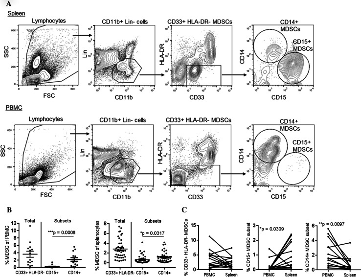
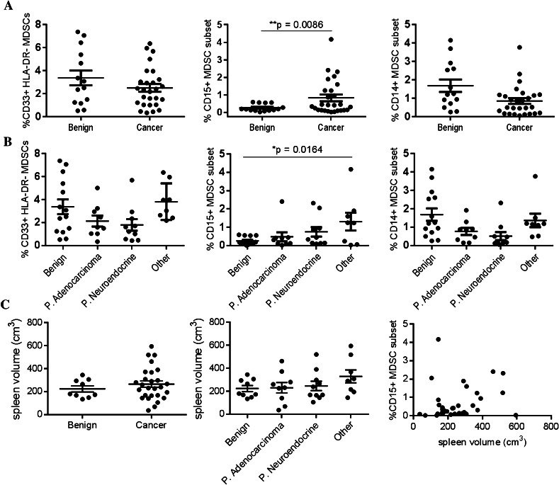
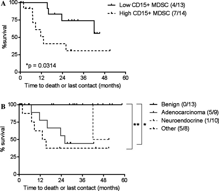
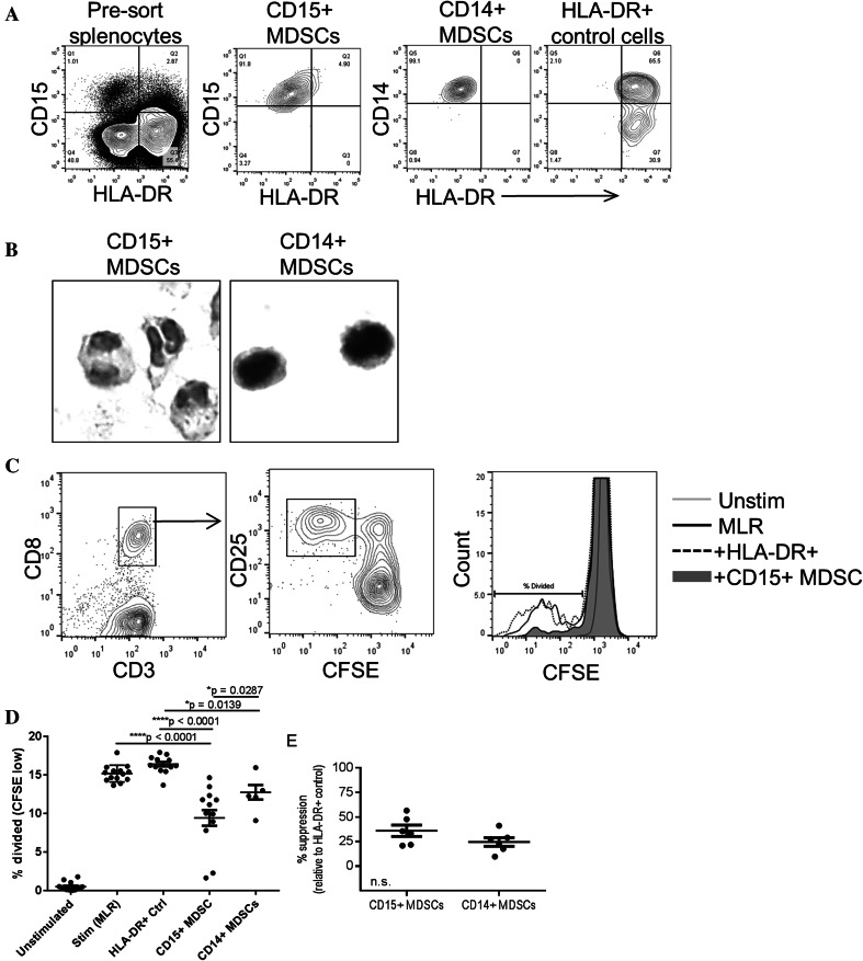
Myeloid-derived suppressor cells (MDSCs) are a heterogeneous population of myeloid cells that are increased in the peripheral blood of cancer patients and limit productive immune responses against tumors. Immunosuppressive MDSCs are well characterized in murine splenic tissue and are found at higher frequencies in spleens of tumor-bearing mice. However, no studies have yet analyzed these cells in parallel human spleens. We hypothesized that MDSCs would be increased in the spleens of human cancer patients, similar to tumor-bearing mice. We compared the frequency and function of MDSC subsets in dissociated human spleen from 16 patients with benign pancreatic cysts and 26 patients with a variety of cancers. We found that total MDSCs (Linneg CD11bpos CD33pos HLA-DRneg), granulocytic MDSCs (additional markers CD14neg CD15pos), and monocytic MDSCs (CD14pos CD15neg) were identified in human spleen. The monocytic subset was the most prominent in both spleen and peripheral blood and the granulocytic subset was expanded in the spleen relative to matched peripheral blood samples. Importantly, the frequency of CD15pos MDSCs in the spleen was increased in patients with cancer compared to patients with benign pancreatic cysts and was associated with a significantly increased risk of death and decreased overall survival. Finally, MDSCs isolated from the spleen suppressed T cell responses, demonstrating for the first time the functional capacity of human splenic MDSCs. These data suggest that the human spleen is a potential source of large quantities of cells with immunosuppressive function for future characterization and in-depth studies of human MDSCs.
Keywords: Cancer; Human spleen; Immunosuppression; MDSCs.
Conflict of interest statement
The authors declare that they have no conflicts of interest.
Figures
Similar articles
-
CD4+ T effector memory cell dysfunction is associated with the accumulation of granulocytic myeloid-derived suppressor cells in glioblastoma patients.Neuro Oncol. 2016 Jun;18(6):807-18. doi: 10.1093/neuonc/nov280. Epub 2015 Nov 17. Neuro Oncol. 2016. PMID: 26578623 Free PMC article.
-
A circulating subpopulation of monocytic myeloid-derived suppressor cells as an independent prognostic/predictive factor in untreated non-small lung cancer patients.J Immunol Res. 2014;2014:659294. doi: 10.1155/2014/659294. Epub 2014 Nov 11. J Immunol Res. 2014. PMID: 25436215 Free PMC article.
-
Human splenic myeloid derived suppressor cells: Phenotypic and clustering analysis.Cell Immunol. 2021 May;363:104317. doi: 10.1016/j.cellimm.2021.104317. Epub 2021 Mar 1. Cell Immunol. 2021. PMID: 33714729 Free PMC article.
-
Myeloid-derived suppressor cells in cancer immunotherapy-clinical perspectives.Life Sci. 2021 Jul 15;277:119627. doi: 10.1016/j.lfs.2021.119627. Epub 2021 May 15. Life Sci. 2021. PMID: 34004256 Review.
-
The CD14+HLA-DRlo/neg Monocyte: An Immunosuppressive Phenotype That Restrains Responses to Cancer Immunotherapy.Front Immunol. 2019 May 22;10:1147. doi: 10.3389/fimmu.2019.01147. eCollection 2019. Front Immunol. 2019. PMID: 31191529 Free PMC article. Review.
Cited by
-
Immunosuppression mediated by myeloid-derived suppressor cells (MDSCs) during tumour progression.Br J Cancer. 2019 Jan;120(1):16-25. doi: 10.1038/s41416-018-0333-1. Epub 2018 Nov 9. Br J Cancer. 2019. PMID: 30413826 Free PMC article. Review.
-
Tumor-Associated Macrophages in Hepatocellular Carcinoma Pathogenesis, Prognosis and Therapy.Cancers (Basel). 2022 Jan 4;14(1):226. doi: 10.3390/cancers14010226. Cancers (Basel). 2022. PMID: 35008390 Free PMC article. Review.
-
Splenic Volume as a Surrogate Marker of Immune Checkpoint Inhibitor Efficacy in Metastatic Non Small Cell Lung Cancer.Cancers (Basel). 2021 Jun 16;13(12):3020. doi: 10.3390/cancers13123020. Cancers (Basel). 2021. PMID: 34208673 Free PMC article.
-
Evaluating the efficacy of enzalutamide and the development of resistance in a preclinical mouse model of type-I endometrial carcinoma.Neoplasia. 2020 Oct;22(10):484-496. doi: 10.1016/j.neo.2020.07.003. Epub 2020 Aug 17. Neoplasia. 2020. PMID: 32818842 Free PMC article.
-
High-Dimensional Analysis of Postsplenectomy Peripheral Immune Cell Changes.Immunohorizons. 2020 Feb 18;4(2):82-92. doi: 10.4049/immunohorizons.1900089. Immunohorizons. 2020. PMID: 32071067 Free PMC article.
References
-
- Mandruzzato S, Brandau S, Britten CM, Bronte V, Damuzzo V, Gouttefangeas C, Maurer D, Ottensmeier C, van der Burg SH, Welters MJ, Walter S. Toward harmonized phenotyping of human myeloid-derived suppressor cells by flow cytometry: results from an interim study. Cancer Immunol Immunother. 2016;65(2):161–169. doi: 10.1007/s00262-015-1782-5. - DOI - PMC - PubMed
-
- Zea AH, Rodriguez PC, Atkins MB, Hernandez C, Signoretti S, Zabaleta J, McDermott D, Quiceno D, Youmans A, O’Neill A, Mier J, Ochoa AC. Arginase-producing myeloid suppressor cells in renal cell carcinoma patients: a mechanism of tumor evasion. Cancer Res. 2005;65(8):3044–3048. - PubMed
-
- Hoechst B, Ormandy LA, Ballmaier M, Lehner F, Kruger C, Manns MP, Greten TF, Korangy F. A new population of myeloid-derived suppressor cells in hepatocellular carcinoma patients induces CD4(+)CD25(+)Foxp3(+) T cells. Gastroenterology. 2008;135(1):234–243. doi: 10.1053/j.gastro.2008.03.020. - DOI - PubMed
-
- Bronte V, Brandau S, Chen SH, Colombo MP, Frey AB, Greten TF, Mandruzzato S, Murray PJ, Ochoa A, Ostrand-Rosenberg S, Rodriguez PC, Sica A, Umansky V, Vonderheide RH, Gabrilovich DI. Recommendations for myeloid-derived suppressor cell nomenclature and characterization standards. Nat Commun. 2016;7:12150. doi: 10.1038/ncomms12150. - DOI - PMC - PubMed
Publication types
MeSH terms
Grants and funding
LinkOut - more resources
Full Text Sources
Other Literature Sources
Research Materials
