Review
. 2017 Apr;8(2):199-212.
doi: 10.1007/s13244-016-0541-2. Epub 2017 Jan 20.

"Unforgettable" - a pictorial essay on anatomy and pathology of the hippocampus

Affiliations
Review

"Unforgettable" - a pictorial essay on anatomy and pathology of the hippocampus

Sven Dekeyzer et al. Insights Imaging. 2017 Apr.

Abstract

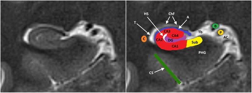
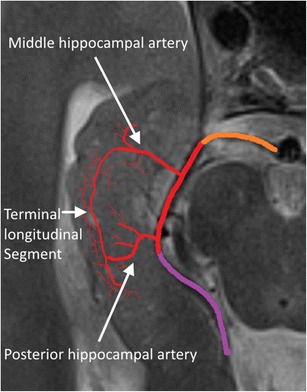
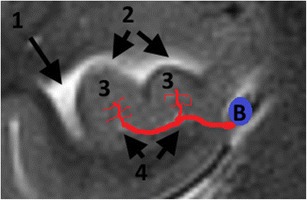
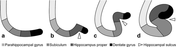
The hippocampus is a small but complex anatomical structure that plays an important role in spatial and episodic memory. The hippocampus can be affected by a wide range of congenital variants and degenerative, inflammatory, vascular, tumoral and toxic-metabolic pathologies. Magnetic resonance imaging is the preferred imaging technique for evaluating the hippocampus. The main indications requiring tailored imaging sequences of the hippocampus are medically refractory epilepsy and dementia. The purpose of this pictorial review is threefold: (1) to review the normal anatomy of the hippocampus on MRI; (2) to discuss the optimal imaging strategy for the evaluation of the hippocampus; and (3) to present a pictorial overview of the most common anatomic variants and pathologic conditions affecting the hippocampus.

Teaching points: • Knowledge of normal hippocampal anatomy helps recognize anatomic variants and hippocampal pathology. • Refractory epilepsy and dementia are the main indications requiring dedicated hippocampal imaging. • Pathologic conditions centered in and around the hippocampus often have similar imaging features. • Clinical information is often necessary to come to a correct diagnosis or an apt differential.

Keywords: Dementia; Epilepsy; Herpes simplex encephalitis; Hippocampus; MRI.


Conflict of interest statement

Martin Wiesmann has the following disclosures:

• Grants: Stryker Neurovascular. Siemens Healthcare

• Personal fees: Stryker Neurovascular. Silkroad Medical. Siemens Healthcare. Bracco

• Non-financial support: Codman Neurovascular. Covidien. Abbott. St. Jude Medical. Phenox. Penumbra. Microvention/Terumo. B. Braun. Bayer. Acandis. ab medica

The other authors have nothing to disclose.

The article received no grants or funding.

Figures

Fig. 1
Fig. 1
Anatomy of the hippocampal formation on 3-T axial T2 (a) and sagittal 3D-MPRAGE images (b). Zoomed-in 3-T coronal T2-weighted images at the level of the hippocampal head (c) and the hippocampal tail (d). The hippocampal body is shown in detail in Fig. 2. 1 = hippocampal head, 2 = hippocampal body, 3 = hippocampal tail, 4 = mesencephalon, 5 = amygdala, 6 = hippocampal digitations, 7 = temporal horn of the lateral ventricle, 8 = uncal recess of the lateral ventricle, 9 = splenium of the corpus callosum, 10 = subsplenial gyri, 11 = crura of the fornices. To easily recognize the different portions of the hippocampus, we can use the mesencephalon (4). The head (1) is located in front of the mesencephalon, the body (2) can be found at the level of the mesencephalon and the tail (3) is posterior to the mesencephalon. The hippocampal head is the only portion of the hippocampus not covered by the choroid plexus (7). The hippocampal head is separated from the amygdala (5) by the uncal recess of the lateral ventricle (7) and is characterized by small digitations separated by small sulci, the digitationes hippocampi (6). At the level of the hippocampal tail, the fimbriae continue posteriorly as the crux of the fornix (11) that slants upwards towards the splenium of the corpus callosum (9) and the hippocampal tail continues as the subsplenial gyri (10).
Fig. 2
Fig. 2
Anatomy of the hippocampal formation at the level of the hippocampal body on 3-T coronal T2. The hippocampal formation consists of the cornu ammonis or hippocampus proper, which can histologically be divided in the four Sommer sectors CA1–CA4, and the dentate gyrus (DG). A small hippocampal cyst (Hs) reflects the location of the largely obliterated hippocampal sulcus. A = alveus, Ac = Ambient cistern, B = basal vein of Rosenthal, C = tail of caudate nucleus, ChF = choroid fissure, CS = collateral suclus, DG = dentate gyrus, P = posterior cerebral artery, PHG = parahippocampal gyrus, Sub = subiculum, T = temporal horn of the lateral ventricle, Tb = transverse fissure of Bichat
Fig. 3
Fig. 3
Arterial supply of the hippocampal body and tail. Orange = P1, red = P2 and purple = P3 segment of the posterior cerebral artery. The anterior hippocampal artery is hidden in the uncal sulcus and is shown in Fig. 4
Fig. 4
Fig. 4
Arterial supply of the hippocampal head. B = basal Rosenthal vein, 1 = temporal horn of the lateral ventricle, 2 = uncal recess of the lateral ventricle, 3 = hippocampal digitations, 4 = uncal sulcus. Both the anterior hippocampal artery, originating from the trunk or branches of the posterior cerebral artery, and the uncal branch of the anterior choroidal artery, dive into the uncal sulcus at the level of the hippocampal head and form anastomoses in the sulci between the hippocampal digitations. Here, only one of both arteries in the uncal sulcus is drawn
Fig. 5
Fig. 5
Embryologic development of the hippocampus (image taken from Atlas klinische Neuroradiologie des Gehirns, Springer Berlin Heidelberg, 2011, p. 23, by Lin, Wiesmann and Brückmann. With permission of Springer ©)
Fig. 6
Fig. 6
Bilateral sulcal remnant cysts (a) and right-sided choroid fissure cyst (b, c). Coronal T2 shows small bilateral cysts at the apex of the hippocampal fold between the dentate gyrus and Ammon’s horn (a). Coronal FLAIR (b) and axial T2-weighted (c) images show a space-occupying cystic lesion, iso-intense to cerebrospinal fluid, at the level of the right choroid fissure
Fig. 7
Fig. 7
Isolated IHI. Coronal 3-T T2-weighted image in a 23-year-old patient with epilepsy shows a normal hippocampus on the right compared to an incompletely inverted hippocampus with an abnormal rounded or pyramidal shape on the left. The left collateral sulcus (asterisk) has a more vertical orientation and is found lateral of the hippocampal body
Fig. 8
Fig. 8
IHI associated with other developmental anomalies. Coronal T2-weighted image in a 22-year-old male patient with mental retardation and epilepsy (a) shows incomplete inversion of the right hippocampus (white asterisk at the collateral sulcus) along with a right perisylvian open lip schizencephaly (white arrows), right perisylvian subependymal heterotopia (white arrowheads) and agenesis of the septum pellucidum. Coronal T2-weighted images in a 25-year-old patient with mental retardation and epilepsy (b) shows agenesis of the corpus callosum and an abnormal globular shape of both hippocampi, corresponding to a bilateral incomplete hippocampal inversion
Fig. 9
Fig. 9
Hippocampal calcifications. Non-enhanced axial CT images angulated parallel to the hippocampal body show bilateral symmetrical calcifications lateral in the hippocampal bodies as a coincidental finding in a 69-year-old female patient with dysarthria
Fig. 10
Fig. 10
Left-sided mesial temporal sclerosis. 3 T coronal T2-weighted (a) and FLAIR images (b) in a 43-year-old patient with medically refractory epilepsy show volume loss, increased signal intensity and blurring of the internal structure of the left hippocampus, compatible with mesial temporal sclerosis
Fig. 11
Fig. 11
Bilateral hippocampal sclerosis and dual pathology. 3-T coronal T2-weighted image in a 35-year-old patient with medically refractory epilepsy shows volume loss and increased signal intensity of the right hippocampus, compatible with mesial temporal sclerosis (a). The left side the hippocampal volume looks normal, but T2 signal intensity is slightly increased in the CA1 and CA2 sectors and there is blurring of the internal structure (a). On follow-up MRI performed 3 years later, there is clear bilateral hippocampal sclerosis with atrophy and increased T2 signal intensity of both hippocampi (b). Axially reconstructed 3D MPRAGE images (c) show multiple periventricular heterotopias (white arrows) along the lateral wall of the left lateral ventricle
Fig. 12
Fig. 12
The Scheltens mesial temporal atrophy scale in coronal 3D-MPRAGE images (images from the Radiology Assistant website with permission – http://www.radiologyassistant.nl)
Fig. 13
Fig. 13
Alzheimer’s disease (a) and frontotemporal dementia (b). 3-T Coronal T2-weighted images in an 84-year-old patient with clinically advanced sporadic Alzheimer dementia show pronounced mesial temporal atrophy corresponding to an MTA score of 4 (a). In comparison, coronal T2-weighted images in a 58-year-old patient with a semantic variant of frontotemporal dementia also shows pronounced bilateral mesiotemporal atrophy (MTA score 4), along with asymmetric pronounced cortical atrophy of the left temporal neocortex (b)
Fig. 14
Fig. 14
Early stage (a–d) versus more advanced (e–h) herpes encephalitis. 44-year-old male patient with fever, headache and acute epileptic seizures. 1.5-T axial T2W (a), FLAIR (b) and diffusion-weighted (c) images with an ADC map (d) show subtle T2-FLAIR hyperintensity in the right amygdala and hippocampus with diffusion restriction. Follow-up MRI performed one week later (e–h) shows more pronounced T2-hyperintense bilateral frontotemporal cortical oedema and diffusion restriction, more pronounced in the right hemisphere. PCR was positive for HSV
Fig. 15
Fig. 15
Herpes encephalitis. On coronal T2-weighted images (a) extensive corticosubcortical oedema is seen in the right temporal and insular lobe. Notice the presence of cortical T2-hypointense abnormalities in the right hippocampus and along the right collateral sulcus, denoting small petechial haemorrhages. On contrast, enhanced T1-weighted images (b), there is extensive right temporal and insular gyral enhancement
Fig. 16
Fig. 16
Limbic encephalitis. 1.5-T coronal FLAIR (a), axial FLAIR (b) and contrast-enhanced T1W images (c) show T2-FLAIR-hyperintense swelling of the left hippocampus and discrete patchy enhancement in the left amygdala. On axial FLAIR on follow-up MRI performed 6 months later (d), the signal abnormalities have disappeared and there is volume loss of the left hippocampus with loss of the normal hippocampal digitations of the hippocampal head
Fig. 17
Fig. 17
Low-grade glioma. 1.5-T axial FLAIR (a), T2W (b) and contrast-enhanced T1W images (c) show a T2-hyperintense, T1-hypointense, non-contrast-enhancing infiltrative mass in the left hippocampus, corresponding to a pathologically proven glioma
Fig. 18
Fig. 18
Cerebral metastasis. 1.5-T coronal contrast-enhanced T1W images show multiple bilateral nodular contrast-enhancing lesions, including one in the right hippocampal body (white arrow). This was a brain metastasized bronchial carcinoma
Fig. 19
Fig. 19
Ganglioglioma. Coronal FLAIR (a), zoomed-in coronal T2 (b) and zoomed-in sagittal contrast-enhanced 3D-MPRAGE (c) show a lobulated T2-FLAIR hyperintense space-occupying lesion in the left amygdala with focal infiltration of the left hippocampal head. The lesion contains a small central cystic (b) as well as a nodular contrast-enhancing component (c)
Fig. 20
Fig. 20
DNET. Axial T2 (a) and zoomed-in coronal T2 at the level of the hippocampal head (b) and amygdala (c) show a multicystic non-enhancing (images not shown) lesion with a bubbly appearance in the cortex and subcortical white matter of the left mesiotemporal lobe infiltrating the left amygdala and hippocampal head
Fig. 21
Fig. 21
Hippocampal infarction at the level of the longitudinal terminal artery segments in posterior cerebral artery stroke. Axial diffusion-weighted images at the level of the hippocampal head (a), body (b) and tail (c) show diffusion restriction laterally in the hippocampus extending from the body to tail at the level of the longitudinal terminal segments, as well as several cortical diffusion-restrictive foci in the left occipital lobe
Fig. 22
Fig. 22
Infarction of the hippocampal head in anterior choroidal artery stroke. Axial diffusion-weighted images at the level of the hippocampal head (a) and the basal ganglia (b) show diffusion restriction in the hippocampal head and in the posterior limb of the internal capsule
Fig. 23
Fig. 23
64-year-old man with transient global amnesia. Axial T2W (a) and diffusion-weighted images (b) with an ADC map (c) show a small T2-hyperintense, diffusion-restrictive focus in the left hippocampal body (white arrows)
Fig. 24
Fig. 24
Status epilepticus. A 30-year-old woman with epilepsy quit her anti-epileptic treatment during pregnancy and was brought to the hospital in status epilepticus. Coronal T2W (a) and axial FLAIR images (b) in the acute phase show extensive cortical oedema in the right temporal lobe with involvement of the right hippocampus as well as in the right insula. There was no diffusion restriction or contrast enhancement (images not shown). Control MRI performed 4 days later after initiation of anti-epileptic treatment shows regression of the oedema on axial FLAIR (c). On follow-up MRI performed one month later, axial FLAIR shows volume loss of the right hippocampus as well as increased signal intensity, reflecting secondary gliotic changes (d). Alternatively, this could also be a preexistent mesial temporal sclerosis. As extensive clinical work-up revealed no other possible cause for the observed cortical oedema, final diagnosis was seizure-induced cortical edema
Fig. 25
Fig. 25
Postictal edema. A 60-year-old woman with stage 4 bronchial carcinoma was admitted to the ER because of generalized epileptic seizures. Contrast-enhanced axial T1 (a) showed a contrast-enhancing mass lesion with extensive perilesional oedema in the left parietal lobe, corresponding to a brain metastasis. An axial T2 image (b) shows increased signal intensity in the left hippocampus and amygdala. Diffusion-weighted images (c) with an ADC map (d) show restricted diffusion laterally in the left hippocampus
Fig. 26
Fig. 26
Presumed carbon monoxide poisoning. A 49-year-old man was found comatose and with an alcohol intoxication in his garage next to his car, which was not running. The patient received an MRI 14 days after admittance to the hospital. Coronal T2-weighted (a,c) and FLAIR images (c) showed T2-hyperintense lesions with central iso- to hypo-intense areas in both pallidi (white arrows) as well as T2 hyper-intensity of both hippocampi from head to tail involving the CA2 sector on the right and the CA1 and CA2 sectors on the left (white arrowheads). On follow-up MRI performed 4 months later, these abnormalities had disappeared (d). The patient suffered from amnesia and the details of what happened exactly remain a mystery to this date. The patient was suicidal and the abnormalities in the pallidi are very suggestive for carbon monoxide poisoning, however, with the hippocampal abnormalities reflecting damage to the especially vulnerable Sommers CA2 sector of the hippocampi
Fig. 27
Fig. 27
Hypoglycemic encephalopathy. A 60-year-old diabetic woman with an old right occipital infarction tried to commit suicide by overdosing on insulin. MRI performed 7 days after being admitted to the intensive care unit shows subtle patchy increased T2 signal in the bilateral caudate and lentiform nuclei (a), which is better appreciated on the zoomed-in axial T2 and FLAIR images (d, e). Increased T2-FLAIR signal intensity and restricted diffusion is also observed in both hippocampi (b, c). Zoomed-in axial T2 images of the hippocampal head nicely illustrate the increased T2 signal (f)

References

    1. Duvernoy HM. The Human Hippocampus: Functional Anatomy, Vascularization and Serial Sections with MRI. 3. Berlin: Springer Verlag; 2005.
    1. von Bechterew W. Demonstration eines Gehirns mit Zerstörung der vorderen und inneren Theile der Himrinde beider Schlafenlappen. Neurologisch Zentralbl. 1900;19:990–999.
    1. Scoville WB, Milner B. Loss of recent memory after bilateral hippocampal lesions. J Neurol Neurosurg Psychiatry. 1957;120:11–21. doi: 10.1136/jnnp.20.1.11. - DOI - PMC - PubMed
    1. Deblaere K, Achten E. Structural magnetic resonance imaging in epilepsy. Eur Radiol. 2008;18:119–129. doi: 10.1007/s00330-007-0710-2. - DOI - PubMed
    1. Barkhof F, Fox NC, Bastos-Leite AJ, Scheltens P. Neuroimaging in dementia. Berlin Heidelberg: Springer-Verlag; 2011.

LinkOut - more resources