. 2017 Jan 21:6:e23006.
doi: 10.7554/eLife.23006.

Using the Volta phase plate with defocus for cryo-EM single particle analysis

Affiliations

Using the Volta phase plate with defocus for cryo-EM single particle analysis

Radostin Danev et al. Elife. .

Abstract

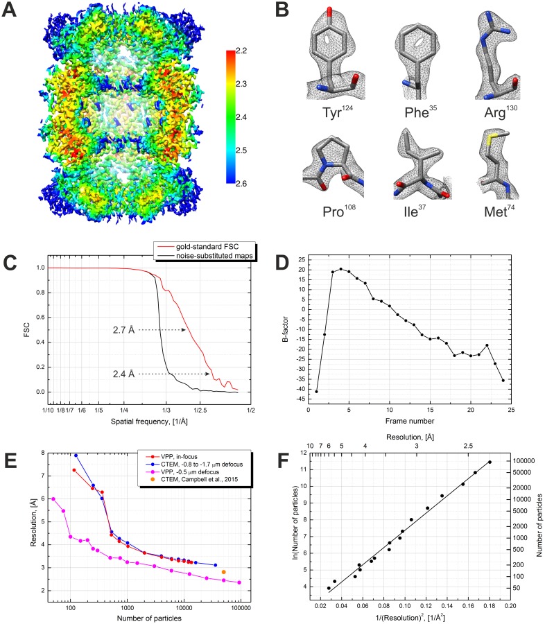
Previously, we reported an in-focus data acquisition method for cryo-EM single-particle analysis with the Volta phase plate (Danev and Baumeister, 2016). Here, we extend the technique to include a small amount of defocus which enables contrast transfer function measurement and correction. This hybrid approach simplifies the experiment and increases the data acquisition speed. It also removes the resolution limit inherent to the in-focus method thus allowing 3D reconstructions with resolutions better than 3 Å.

Keywords: biophysics; cryo-EM; none; phase plate; proteasome; structural biology.


Conflict of interest statement

RD: RD is a co-inventor in US patent US9129774 B2 "Method of using a phase plate in a transmission electron microscope". WB: WB is on the Scientific Advisory Board of FEI Company. The other author declares that no competing interests exist.

Figures

Figure 1.
Figure 1.. Volta phase plate with defocus cryo-EM dataset of 20S proteasome.
(A) Representative image of 20S proteasomes in ice, defocus 500 nm. (B) Power spectrum of the image in (A) showing contrast transfer function rings (Thon rings). To enhance the visibility of Thon rings, the power spectrum was calculated as the sum of the power spectra of individual movie frames (McMullan et al., 2015). (C) Phase shift history throughout the dataset. The phase shift gradually increases until the phase plate is moved to a new position where it suddenly drops and starts to raise again. (D) Histogram illustrating the phase shift distribution. (E) Defocus history throughout the dataset. The target defocus was changed after ~200 images from 500 nm to 300 nm. (F) Histograms illustrating the defocus distributions. Scale bar: 50 nm. DOI: http://dx.doi.org/10.7554/eLife.23006.002
Figure 2.
Figure 2.. Effects of the Volta phase plate phase shift on the image appearance and the contrast transfer function.
(A) Examples of images at low (0.1 π), optimal (0.5 π) and high (0.9 π) phase shifts. (B) Simulated contrast transfer functions (CTF) at 500 nm defocus and different phase shifts (top). Relative phase shift (PS) profile of the Volta phase plate (bottom). The black dashed line represents a CTF of an ideal (delta function) phase plate with 0.9 π phase shift. (C) Phase shift histograms before (gray) and after (red) 3D classification of the particles. Particles with low (<0.2 π) and high (>0.8 π) phase shifts were predominantly rejected. The blue line (right vertical axis) is the particle retention (after vs before 3D classification). Scale bar: 20 nm. DOI: http://dx.doi.org/10.7554/eLife.23006.003
Figure 3.
Figure 3.. Results from the 3D reconstruction of the 20S proteasome dataset with Relion particle polishing.
(A) Cross-section of the 3D map colored according to the local resolution. (B) Examples of sidechain details. (C) Fourier shell correlation (FSC) plots indicating a resolution of 2.4 Å according to the gold-standard FSC = 0.143 criterion. (D) Per-frame B-factors calculated during the particle polishing. (E) Resolution as a function of the number of particles measured using random particle subsets. Also shown is a data point from Campbell et al. (2015). (F) Same data as in (E) but with logarithmic and squared reciprocal axes. The slope of the linear fit indicates an overall B-factor of 103 Å2. DOI: http://dx.doi.org/10.7554/eLife.23006.004

References

    1. Campbell MG, Cheng A, Brilot AF, Moeller A, Lyumkis D, Veesler D, Pan J, Harrison SC, Potter CS, Carragher B, Grigorieff N. Movies of ice-embedded particles enhance resolution in electron cryo-microscopy. Structure. 2012;20:1823–1828. doi: 10.1016/j.str.2012.08.026. - DOI - PMC - PubMed
    1. Campbell MG, Veesler D, Cheng A, Potter CS, Carragher B. 2.8 å resolution reconstruction of the Thermoplasma acidophilum 20S proteasome using cryo-electron microscopy. eLife. 2015;4:e06380. doi: 10.7554/eLife.06380. - DOI - PMC - PubMed
    1. Cheng Y, Grigorieff N, Penczek PA, Walz T. A primer to single-particle cryo-electron microscopy. Cell. 2015;161:438–449. doi: 10.1016/j.cell.2015.03.050. - DOI - PMC - PubMed
    1. Chua EY, Vogirala VK, Inian O, Wong AS, Nordenskiöld L, Plitzko JM, Danev R, Sandin S. 3.9 å structure of the nucleosome core particle determined by phase-plate cryo-EM. Nucleic Acids Research. 2016;44:8013–8019. doi: 10.1093/nar/gkw708. - DOI - PMC - PubMed
    1. Danev R, Baumeister W. Cryo-EM single particle analysis with the Volta phase plate. eLife. 2016;5:e13046. doi: 10.7554/eLife.13046. - DOI - PMC - PubMed

Publication types

MeSH terms

LinkOut - more resources