. 2017 Jan 21;5(1):8.
doi: 10.1186/s40478-017-0411-2.

Locus coeruleus cellular and molecular pathology during the progression of Alzheimer's disease

Affiliations

Locus coeruleus cellular and molecular pathology during the progression of Alzheimer's disease

Sarah C Kelly et al. Acta Neuropathol Commun. .

Abstract

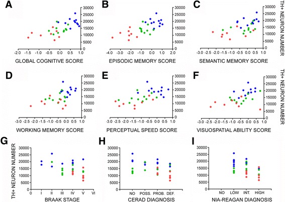
A major feature of Alzheimer's disease (AD) is the loss of noradrenergic locus coeruleus (LC) projection neurons that mediate attention, memory, and arousal. However, the extent to which the LC projection system degenerates during the initial stages of AD is still under investigation. To address this question, we performed tyrosine hydroxylase (TH) immunohistochemistry and unbiased stereology of noradrenergic LC neurons in tissue harvested postmortem from subjects who died with a clinical diagnosis of no cognitive impairment (NCI), amnestic mild cognitive impairment (aMCI, a putative prodromal AD stage), or mild/moderate AD. Stereologic estimates of total LC neuron number revealed a 30% loss during the transition from NCI to aMCI, with an additional 25% loss of LC neurons in AD. Decreases in noradrenergic LC neuron number were significantly associated with worsening antemortem global cognitive function as well as poorer performance on neuropsychological tests of episodic memory, semantic memory, working memory, perceptual speed, and visuospatial ability. Reduced LC neuron numbers were also associated with increased postmortem neuropathology. To examine the cellular and molecular pathogenic processes underlying LC neurodegeneration in aMCI, we performed single population microarray analysis. These studies revealed significant reductions in select functional classes of mRNAs regulating mitochondrial respiration, redox homeostasis, and neuritic structural plasticity in neurons accessed from both aMCI and AD subjects compared to NCI. Specific gene expression levels within these functional classes were also associated with global cognitive deterioration and neuropathological burden. Taken together, these observations suggest that noradrenergic LC cellular and molecular pathology is a prominent feature of prodromal disease that contributes to cognitive dysfunction. Moreover, they lend support to a rational basis for targeting LC neuroprotection as a disease modifying strategy.

Keywords: Alzheimer’s disease; Gene expression; Locus coeruleus; Mild cognitive impairment; Neurodegeneration; Norepinephrine.


Figures

Fig. 1
Fig. 1
LC cell loss in aMCI. a Representative TH-ir neuron and fiber staining in dorsal pontine tissue harvested from NCI, aMCI, and mild/moderate AD subjects. b Unbiased stereological cell counts revealed a significant ~30% decrease in the number of LC neurons in aMCI compared to NCI cases. There was a ~50% loss of LC neurons in the AD group compared to NCI. **, p < 0.01, ***, p < 0.001 compared to NCI, via one-way ANOVA with Bonferroni post hoc testing. There was also a significant ~25% difference in neuronal cell counts between the aMCI and AD groups (p < 0.05). me5 = mesencephalic tract of 5. Scale Bar = 100 μm
Fig. 2
Fig. 2
TH-ir LC neuron number correlates with multiple measures of antemortem cognition and postmortem neuropathology during the progression of AD. Scatterplots show significant relationships between reductions in LC neurons and a the GCS for each individual (r = 0.7, p < 0.0001), as well as poorer performance on composite scores of b episodic memory (r = 0.71, p < 0.0001), c semantic memory (r = 0.598, p = 0.0008), d working memory (r = 0.6, p = 0.0006), e perceptual speed (r = 0.66, p < 0.0001), and f visuospatial ability (r = 0.53, p = 0.003). There were also significant negative correlations between TH+ neuron number and increasing neuropathology as defined by g Braak (r = −0.46, p = 0.01), h CERAD (r = −0.41, p = 0.03), and i NIA-Reagan (r = −0.45, p = 0.01) diagnostic criteria. All associations were tested using Spearman rank correlation analysis. Abbreviations: TH+ (TH-positive), POSS. (possible), PROB. (probable), DEF. (definite), INT. (intermediate). Symbols: NCI (blue-filled circle), aMCI (green-filled circle), AD (red-filled circle)
Fig. 3
Fig. 3
Single LC neuron expression profiling reveals alterations in mRNAs regulating mitochondrial and neuritic function during AD progression. a Primary custom microarray data showing hybridization signal intensities of select mRNA transcripts within TH-ir LC neurons in NCI and aMCI. Abbreviations: cytochrome C1 (Cytc1), glutathione peroxidase (Gpx1), glucose transporter 3 (Glut3), phosphofructokinase-liver isozyme (Pfkl), microtubule associated binding protein 1a (Map1a), and neurofilament heavy (Nfh), medium (Nfm), or light (Nfl) chain subunits. b Heatmap shows significant decreases in transcripts regulating mitochondrial fucntion in LC neurons in aMCI and AD cases compared to NCI, including Cytc1, nuclear respiratory factor 1 (Nrf1), superoxide dismutase 2 (Sod2) and Gpx1, whereas Pfkl and platelet (Pfkp) isozymes were up-regulated in mild AD (red to green = decreasing expression levels). Additional abbreviations: superoxide dismutase 1 (Sod1), glutathione transferase (Gst), cytochrome p450 11a (Cyp11a), and pfk-muscle (Pfkm). c Heatmap shows significant decreases in transcripts regulating cytoskeletal/structural plasticity in LC neurons in aMCI and AD cases compared to NCI, including Map1b, Nfh, integrin 3 (Itga3), netrin 1 (Ntn1), utrophin (Utrn), and synaptopodin (Synpo), whereas calpain 1 (Capn1, m-calpain) and calpain 2 (Capn2, μ-calpain) were up-regulated in mild AD (red to green = decreasing expression levels). Additional mRNAs: mitochondrial associated protein 2 (Map2), and synaptojanin (Synj). a, NCI > aMCI, AD, p < 0.01; b, NCI > aMCI, AD, p < 0.001; c, AD > NCI, aMCI, p < 0.01
Fig. 4
Fig. 4
Alterations in select transcripts isolated from noradrenergic LC neurons correlate with global cognition during the progression of AD. Scatterplots show significant relationships between decreasing levels of a Cytc1 (r = 0.64, p < 0.0001), b Nrf1 (r = 0.57, p = 0.0003), c Map1b (r = 0.59, p = 0.0006), and d Synpo (r = 0.65, p < 0.0001) and worsening GCS score, via Spearman rank correlation analysis. Symbols: NCI (blue-filled circle), aMCI (green-filled circle), AD (red-filled circle)

References

    1. Adolfsson R, Gottfries CG, Roos BE, Winblad B. Changes in the brain catecholamines in patients with dementia of Alzheimer type. Br J Psychiatry. 1979;135:216–223. doi: 10.1192/bjp.135.3.216. - DOI - PubMed
    1. Albert MS, Dekosky ST, Dickson D, Dubois B, Feldman HH, Fox NC, Gamst A, Holtzman DM, Jagust WJ, Petersen RC, Snyder PJ, Carrillo MC, Thies B, Phelps CH. The diagnosis of mild cognitive impairment due to Alzheimer’s disease: Recommendations from the National Institute on Aging-Alzheimer’s Association workgroups on diagnostic guidelines for Alzheimer’s disease. Alzheimers Dement. 2011;7:270–279. doi: 10.1016/j.jalz.2011.03.008. - DOI - PMC - PubMed
    1. Alldred MJ, Che S, Ginsberg SD. Terminal continuation (TC) RNA amplification without second strand synthesis. J Neurosci Methods. 2009;177:381–385. doi: 10.1016/j.jneumeth.2008.10.027. - DOI - PMC - PubMed
    1. Alldred MJ, Duff KE, Ginsberg SD. Microarray analysis of CA1 pyramidal neurons in a mouse model of tauopathy reveals progressive synaptic dysfunction. Neurobiol Dis. 2012;45:751–762. doi: 10.1016/j.nbd.2011.10.022. - DOI - PMC - PubMed
    1. Arendt T. Alzheimer’s disease as a loss of differentiation control in a subset of neurons that retain immature features in the adult brain. Neurobiol Aging. 2000;21:783–796. doi: 10.1016/S0197-4580(00)00216-5. - DOI - PubMed

MeSH terms