. 2017 Feb;134(1):1-9.
doi: 10.1007/s10633-017-9573-2. Epub 2017 Jan 21.

ISCEV Standard for clinical electro-oculography (2017 update)

Affiliations

ISCEV Standard for clinical electro-oculography (2017 update)

Paul A Constable et al. Doc Ophthalmol. 2017 Feb.

Erratum in

Abstract

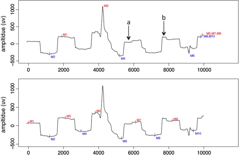
The clinical electro-oculogram (EOG) is an electrophysiological test of the outer retina and retinal pigment epithelium (RPE) in which changes in the electrical potential across the RPE are recorded during successive periods of dark and light adaptation. This document presents the 2017 EOG Standard from the International Society for Clinical Electrophysiology of Vision (ISCEV: www.iscev.org ). This standard has been reorganized and updated to include an explanation of the mechanism of the EOG, but without substantive changes to the testing protocol from the previous version published in 2011. It describes methods for recording the EOG in clinical applications and gives detailed guidance on technical requirements, practical issues and reporting of results with the main clinical measure (the Arden ratio) now termed the light peak:dark trough ratio. The standard is intended to promote consistent quality of testing and reporting within and between clinical centers.

Keywords: Arden ratio; Clinical electrophysiology; Electro-oculogram (EOG); Fast oscillation (FO); ISCEV Standards; Light adaptation; Light peak:dark trough ratio; Retinal pigment epithelium (RPE).


Conflict of interest statement

The authors declare no conflict of interests. Statement of human rights This article does not contain any studies with human participants performed by any of the authors. Statement on the welfare of animals This article does not contain any studies with animals performed by any of the authors. Informed consent As this article does not contain any studies with human participants performed directly by any of the authors, the concept of informed consent is not applicable.

Figures

Fig. 1
Fig. 1
Upper trace shows filtering with 0.1–30 Hz, lower trace with post hoc DC restoration by digital integration, rendering it similar to direct DC recording. DC recording (or restoration) makes it easier to perform the plateau measurements
Fig. 2
Fig. 2
Examples of 10-s saccadic records with a blink artifact at approximately 4000 ms. Arrow a indicates an initial undershoot and arrow b an overshoot of the fixation target visible by the step in the plateau of the EOG recording (upper trace). The lower trace shows the manual placing of markers at the peak and trough of the EOG recording as the eye performs horizontal saccades for 10 s at 1-s intervals
Fig. 3
Fig. 3
Recording electrode positions located near the inner and outer canthi of each eye. As the eyes perform horizontal saccades: left (a) then right (b), the amplitude of the standing potential is recorded across the active electrodes
Fig. 4
Fig. 4
Upper figure shows the raw standing potential values for an EOG with 15 min of dark (black circles) and 15 min of light (white circles). Smoothing of the data helps to define the DT and LP amplitudes from which to calculate the light peak:dark trough ratio (lower figure). Computer algorithms or fitting with a spline rule may also be utilized
Fig. 5
Fig. 5
Typical fast oscillation recording from a normal eye with six cycles of light and dark intervals (75 s each). There is a light trough (LT) and a dark peak (DP). In this case, amplitude measurements were derived from the fit of the sine wave to the raw data. The FO ratio is computed from the amplitude of the DP divided by the amplitude of the LT (1.3)

References

    1. Marmor MF, Zrenner E. Standard for clinical electro-oculography. International Society of Clinical Electrophysiology of Vision. Doc Ophthalmol. 1993;85(2):115–124. doi: 10.1007/BF01371127. - DOI - PubMed
    1. Marmor MF, Brigell MG, McCulloch DL, Westall CA, Bach M. ISCEV Standard for clinical electro-oculography (2010 update) Doc Ophthalmol. 2011;122(1):1–7. doi: 10.1007/s10633-011-9259-0. - DOI - PubMed
    1. Brigell M, Bach M, Barber C, Moskowitz A, Robson J. Guidelines for calibration of stimulus and recording parameters used in clinical electrophysiology of vision. Doc Ophthalmol. 2003;107(2):185–193. doi: 10.1023/A:1026244901657. - DOI - PubMed
    1. McCulloch DL, Marmor MF, Brigell MG, Hamilton R, Holder GE, Tzekov R, Bach M. ISCEV Standard for full-field clinical electroretinography (2015 update) Doc Ophthalmol. 2015;130(1):1–12. doi: 10.1007/s10633-014-9473-7. - DOI - PubMed
    1. Hood DC, Bach M, Brigell M, Keating D, Kondo M, Lyons JS, Marmor MF, McCulloch DL, Palmowski-Wolfe AM. ISCEV Standard for clinical multifocal electroretinography (2011 edition) Doc Ophthalmol. 2012;124(1):1–13. doi: 10.1007/s10633-011-9296-8. - DOI - PMC - PubMed