Review
. 2017 Mar;40(3):138-150.
doi: 10.1016/j.tins.2016.12.004. Epub 2017 Jan 19.

A GluD Coming-Of-Age Story

Affiliations
Review

A GluD Coming-Of-Age Story

Michisuke Yuzaki et al. Trends Neurosci. 2017 Mar.

Abstract

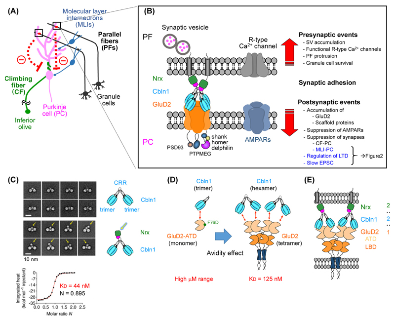
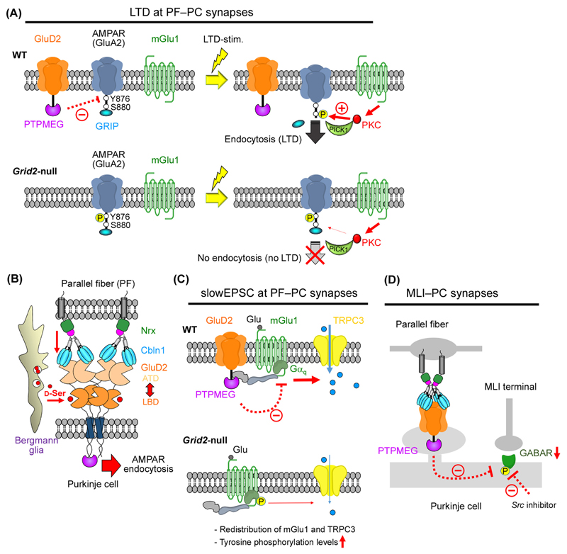
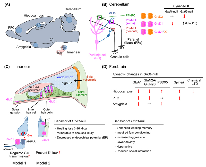
The GluD1 and GluD2 receptors form the GluD ionotropic glutamate receptor (iGluR) subfamily. Without known endogenous ligands, they have long been referred to as 'orphan' and remained enigmatic functionally. Recent progress has, however, radically changed this view. Both GluD receptors are expressed in wider brain regions than originally thought. Human genetic studies and analyses of knockout mice have revealed their involvement in multiple neurodevelopmental and psychiatric disorders. The discovery of endogenous ligands, together with structural investigations, has opened the way towards a mechanistic understanding of GluD signaling at central nervous system synapses. These studies have also prompted the hypothesis that all iGluRs, and potentially other neurotransmitter receptors, rely on the cooperative binding of extracellular small-molecule and protein ligands for physiological signaling.


Figures

Figure 1
Figure 1. GluD2 as a synaptic organizer.
(A) Synapses regulated by GluD2 in cerebellar Purkinje cells (PCs). Postsynaptic GluD2 regulates parallel fiber (PF)–PC synapses directly (boxes), molecular layer interneuron (MLI)–PC and climbing fiber–PC synapses indirectly (dotted lines with minus signs). (B) GluD2 signaling at PF–PC synapses. GluD2 binds to Cbln1 and its presynaptic receptor neurexin (Nrx) on PFs and regulates synaptic adhesion as well as pre- (up arrow) and post-synaptic (down arrow) events. (C) The Nrx–Cbln1 interaction. Negative-stain electron microscopic class averages of Cbln1 alone (upper panels) and Cbln1 with Nrx (bottom panels) illustrate the dimer-of-trimers arrangement. Yellow arrows indicate the suggested position of Nrx binding to the cysteine-rich region (CRR) of Cbn1. Isothermal titration calorimetric analyses revealed the stoichiometry (N) and affinity (KD) of this interaction. Reproduced from [28] with permission. (D) The Cbln1–GluD2 interaction. The weak, high micromolar, binding between Cbln1 trimer and the monomeric amino-terminal domain (ATD) of GluD2 (left; minimal interaction unit) is enhanced to an apparent KD of 125 nM) by the avidity effect in (right; oligomeric interaction). (E) The transsynaptic triad consisting of neurexin-Cbln1-GluD2 with 2 (monomers): 2 (hexamers): 1 (tetramer) stoichiometry
Figure 2
Figure 2. GluD2 as a regulator of functional synaptic transmission.
(A) A model for the GluD2 function in long-term depression (LTD) at PF–Purkinje cell (PC) synapses [42]. In wild-type (WT) mice (top), GluD2 maintains low phosphorylation levels at tyrosine 876 (Y876) of the GluA2 AMPA receptor subunit (dotted line with minus sign) through PTPMEG, a protein tyrosine phosphatase which binds to the GluD2 C-terminus. An LTD-inducing stimulation (LTD-stim.) activates mGlu1 to further reduce Y876 phosphorylation, allowing PKC to phosphorylate serine 880 (S880) of GluA2 (thick arrow with plus sign), a crucial step to replace the AMPA receptor anchoring protein from GRIP to PICK1 for AMPA receptor endocytosis during LTD. In Grid2-null mice, PTPMEG fails to dephosphorylate Y876 of GluA2, thereby impairing S880 phosphorylation (thin arrow) and the LTD. (B) Allosteric interactions between the amino-terminal domain (ATD) and the ligand-binding domain (LBD) of GluD2. The interaction with Cbln1 anchors ATD to the presynaptic site via Nrx (down arrow), and allows the conformational change induced by d-serine binding (right arrow) to the LBD to be transmitted to postsynaptic sites to induce AMPA receptor endocytosis [28]. (C) A model for the GluD2 function in slow excitatory postsynaptic currents (slowEPSCs). Burst stimulation of parallel fibers activates mGlu1 and Gαq to induce slowEPSCs, which are mainly mediated by TRPC3 in adult wild-type mice (top). Low tyrosine phosphorylation levels in PCs, which are partly mediated by PTPMEG (dotted line with minus sign), increase slowEPSC amplitudes by unknown mechanisms. In Grid2-null mice, slowEPSCs are reduced because mGlu1, which is anchored at the perisynaptic site via GluD2 and its interacting proteins (shown as shaded irregular structures), and TRPC3 are redistributed [45]. Alternatively, tyrosine phosphorylation levels are increased [42] by the loss of PTPMEG associated with GluD2 (bottom). (D) A model for the GluD2 function in inhibitory synapses formed between molecular layer interneuron (MLI) and Purkinje cells. Cbln1-GluD2 signaling likely suppresses MLI–PC inhibitory responses by lowering protein tyrosine phosphorylation levels via PTPMEG, anchored to the C-terminus of GluD2 in wild-type PCs [40]. A Src inhibitor reduces increased inhibitory responses in Grid2-null PCs.
Figure 3
Figure 3. GluD1 signaling in various brain regions.
(A) Expression of GluD1 in the forebrain, cerebellum and inner ear in adult mice. (B) GluD1 signaling in the cerebellar cortex. GluD1 is expressed at parallel fiber (PF)–molecular interneuron (MLI) synapses, especially those forming on MLI soma. GluD2 is highly expressed at PF–Purkinje cell (PC) synapses and weakly at MLI-PC synapses forming on MLI dendrites. In Grid1-null mice, PF–MLI synapses decrease significantly. Conversely, somatic PF–MLI synapses increase in Grid2-null mice, probably because of compensatory increase in GluD1 expression [12]. (C) GluD1 signaling in the inner ear. Schematic drawing of the organ of Corti indicating GluD1 expression in the inner hair cells, outer hair cells and spiral ganglia (top) [5]. Two models illustrate the cause of hearing loss in Grid1-null mice (bottom). GluD1 expressed in the afferent fibers may regulate glutamatergic synaptic transmission (model 1). GluD1 may serve as a cell adhesion molecule to maintain the endocochleal potential, which helps drive receptor currents into cochlear hair cells, by restricting K+ leakage from hair cell areas (model 2). (D) GluD1 signaling in the forebrain. Changes in the synaptic proteins, spine numbers and chemical LTD induced by a mGlu1 agonist in three forebrain regions (top) are shown, together with behavioral phenotypes (bottom) of Grid1-null mice. ↓, reduced; ↑, increased; →, no changes.

Similar articles

Cited by

References

    1. Yuzaki M. The delta2 glutamate receptor: 10 years later. Neurosci Res. 2003;46:11–22. - PubMed
    1. Hirano T. Cerebellar regulation mechanisms learned from studies on GluRδ2. Mol Neurobiol. 2006;33:1–15. - PubMed
    1. Mandolesi G, et al. An orphan ionotropic glutamate receptor: the delta2 subunit. Neuroscience. 2009;158:67–77. - PubMed
    1. Schmid SM, Hollmann M. Bridging the synaptic cleft: lessons from orphan glutamate receptors. Sci Signal. 2010;3:pe28. - PubMed
    1. Gao J, et al. Orphan Glutamate Receptor 1 Subunit Required for High-Frequency Hearing. Mol Cell Biol. 2007;27:4500–4512. - PMC - PubMed

Publication types

Substances