. 2017 Feb 2;65(3):447-459.e6.
doi: 10.1016/j.molcel.2016.12.011. Epub 2017 Jan 19.

Interdomain Communication of the Chd1 Chromatin Remodeler across the DNA Gyres of the Nucleosome

Affiliations

Interdomain Communication of the Chd1 Chromatin Remodeler across the DNA Gyres of the Nucleosome

Ilana M Nodelman et al. Mol Cell. .

Abstract

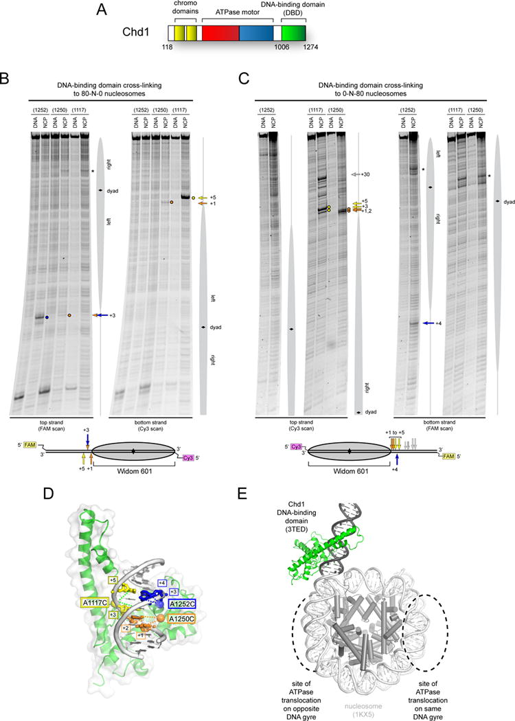
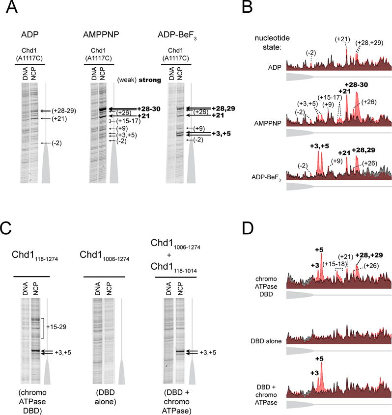
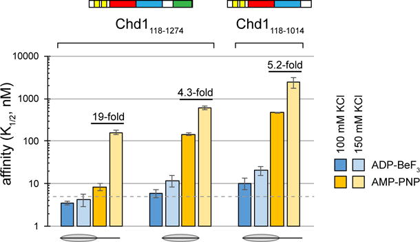
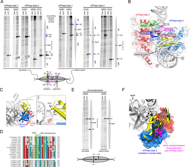
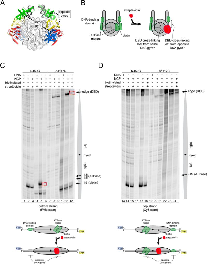
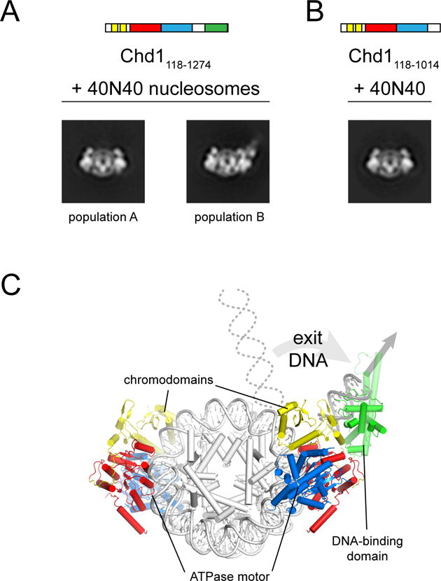
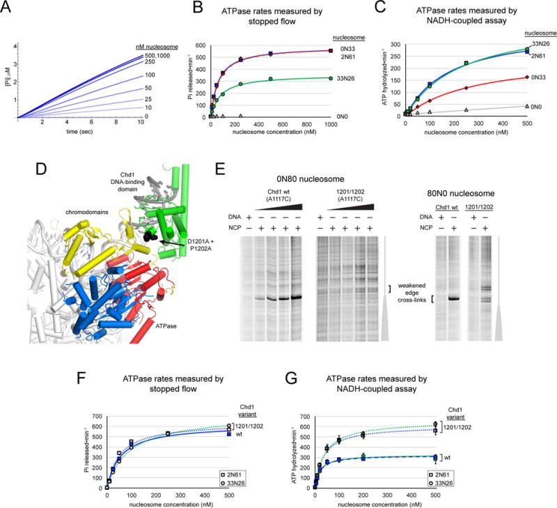
Chromatin remodelers use a helicase-like ATPase motor to reposition and reorganize nucleosomes along genomic DNA. Yet, how the ATPase motor communicates with other remodeler domains in the context of the nucleosome has so far been elusive. Here, we report for the Chd1 remodeler a unique organization of domains on the nucleosome that reveals direct domain-domain communication. Site-specific cross-linking shows that the chromodomains and ATPase motor bind to adjacent SHL1 and SHL2 sites, respectively, on nucleosomal DNA and pack against the DNA-binding domain on DNA exiting the nucleosome. This domain arrangement spans the two DNA gyres of the nucleosome and bridges both ends of a wrapped, ∼90-bp nucleosomal loop of DNA, suggesting a means for nucleosome assembly. This architecture illustrates how Chd1 senses DNA outside the nucleosome core and provides a basis for nucleosome spacing and directional sliding away from transcription factor barriers.

Keywords: CHD remodeler; DNA unwrapping; ISWI remodeler; SF2 ATPase; SHL2; chromatin remodeling; chromodomains; nucleosome assembly; nucleosome sliding; superhelical location 2.


Figures

Figure 1
Figure 1. The Chd1 DNA-binding domain preferentially binds DNA in a specific orientation and position at the edge of the nucleosome
(A) Schematic of Chd1 domain architecture. (B) and (C) Cross-linking of single cysteine variants within the Chd1 DNA-binding domain (DBD) to 80N0 and 0N80 nucleosomes or naked DNA in the presence of ADP∙BeF3. Lower schematics provide a summary of cross-linking positions, with the numbering referring to the distances from the nucleosome edge. Cross-linking sites reported here were observed in six or more independent reactions (n ≥ 6). (D) Cross-links are consistent with a unique position and orientation of the DBD on DNA. The crystal structure of the Chd1 DBD bound to DNA (PDB code 3TED; (Sharma et al., 2011)) is shown, highlighting positions of cysteine substitutions as colored spheres and the corresponding cross-linked bases as colored sticks. (E) A model of the Chd1 DBD at the edge of the nucleosome. Ideal B-form DNA (gray) was added to one end of the nucleosome crystal structure (1KX5, (Davey et al., 2002)), and the Chd1 DBD was positioned by docking DNA from the crystal structure (3TED) to match cross-linking as shown in (C). See also Figure S1.
Figure 2
Figure 2. Cross-linking of the Chd1 DNA-binding at the nucleosome edge requires the chromo-ATPase
(A) The nucleotide-bound state of the remodeler biases the distribution of cross-links made by the DBD. Cross-linking reactions were carried out using 0N80 nucleosomes (n = 6). (B) Intensity profiles of gel lanes shown in (A). Nucleosome cross-linking profiles (light red) are aligned with the background DNA-only cross-linking (gray). (C) The chromo-ATPase is necessary for cross-linking of the DBD at the nucleosome edge and can facilitate cross-linking in trans. Cross-linking was carried out with the naturally connected chromo-ATPase-DBD (left), the isolated DBD (center), and the chromo-ATPase added to the DBD in trans (right) (n = 5). (D) Intensity profiles of gel lanes shown in (C), as described in (B). See also Figure S2.
Figure 3
Figure 3. The DBD most strongly contributes to nucleosome binding under AMP-PNP conditions when flanking DNA is available
0N63 and 0N0 nucleosomes were used for AMP-PNP conditions, and 2N61 and 0N0 nucleosomes for ADP·BeF3 conditions. The dotted line marks 5 nM, which is near the limit for this assay. Error bars represent the standard deviations of three or more measurements. See also Table S1.
Figure 4
Figure 4. The Chd1 chromodomains and ATPase motor sit in unique positions and orientations on nucleosomal DNA
(A) Single-cysteine variants in both ATPase lobes of Chd1 form specific cross-links to nucleosomal DNA. Cross-linking reactions shown were performed in the presence of AMP-PNP (E493C) or ADP·BeF3 (all others) with either 10N70 nucleosomes (N459C, E493C) or 29N19 nucleosomes (N650C, V721C, G743C). The lower schematic summarizes the cross-linking positions on the nucleosome, with the numbering representing the distances from the dyad in either the 5′ direction (negative) or 3′ direction (positive). (n ≥ 4) (B) A model for the Chd1 ATPase motor in a closed, nucleotide-bound state, docked onto the nucleosome crystal structure (1KX5; (Davey et al., 2002)) to match cysteine-substituted positions (spheres) with cross-linked nucleotides (sticks). This orientation shows a view from the histone-interacting side of one SHL2 site, with the histones not shown for clarity. (C) Docking the chromo-ATPase crystal structure onto the nucleosome suggests a potential DNA-interacting loop on the chromodomains. The same docking of the ATPase motor is shown as in (B), but from a view showing the face of the nucleosome disk, with the chromodomains positioned relative to the first ATPase lobe as in the 3MWY structure. (D) Sequence alignment shows high conservation of the predicted DNA-interacting loop of the chromodomains. The upper sequences show Chd1 orthologs, whereas the lower group is from the related CHD6-9 clade. Although the CHD6-9 group does not maintain the highly conserved Gly in the middle of this loop, additional basic residues on either side of this loop segment are conserved. (E) The Chd1 chromodomains specifically contact nucleosomal DNA. Cross-linking reactions with the G238C variant were carried out in ADP·BeF3 with 29N19 nucleosomes (n = 4). (F) A predicted shift of chromodomains on the nucleosome. The position of the chromodomains from the crystal structure (3MWY, charcoal) sterically overlaps with the second ATPase lobe in the modeled closed state (magenta). A shift of the chromodomains by ~23° (yellow) relieves steric clash and places G238 close to cross-linking sites on nucleosomal DNA. See also Figures S3 and S4.
Figure 5
Figure 5. The Chd1 chromo-ATPase and DNA-binding domain interact across the gyres of the nucleosome
(A) Model of Chd1 domains docked on the nucleosome crystal structure (1KX5, (Davey et al., 2002)) based on cross-linking. (B) Experimental design for determining which Chd1 domains interact on the nucleosome. Targeting streptavidin via biotin should block the ATPase motor at only one SHL2 site. The streptavidin block should reveal which DBD depends on this ATPase motor. (C) Blocking the ATPase motor at one SHL2 site disrupts cross-linking of the DBD on the opposite DNA gyre of the nucleosome. Chd1 was added to 29N19 nucleosomes at a 1:1 molar ratio. Dotted red boxes highlight cross-links that are specifically lost upon addition of streptavidin to biotinylated nucleosomes. Schematic below illustrates the relative positioning of domains detected by cross-linking. (D) The ATPase motor remaining on the nucleosome stabilizes the DBD on the opposite DNA gyre. The same cross-linking reactions shown in (B) but following cross-linking to the other (non-biotinylated) DNA strand (n = 4). See also Figure S5.
Figure 6
Figure 6. The DNA-binding domain stably packs against the Chd1 chromo-ATPase on the nucleosome
(A) Class averages from negative stain EM representing the two major populations Chd1118–1274-nucleosome complexes observed in a face-on view. Chd1118–1274 was added to 40N40 nucleosomes at a 2:1 ratio in ADP·BeF3. (B) Class average from negative stain EM representing the only 2:1 Chd1118–1014-nucleosome population observed in face-on view. Chd1118–1014 was added to 40N40 nucleosomes at a 2:1 ratio in ADP·BeF3. (C) A model for Chd1 domain organization on the nucleosome based on cross-linking and negative stain EM.
Figure 7
Figure 7. The presence of exit DNA, communicated via the DNA-binding domain, reduces ATPase activity of Chd1
(A) Chd1 ATPase activity measured by a stopped flow phosphate binding assay. Shown is a representative titration with 2N61 nucleosomes, with 3–7 technical replicates averaged for each trace. (B) End-positioned nucleosomes stimulate the Chd1 ATPase more than nucleosomes with flanking DNA on both sides. Stopped flow ATPase activities were calculated from a 2–3 sec window after mixing. Michaelis Menten fits yielded kcat values of 592 ± 17 min−1 (0N33 nucleosomes, red diamonds), 587 ± 8 min−1 (2N61 nucleosomes, blue squares), and 360 ± 50 min−1 (33N26 nucleosomes, green circles). (C) ATPase activities measured by an NADH-coupled assay. Michaelis Menten fits to the data yielded kcat values of 371 ± 10 min−1 (2N61 nucleosomes, blue squares), 409 ± 13 min−1 (33N26 nucleosomes, green circles), and 260 ± 30 min−1 (0N33 nucleosomes, red diamonds). The titration series for 0N0 nucleosomes (gray triangles) is shown with a linear fit. (D) A view of the domain-docked model of Chd1 on the nucleosome that highlights the position of residues Asp1201 and Pro1202 (black spheres), which lie at the interface with the chromo-ATPase. (E) Cross-linking of the DBD (A1117C) to the nucleosome edge is lost (0N80) or weakened (80N0) with the D1201A/P1202A substitution. DNA and nucleosome concentrations were 150 nM, and Chd1 concentrations were 600 nM (for DNA alone), 75 nM, 150 nM, 300 nM, and 600 nM for 0N80 and 450 nM for 80N0. (F) The D1201A/P1202A variant shows similar ATPase activation as wild type on end-positioned nucleosomes, yet unlike wild type is insensitive to the presence of exit-side DNA. Michaelis Menten fits yielded kcat values of 641 ± 9 min−1 (2N61 with D1201A/P1202A variant), 618 ± 6 min−1 (2N61 for wildtype Chd1), and 718 ± 8 min−1 (33N26 with D1201A/P1202A variant). (G) ATP hydrolysis activity of the D1201A/P1202A variant of Chd1 is insensitive to the presence of flanking DNA. Michaelis Menten fits yielded kcat values of 610 ± 50 min−1 and 660 ± 30 min−1 for 2N61 (squares) and 33N26 (circles) nucleosomes, respectively, compared with 320 ± 20 min−1 and 320 ± 40 min−1 values for wildtype Chd1. Data reported in (B), (C), and (G) are averages of three or more independent experiments, with error bars (sometimes obscured by symbols) indicating ±S.D.; data points in (F) are averages from duplicate independent experiments, with the value ranges obscured by the symbols.

References

    1. Brune M, Hunter JL, Corrie JE, Webb MR. Direct, real-time measurement of rapid inorganic phosphate release using a novel fluorescent probe and its application to actomyosin subfragment 1 ATPase. Biochemistry. 1994;27:8262–8271. - PubMed
    1. Clapier CR, Cairns BR. Regulation of ISWI involves inhibitory modules antagonized by nucleosomal epitopes. Nature. 2012;7428:280–284. - PMC - PubMed
    1. Dang W, Bartholomew B. Domain architecture of the catalytic subunit in the ISW2-nucleosome complex. Mol Cell Biol. 2007;23:8306–8317. - PMC - PubMed
    1. Davey CA, Sargent DF, Luger K, Maeder AW, Richmond TJ. Solvent mediated interactions in the structure of the nucleosome core particle at 1.9 a resolution. J Mol Biol. 2002;5:1097–1113. - PubMed
    1. Dechassa ML, Hota SK, Sen P, Chatterjee N, Prasad P, Bartholomew B. Disparity in the DNA translocase domains of SWI/SNF and ISW2. Nucleic Acids Res. 2012;10:4412–4421. - PMC - PubMed

LinkOut - more resources