. 2017 Jan 26;168(3):427-441.e21.
doi: 10.1016/j.cell.2016.12.044. Epub 2017 Jan 19.

ApoE2, ApoE3, and ApoE4 Differentially Stimulate APP Transcription and Aβ Secretion

Affiliations

ApoE2, ApoE3, and ApoE4 Differentially Stimulate APP Transcription and Aβ Secretion

Yu-Wen Alvin Huang et al. Cell. .

Abstract

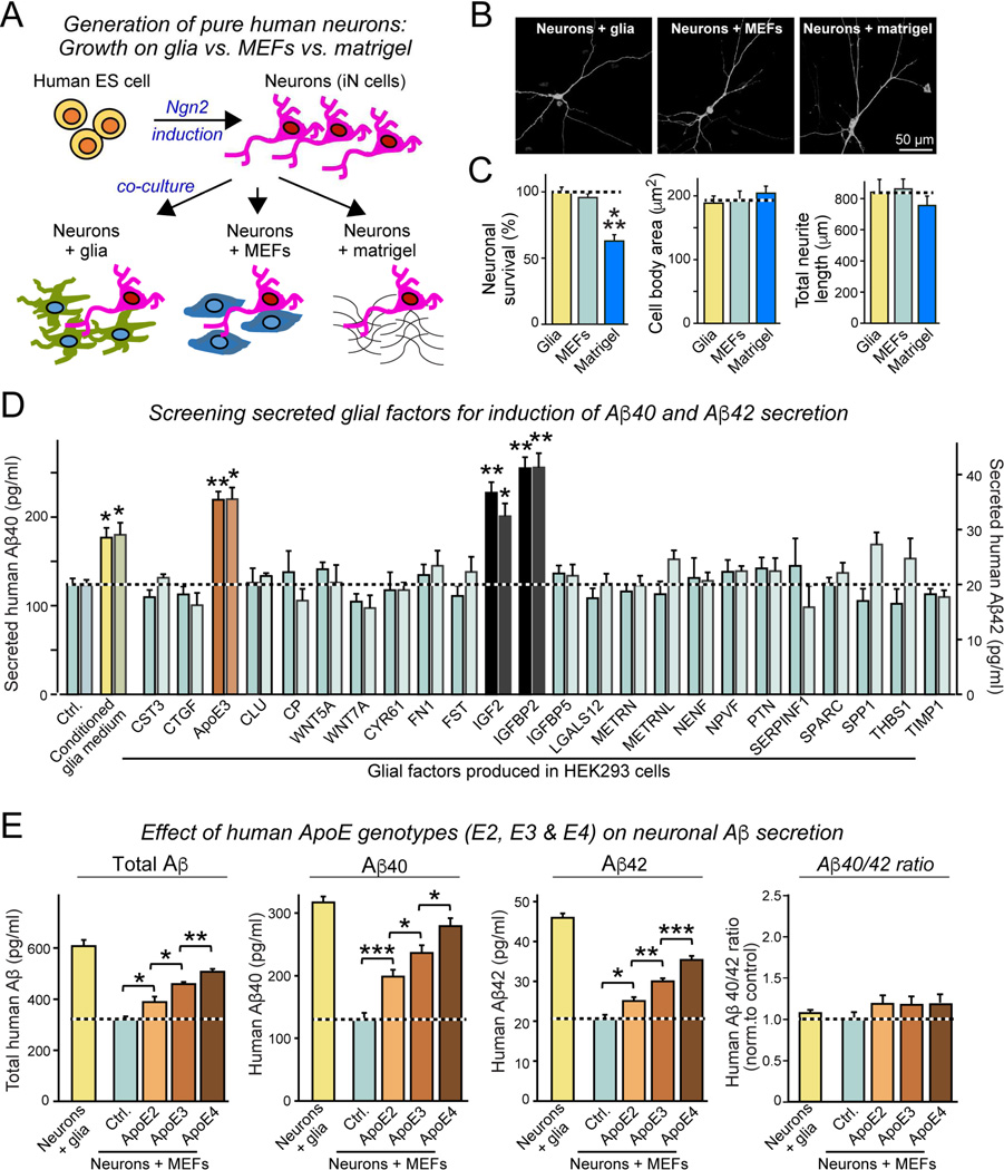
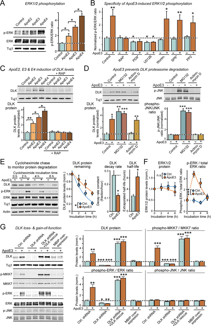
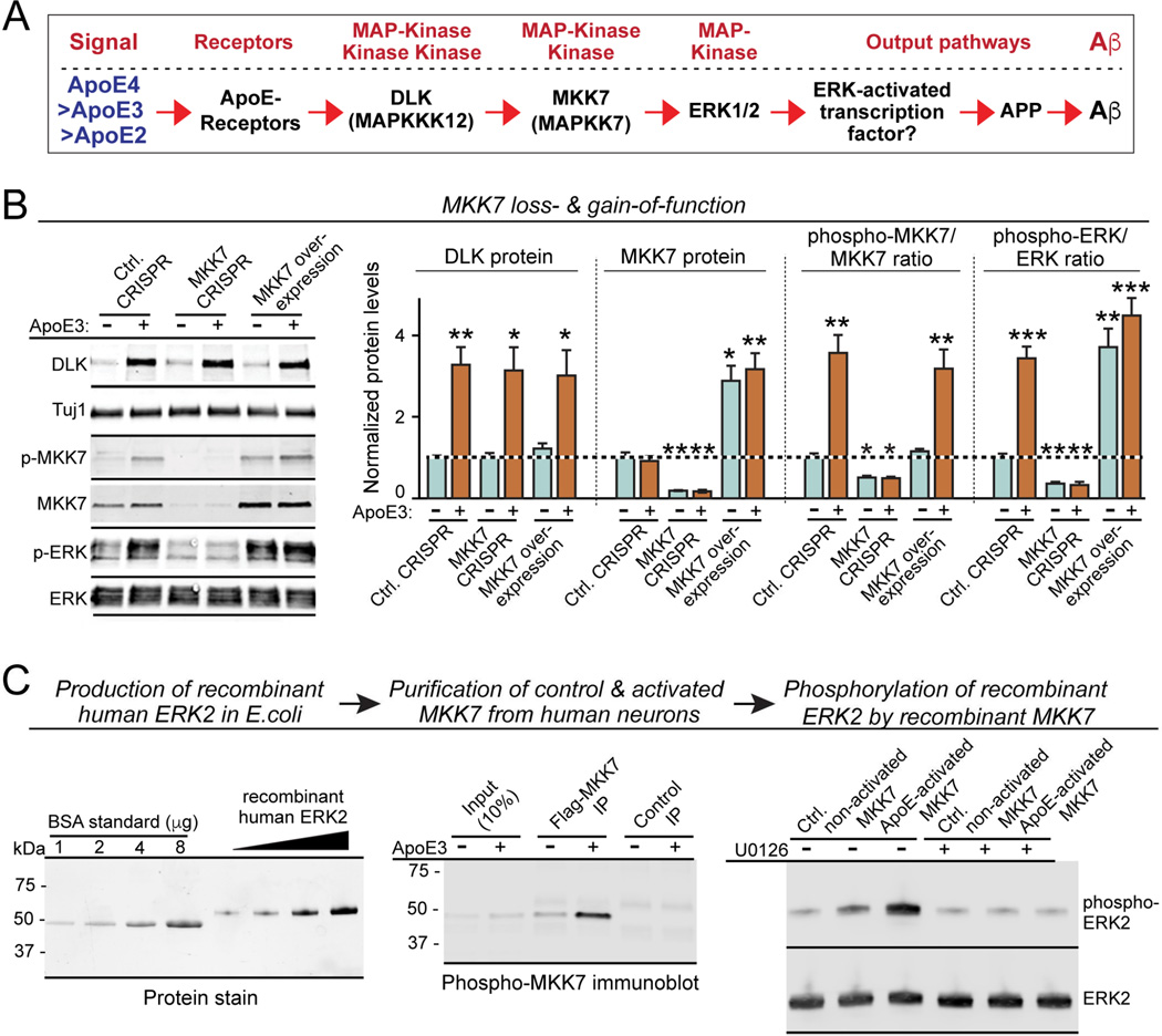
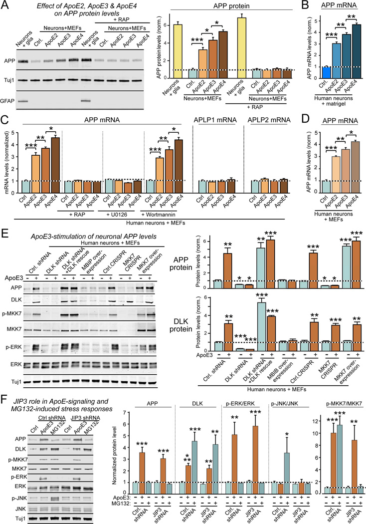
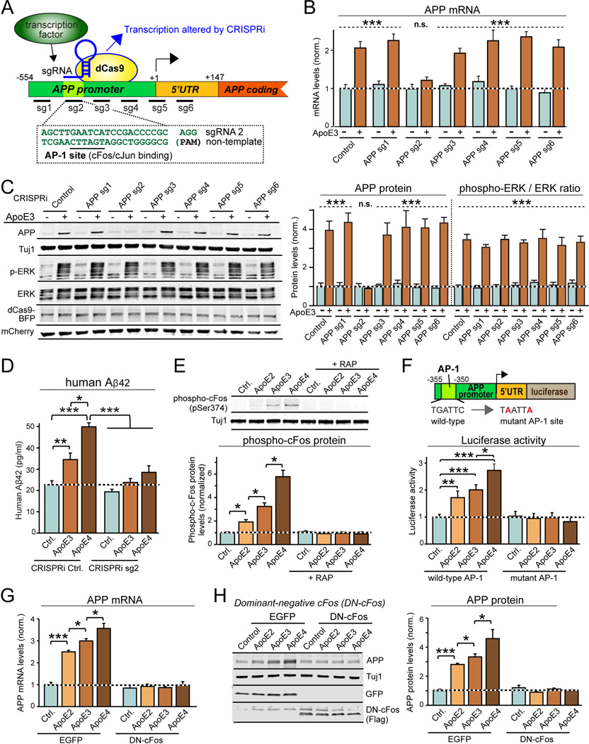
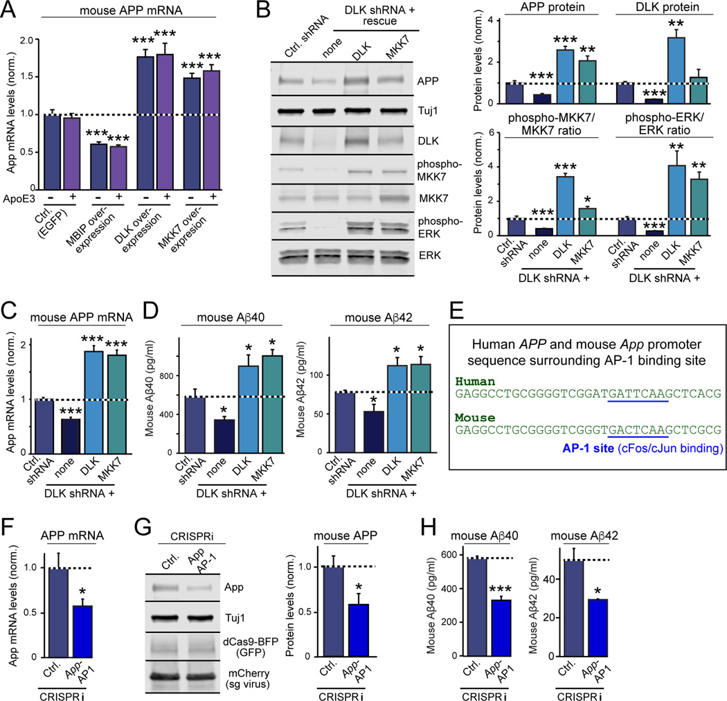
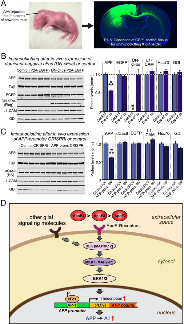
Human apolipoprotein E (ApoE) apolipoprotein is primarily expressed in three isoforms (ApoE2, ApoE3, and ApoE4) that differ only by two residues. ApoE4 constitutes the most important genetic risk factor for Alzheimer's disease (AD), ApoE3 is neutral, and ApoE2 is protective. How ApoE isoforms influence AD pathogenesis, however, remains unclear. Using ES-cell-derived human neurons, we show that ApoE secreted by glia stimulates neuronal Aβ production with an ApoE4 > ApoE3 > ApoE2 potency rank order. We demonstrate that ApoE binding to ApoE receptors activates dual leucine-zipper kinase (DLK), a MAP-kinase kinase kinase that then activates MKK7 and ERK1/2 MAP kinases. Activated ERK1/2 induces cFos phosphorylation, stimulating the transcription factor AP-1, which in turn enhances transcription of amyloid-β precursor protein (APP) and thereby increases amyloid-β levels. This molecular mechanism also regulates APP transcription in mice in vivo. Our data describe a novel signal transduction pathway in neurons whereby ApoE activates a non-canonical MAP kinase cascade that enhances APP transcription and amyloid-β synthesis.

Keywords: APP; Alzheimer’s disease; ApoE; Aβ; CRISPR; CRISPRi; DLK; MAP kinase signaling; amyloid precursor protein; apolipoprotein E; beta amyloid; cFos; dual leucine-zipper kinase; transcription.


Figures

Figure 1
Figure 1. ApoE stimulates Aβ-production with an ApoE4>ApoE3>ApoE2 potency rank order in human neurons cultured in the absence of glia or serum
(A) Experimental design. Human neurons (iN cells) were generated from H1 ES cells by forced expression of neurogenin-2 (Ngn2), and cultured either on mouse glia (green) which secrete copious amounts of ApoE, on murine embryonic fibroblasts (MEFs) which secrete no ApoE (blue), or on matrigel (black lines). (B) Representative images of human neurons at day 10 after induction (D10) cultured on glia, MEFs, or matrigel, and sparsely labeled by EGFP transfection. (C) Survival, soma size, and neurite length of human neurons cultured on glia, MEFs, or matrigel. (D) Screening 24 secreted proteins that are abundantly produced by cultured mouse glia reveals three factors (ApoE, IGF2, and IGFBP2) that induce Aβ40 and Aβ42 secretion from human neurons cultured on MEFs. Various factors were produced as human proteins in HEK293 cells (names reflect gene symbols), and added to human neurons on MEFs at D10. Media from treated neurons were analyzed by ELISA at D12. (E) ApoE2, ApoE3, and ApoE4 (all at 10 µg/ml; from D10-12) differentially stimulate Aβ40 and Aβ42 secretion by human neurons cultured on MEFs with an ApoE4>ApoE3>ApoE2 potency rank order, thereby partially rescuing the decrease in Aβ40 and Aβ42 secretion when neurons are co-cultured with MEFs instead of glia. Summary graphs show total concentrations of Aβ (left), Aβ40 (left center), and Aβ42 (right middle) in the medium, and of the Aβ42/Aβ40 ratio (right) measured by ELISA at D12. For cellular Aβ, Aβ40, and Aβ42 levels and for similar results for neurons generated from iPS cells, see Fig. S1. Data are means ± SEM (n≥3 independent experiments); statistical significance (*, p<0.05, **, p<0.01; ***, p<0.001) was evaluated with one-way ANOVA and Tukey’s post hoc multiple comparisons. For additional data and controls, see Fig. S1
Figure 2
Figure 2. ApoE stimulates a non-canonical DLK→MKK7→ERK1/2 MAP-kinase signalling cascade in human neurons
(A) ApoE (10 µg/ml, applied at D10 for 2 h) activates ERK1/2 phosphorylation in human neurons cultured on MEFs with an ApoE4>ApoE3>ApoE2 potency rank order (left, representative immunoblots; right, summary graphs). (B) ApoE3-induced ERK1/2-phosphorylation is abolished by the ApoE-receptor blocker RAP and by inhibitors of MEKs (PD9* [PD98059] and U0126; both 50 µM), but not by inhibitors of JNK (SP6* [SP600125]; 25 µM), PI3K kinase (Wortmannin; 0.1 µM), or Src kinase (PP2; 10 µM). Drugs were applied 30 min before a 2 h ApoE3 (10 µg/ml) incubation at D10. For additional data, see Fig. S2. (C) ApoE2, ApoE3, and ApoE4 cause a rapid, 2–3 fold increase in DLK in human neurons cultured on MEFs with an ApoE4>ApoE3>ApoE2 potency rank order; recombinant RAP that blocks ApoE-receptor binding also blocks ApoE-induced increases in DLK (left, representative immunoblots; right, summary graphs from neurons at D10 treated for 2 h with 10 µg/ml ApoE). (D) Proteasome inhibitor MG132 (10 µM, applied for 2 h at D10) increases DLK in human neurons cultured on MEFs similar to ApoE3 (10 µg/ml), thereby occluding the effect of ApoE3 (left); in addition, MG132 stimulates JNK phosphorylation (right). The transcription inhibitor actinomycin D (1 µg/ml) has no effect on the ApoE-induction of DLK (left) or JNK phosphorylation (right). (E) ApoE3 significantly slows the rapid turnover of DLK protein (measured by immunoblotting after addition of the protein synthesis inhibitor cycloheximide (0.1 g/l); left, representative blot; center, summary plots of the fraction of DLK remaining as a function of time after cycloheximide addition; right, summary graphs of the DLK decay rates and calculated half-lives as a function of ApoE3). (F) ApoE3 has no effect on ERK1/2 protein levels that are stable, but ApoE3-stimulated ERK1/2 phosphorylation decays in parallel with DLK protein levels after protein synthesis inhibition (left, summary plots of the fraction of ERK1/2 remaining as a function of time after cycloheximide addition; right, summary plot of the phospho-ERK/total ERK ratio). (G) ApoE3 strongly stimulates MKK7 and ERK1/2 phosphorylation but not JNK phosphorylation in human neurons cultured on MEFs (at D10); DLK knockdown with an shRNA or DLK inhibition by MBIP overexpression block ApoE3-induced MKK7 and ERK1/2 phosphorylation, whereas DLK overexpression constitutively activates MKK7 and ERK1/2 phosphorylation (left, representative immunoblots; right, summary graphs). Data are means ± SEM (n≥3 independent experiments); statistical significance (*, p<0.05, **, p<0.01; ***, p<0.001) was evaluated with one-way ANOVA and Tukey’s post-hoc test in pairwise comparisons in A and C and comparisons to control in B and D, and with two-way ANOVA in E and F.
Figure 3
Figure 3. Validation of the non-canonical ApoE-stimulated DLK→MKK7→ERK1/2 MAP-kinase cascade that excludes JNK activation
(A) Diagram of the proposed ApoE-induced non-canonical MAP-kinase signaling pathway. (B) MKK7 inactivation by CRISPR blocks ApoE3 induction of ERK1/2 phosphorylation, while MKK7 overexpression constitutively activates ERK1/2 phosphorylation independent of ApoE3. Data are means ± SEM (n≥3 independent experiments); statistical significance (*, p<0.05, **, p<0.01; ***, p<0.001) was evaluated with one-way ANOVA and Tukey’s post-hoc test, comparing all conditions to the control without ApoE treatment. For additional data and reagent validation, see Fig. S2. (C) In vitro kinase assay with purified recombinant proteins demonstrates that ApoE3-activated MKK7 directly phosphorylates ERK2. Recombinant human ERK2 was produced in E. coli (left, stain-free SDS-polyacrylamide gel visualized by UV illumination), and naïve or ApoE-activated MKK7 was immunopurified from human neurons that overexpressed Flag-tagged MKK7 and were treated at D10 for 2 h with control or ApoE medium (center; phospho-MKK7 immunoblot). Recombinant ERK2 was then incubated for 30 min at 30 oC in the absence (test) and presence of the MEK inhibitor U0126 (50 µM; used as a further control) with Flag-beads containing immunoprecipitated control or ApoE-activated MKK7, or with control HA beads. Samples were analyzed by immunoblotting (right).
Figure 4
Figure 4. ApoE increases APP expression 3-4-fold with an ApoE4>ApoE3>ApoE2 potency rank order by activating the DLK→MKK7→ERK1/2 MAP-kinase pathway
(A) Human neurons synthesize ~5-fold less APP when cultured on MEFs instead of glia; addition of ApoE2, ApoE3, or ApoE4 (each 10 µg/ml applied from D10-12) stimulates APP synthesis in human neurons on MEFs with a ApoE4>ApoE3>ApoE2 potency rank order, which is blocked by the ApoE-receptor antagonist RAP (left, representative immunoblots; right, summary graphs; see also Fig. S5). (B) ApoE2, ApoE3, and ApoE4 increase APP mRNA levels 3–4 fold with a ApoE4>ApoE3>ApoE2 potency rank order in human neurons cultured on matrigel only. (C) ApoE2, ApoE3, and ApoE4 increase only APP, but not APLP1 or APLP2 mRNA levels in human neurons on MEFs with an ApoE4>ApoE3>ApoE2 potency rank order; ApoE-induced APP mRNA increase is inhibited by RAP and the MAP-kinase inhibitor U0126, but not by the PI3K inhibitor Wortmannin. (D) Recombinant cholesterol-free ApoE2, ApoE3, and ApoE4 produced in bacteria stimulate APP mRNA levels in human neurons on MEFs similar to recombinant ApoE2, ApoE3, and ApoE4 produced in HEK293 cells. (E) Inhibition of DLK by shRNAs or MBIP blocks ApoE3-induced increases in APP protein levels, while DLK and MKK7 overexpression during rescue experiments constitutively increases APP protein levels independent of ApoE3. Note that DLK protein levels were not affected by MKK7 manipulations (left, representative blots; right, summary graphs). (F) Knockdown of the JNK scaffold JIP3 has no effect on ApoE3-induced activation of the DLK→MKK7→ERK1/2 signal transduction cascade, but blocks induction of the JNK MAP-kinase cascade during the stress response to MG132. Human neurons on MEFs were infected with lentiviruses expressing a control shRNA or a JIP3 shRNA at D4, treated with control medium, ApoE3 (10 µg/ml), or MG132 (0.1 g/l) for 2 hours at D10, and analyzed by immunoblotting (left, representative blots; right, summary graphs). Data are means ± SEM (n≥3 independent experiments for all bar graphs); statistical significance (*, p<0.05, **, p<0.01; ***, p<0.001) was evaluated with one-way ANOVA and Tukey’s post-hoc test in pairwise comparison (A–D, F) or comparisons to controls (E). For further data and controls, see Fig. S4, S5.
Figure 5
Figure 5. ApoE-mediated activation of cFos-containing transcription factor AP-1 stimulates APP-gene transcription
(A) CRISPRi strategy to identify APP promoter sequences required for ApoE-stimulation of APP-transcription using guide RNAs (sg1 to sg6) covering the proximal human APP-promoter (note that sg2 targets conserved AP-1 binding site). (B) CRISPRi of AP-1 binding sequence in human APP-promoter blocks ApoE3-induced increase in APP mRNA levels. Human neurons on MEFs were infected at D4 with lentiviruses co-expressing BFP-tagged dCas9, various sgRNAs, and mCherry. Neurons were treated at D10 with ApoE3 (10 µg/ml), and APP mRNA levels were measured at D12. (C) CRISPRi of AP-1 binding sequence in human APP-promoter blocks ApoE3-induced increase in APP protein but not in ERK1/2-phosphorylation. Experiments were performed as for B, except that neurons were analyzed by quantitative immunoblotting (left, representative blots; right, summary graph). (D) CRISPRi of AP-1 binding sequence in human APP-promoter suppresses ApoE-induced, but not basal Aβ42 secretion from human neurons on MEFs. Experiments were performed as described for (B). (E) ApoE activates cFos phosphorylation in human neurons co-cultured with MEFs with an ApoE4>ApoE3>ApoE2 rank potency order; cFos phosphorylation is blocked by the ApoE-receptor antagonist RAP (top, representative immunoblots; bottom, summary graphs). (F) AP-1 binding site in human APP-promoter mediates ApoE stimulation of APP-gene transcription. Human neurons cultured on MEFs were infected at D4 with lentiviruses containing APP-promoter-driven firefly luciferase and constitutively expressed renilla luciferase (internal control), treated with ApoE (10 µg/ml) at D10, and analyzed at D12 (top, schematic of promoter reporter construct; bottom, summary graph of luciferase expression normalized to the renilla luciferase control). (G & H) Dominant-negative cFos (DN-cFos) suppresses ApoE-induction of APP mRNA levels (G) and APP protein (H). Data are means ± SEM (n≥3 independent experiments for all bar graphs); statistical significance (*, p<0.05, **, p<0.01; ***, p<0.001; n.s., not significant) was evaluated with one-way ANOVA with Tukey’s post-hoc test or Student’s t-test. In (B) and (C), the difference between –ApoE3 and +ApoE3 is significant for control, sg1 and 3–6 groups (p<0.001 as indicated), but not for sg2 (n.s.). For additional data, see Fig. S6.
Figure 6
Figure 6. The DLK- and AP-1-dependent signaling pathway controlling App gene transcription and Aβ-synthesis is conserved in mouse neurons
All experiments were carried out in dissociated mixed mouse neuron/glia cultures in which endogenous glia factors maximally stimulate ApoE-dependent signaling pathways. (A) Exogenous ApoE3 has no effect on APP mRNA levels in neuron/glia cultures from mouse cortex, but inhibition of DLK by MBIP overexpression decreases APP mRNA levels, whereas DLK or MKK7 overexpression increase APP mRNA levels. Neuron/glia cultures were transduced with lentiviruses at DIV4, treated with ApoE3 (10 µg/ml) at DIV10, and analyzed by RT-PCR at DIV12. (B) DLK knockdown decreases APP and DLK protein levels in neuron/glia cultures from mouse hippocampus, and additionally suppresses steady-state phosphorylation of ERK1/2 and MKK7, whereas rescue overexpression of either DLK or MKK7 increases APP protein levels and MKK7 and ERK1/2 phosphorylation. (C & D) DLK knockdown decreases APP mRNA levels (C) and Aβ secretion (D) in neuron/glia cultures from mouse hippocampus, whereas rescue overexpression of either DLK or MKK7 increases APP mRNA levels (C) and Aβ40 and Aβ42 secretion (D). (E) Alignment of the human APP- and murine App-promoter sequences containing the AP-1 binding site demonstrates a high degree of conservation. (F & G) CRISPRi-mediated inhibition of AP-1 binding to the App-promoter in neuron/glia cultures from mouse hippocampus suppresses APP mRNA (F) and protein levels (G). (H) CRISPRi-mediated inhibition of AP-1 binding to the App-promoter decreases Aβ40 and Aβ42 secretion in neuron/glia cultures from mouse hippocampus. Data are means ± SEM (n≥3 independent experiments for all bar graphs); statistical significance (*, p<0.05, **, p<0.01; ***, p<0.001) was evaluated with one-way ANOVA and comparing to control with Tukey’s post-hoc multiple comparisons [(A) to (D)] and Student’s t test [(F) to (H)]. For additional data, see Fig. S7.
Figure 7
Figure 7. cFos-dependent signaling pathway regulates mouse App gene transcription in vivo
(A) Experimental design. AAVs (encoding EGFP alone or EGFP and dominant-negative cFos (DN-cFos); and EGFP plus dCAS9 with either a control guide RNA or a guide RNA directed to the App promoter AP-1 binding site) were stereotactically injected into the cortex of anesthetized newborn mice (left), and cortex expressing EGFP was analyzed at P7-P8 (right). (B & C) Suppression of cFos-signaling using DN-cFos (A; n = 5 mice for test and control) or CRISPRi of the AP-1 binding sequence of the App promoter (B; n = 6 mice for test and control) selectively decreases APP expression in vivo (for mRNA measurements, see Fig. S7). Data are means ± SEM (n.d., not detectable); statistical significance was evaluated with Student’s t-test (***, p<0.001). (D) Schematic of the ApoE-signaling pathway that controls APP transcription and Aβ production via activation of the DLK MAP-kinase cascade. See text for details. ApoE is proposed to increase AD risk by causing an incremental chronic increase in APP abundance and Aβ secretion, with ApoE4 being more, and ApoE2 being less efficacious than ApoE3 in a parallel to their effects on AD risk.

References

    1. Brouwers N, Sleegers K, Engelborghs S, Bogaerts V, Serneels S, Kamali K, Corsmit E, De Leenheir E, Martin JJ, De Deyn PP, et al. Genetic risk and transcriptional variability of amyloid precursor protein in Alzheimer’s disease. Brain. 2006;129:2984–2991. - PubMed
    1. Castellano JM, Kim J, Stewart FR, Jiang H, DeMattos RB, Patterson BW, Fagan AM, Morris JC, Mawuenyega KG, Cruchaga C, et al. Human apoE isoforms differentially regulate brain amyloid-β peptide clearance. Sci. Transl. Med. 2011;3:89ra57. - PMC - PubMed
    1. Chen M, Maloney JA, Kallop DY, Atwal JK, Tam SJ, Baer K, Kissel H, Kaminker JS, Lewcock JW, Weimer RM, et al. Spatially coordinated kinase signaling regulates local axon degeneration. J. Neurosci. 2012;32:13439–13453. - PMC - PubMed
    1. Christ A, Christa A, Kur E, Lioubinski O, Bachmann S, Willnow TE, Hammes A. LRP2 is an auxiliary SHH receptor required to condition the forebrain ventral midline for inductive signals. Dev. Cell. 2012;22:268–278. - PubMed
    1. De Strooper B, Karran E. The cellular phase of Alzheimer’s disease. Cell. 2016;164:603–615. - PubMed

Publication types