. 2017 Feb 8;93(3):606-615.e3.
doi: 10.1016/j.neuron.2016.12.020. Epub 2017 Jan 19.

GLUT4 Mobilization Supports Energetic Demands of Active Synapses

Affiliations

GLUT4 Mobilization Supports Energetic Demands of Active Synapses

Ghazaleh Ashrafi et al. Neuron. .

Abstract

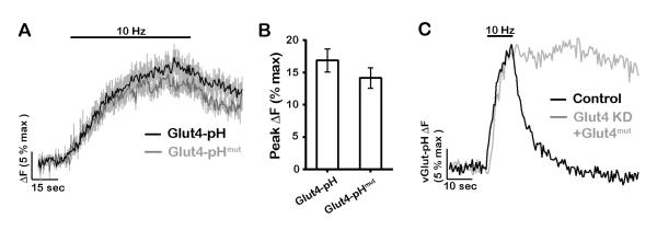
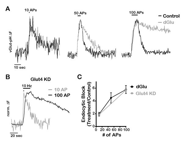
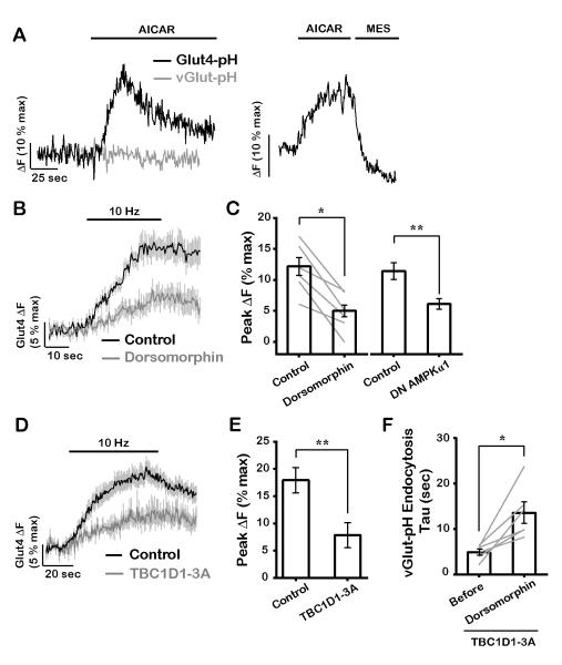
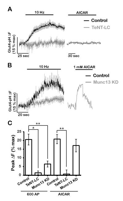
The brain is highly sensitive to proper fuel availability as evidenced by the rapid decline in neuronal function during ischemic attacks and acute severe hypoglycemia. We previously showed that sustained presynaptic function requires activity-driven glycolysis. Here, we provide strong evidence that during action potential (AP) firing, nerve terminals rely on the glucose transporter GLUT4 as a glycolytic regulatory system to meet the activity-driven increase in energy demands. Activity at synapses triggers insertion of GLUT4 into the axonal plasma membrane driven by activation of the metabolic sensor AMP kinase. Furthermore, we show that genetic ablation of GLUT4 leads to an arrest of synaptic vesicle recycling during sustained AP firing, similar to what is observed during acute glucose deprivation. The reliance on this biochemical regulatory system for "exercising" synapses is reminiscent of that occurring in exercising muscle to sustain cellular function and identifies nerve terminals as critical sites of proper metabolic control.

Keywords: GLUT4; glucose transport; glycolysis; neuronal metabolism; presynaptic function; vesicle cycle.


Figures

Figure 1
Figure 1. The glucose transporter Glut4 is expressed in the brain and is present at nerve terminals
(A-D) Immunohistochemical staining of adult mouse brain slices with antibodies against Glut4 (red), and (A, C) the presynaptic marker vGlut1, or (B, D) the Purkinje cell marker calbindin (green). Hoechst nuclear staining is shown in blue. Enlarged images of the cyan boxes in A and B show that (C) Glut4 is enriched in the synaptic-rich stratum radiatum, and (D) expressed at lower levels in Purkinje cells (arrowhead). DG, dentate gyrus; GL, granule layer; ML, molecular layer; PC: Purkinje cells; SP, stratum pyramidale; SR, stratum radiatum. CA1 and CA3 are hippocampal regions. (E-G) Immunostaining of dissociated rat hippocampal neurons with antibodies against Glut4 (red) and (E, F) the presynaptic marker synapsin, or (G) pHluorin/GFP of vGlut-pH (green) in neurons expressing Glut4 shRNA. (E) Glut4 is broadly expressed in somato-dendritic regions, (F) but also co-localizes with synapsin at nerve terminals (arrowheads) as shown in the enlarged image of the cyan box in D. (G-H) Glut4 immunofluorescence is reduced (by 70%, see STAR Methods) in neurons transfected with Glut4 shRNA and vGlut-pH, indicating the specificity of Glut4 antibody. (G) Large arrowheads point to the soma and (H) small arrowheads to boutons of the transfected neuron. Scale bars: (A, B) 50 μm, (C-H) 5 μm. See also Figure S1.
Figure 2
Figure 2. Neuronal firing drives Glut4 vesicles to the presynaptic plasma membrane
(A-D) Neurons expressing Glut4-pH were electrically stimulated with 600 AP. (A) Glut4-pH (pseudocolor) and the synaptic vesicle marker vGlut-mO (red) before and after stimulation. Neutralization of Glut4-pH vesicles with NH4Cl (white) reveals total axonal pool. (B) Average trace of Glut4-pH (n = 12 cells) with 600 AP-stimulation with the inset showing response after the first 100 AP. ΔF values were normalized to maximal ΔF from NH4Cl treatment. Error bars are shown in gray and are SEM. (C) a sample trace where stimulation was followed by quenching of extracellular pHluorin with MES acid and neutralization of Glut4-pH vesicles with NH4Cl. (D) Average surface fraction of Glut4-pH before and after stimulation (% total). Before: 7 ± 2, after 600 AP: 23 ± 4; n = 11 cells. (E-G) Glut3-pH does not mobilize at nerve terminals in response to activity. (E) Pseudocolor images of Glut3-pH in axons co-expressing the synaptic vesicle marker VAMP-mCherry (red) before and after stimulation with 600 AP (10 Hz). (F) Sample trace of Glut3-pH fluorescence (in arbitrary units) in response to stimulation. (G) Average surface fraction of Glut3-pH before and after stimulation (% toal). Before: 97 ± 1, after 600 AP: 94 ± 5; n = 5 cells. Scale bars (A, E), 5 μm. All data are shown as mean ± SEM. See also Figure S2 and S3.
Figure 3
Figure 3. Glut4 vesicles are distinct from synaptic vesicles
(A) The pH of axonal Glut4 vesicle measured from responses to acid quenching and neutralization with NH4Cl. The box and whisker plot represents median (line), 25th-75th percentile (box), and min-max (whisker). Mean pH: 6.1 ± 0.1; n = 9 cells. (B) Sample traces from boutons co-expressing Glut4-pH and vGlut-mO stimulated with 600 AP (20 Hz). (C) Decay half-time (sec) of Glut4-pH and vGlut-pH (in separate cells) after stimulation with 600 AP (10 Hz): Glut4-pH: 101 ± 17, vGlutpH: 6 ± 1; n = 7-10 cells per condition. *** P<0.001.
Figure 4
Figure 4. Glut4 is required for synaptic vesicle recycling following bursts of AP firing
(A-D) Sample vGlut-pH traces in response to 100 AP (10 Hz) in (A) control, Glut4 KD, and Glut4 KD neurons expressing shRNA-resistant Glut4-RFP, or (B) in Glut4 KD neurons where stimulation was followed by quenching of extracellular pHluorin with MES acid. (C) Average endocytic block measured as the fraction of vGlut-pH signal remaining at two endocytic time constants (2⊺) of the control at the end of stimulation. Control: 0.15 ± 0.03, Glut4 KD: 0.7 ± 0.1, rescue: 0.26 ± 0.07; n = 13-17 cells. (D) Average exocytosis of vGlut-pH measured as ΔF at the end of 100 AP normalized to ΔFNH4Cl (% max). Control: 23 ± 4, Glut4 KD: 25 ± 4; n= 13-15 cells. All error bars are SEM. **** P<0.0001. See also Figure S4.
Figure 5
Figure 5. The glucose transport activity of Gut4 is essential for synaptic vesicle recycling
(A-B) Glut4 mutant defective in glucose transport is recruited to synaptic surface similar to wildtype. (A) Average traces of wildtype and mutant Glut4-pH stimulated with 600 AP (10Hz). Error bars are shown in gray. (B) Average peak ΔF of Glut4-pH (% max) in response to 600 AP. Control data are the same as in Fig. 2B. Control: 17 ± 2, Glut4-pHmut: 14 ± 2; n = 6 - 16 cells. (C) Sample vGlut-pH traces in response to 100 AP (10 Hz) in control or Glut4 KD neurons expressing shRNA-resistant Glut4 mutant. All error bars are SEM.
Figure 6
Figure 6. Energetic requirement for glycolysis and glucose uptake increases with duration of activity
(A) Normalized vGlut-pH traces in response to 10, 50 and 100 AP (10Hz) before and 5 min after incubation with dGlu. (B) Normalized vGlut-pH traces of Glut4 KD neurons stimulated with 10 or 100 AP (10 Hz). (C) Average endocytic block for varying AP trains measured as the fraction of the maximal fluorescence remaining after 2 endocytic time constants in treated (dGlu or Glut4 KD) compared to control conditions, n = 4-21 cells per condition. All error bars are SEM.
Figure 7
Figure 7. AMP kinase mediates mobilization of Glut4 at presynaptic boutons
(A) Representative responses of (left) Glut4-pH and vGlut-pH to treatment with 1 mM AICAR, or (right) AICAR treatment of Glut4-pH, immediately followed by MES acid quench. (B) Average traces of Glut4-pH stimulated with 300 AP (10 Hz) before and 25 minutes after incubation with 10 μM dorsomorphin, an AMPK inhibitor. (C) Average Glut4 peak ΔF (% max) in response to 300 AP before and after dorsomorphin treatment, or 600 AP with or without expression of dominant negative (DN) AMPKα1. Due to the reduction of ΔFmax values with the expression of DN AMPKα1, all ΔFmax values were normalized to the control (see Material and Methods). Control: 12 ± 1, dorsomorphin: 5 ± 1, control: 11 ± 1, DN AMPKα1: 5.9 ± 0.8; n = 6-8 cells. (D) Average Glut4-pH traces in response to 600 AP (10 Hz) in control neurons or neurons expressing TBC1D1-3A in which putative AMPK phosphorylation sites were mutated. (E) Average maximal ΔF (% max) in response to 600 AP in control and TBC1D1-3A-expressing neurons. Control data are the same as in Fig. 2B. Control: 18 ± 2, TBC1D1-3A: 8 ± 2; n = 8-12 cells. (F) Endocytosis time constant (sec) of vGlut-pH in neurons expressing TBC1D1-3A, stimulated with 600 AP (10Hz) before and after 30 minutes of dorsomorphin treatment. Before: 4.9 ± 0.7, dorsomorphin: 14 ± 2; n = 6 cells. Error bars in graphs are shown in gray (B and D). All error bars are SEM. * P< 0.05, ** P< 0.01. See also Figure S5.
Figure 8
Figure 8. Glut4 and synaptic vesicles use distinct machinery for exocytosis
(A) Expression of tetanus toxin light chain (TeNT-LC) blocks Glut4 exocytosis membrane in response to electrical stimulation (600 AP, 10 Hz) and AICAR treatment. (B) Munc13 KD does not block AICAR-driven Glut4 exocytosis while it only partially inhibits activity-driven exocytosis (600 AP, 10 Hz). (C) Average peak ΔF (% max) in response to 600 AP or AICAR in the same genotypes as shown in A and B. 600 AP (control: 20 ± 3, TeNT-LC: 1 ± 1, Munc13 KD: 6 ± 2); AICAR (control: 20 ± 3, TeNT-LC: 0 ± 2, Munc13 KD: 17 ± 4); n = 3-13 cells. Error bars in graphs are shown in gray (A and B). All error bars are SEM. See also Figure S6.

References

    1. Abbott MA, Wells DG, Fallon JR. The insulin receptor tyrosine kinase substrate p58/53 and the insulin receptor are components of CNS synapses. J Neurosci. 1999;19:7300–7308. - PMC - PubMed
    1. Abel ED, Kaulbach HC, Tian R, Hopkins JC, Duffy J, Doetschman T, Minnemann T, Boers ME, Hadro E, Oberste-Berghaus C, et al. Cardiac hypertrophy with preserved contractile function after selective deletion of GLUT4 from the heart. J Clin Invest. 1999;104:1703–1714. - PMC - PubMed
    1. Balaji J, Armbruster M, Ryan TA. Calcium control of endocytic capacity at a CNS synapse. J Neurosci. 2008;28:6742–6749. - PMC - PubMed
    1. Balaji J, Ryan TA. Single-vesicle imaging reveals that synaptic vesicle exocytosis and endocytosis are coupled by a single stochastic mode. Proc Natl Acad Sci U S A. 2007;104:20576–20581. - PMC - PubMed
    1. Burchfield JG, Lu J, Fazakerley DJ, Tan SX, Ng Y, Mele K, Buckley MJ, Han W, Hughes WE, James DE. Novel systems for dynamically assessing insulin action in live cells reveals heterogeneity in the insulin response. Traffic. 2013;14:259–273. - PubMed