. 2017 Mar 2;20(3):345-359.e5.
doi: 10.1016/j.stem.2016.12.006. Epub 2017 Jan 19.

Pericytes of Multiple Organs Do Not Behave as Mesenchymal Stem Cells In Vivo

Affiliations

Pericytes of Multiple Organs Do Not Behave as Mesenchymal Stem Cells In Vivo

Nuno Guimarães-Camboa et al. Cell Stem Cell. .

Abstract

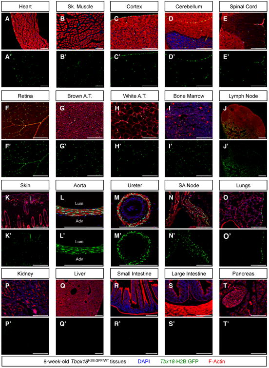
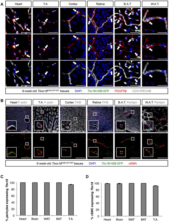
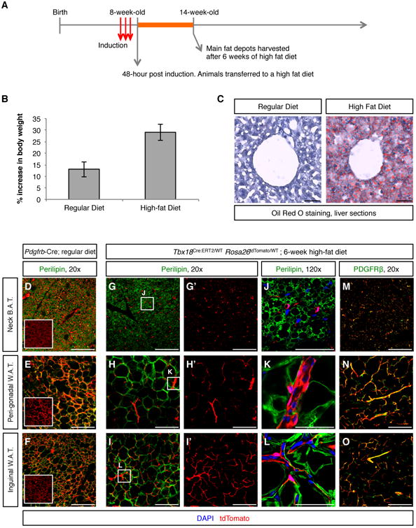
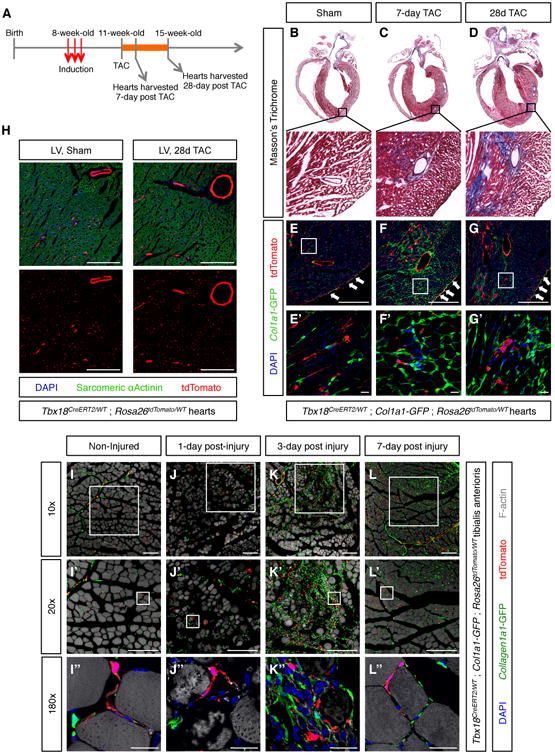
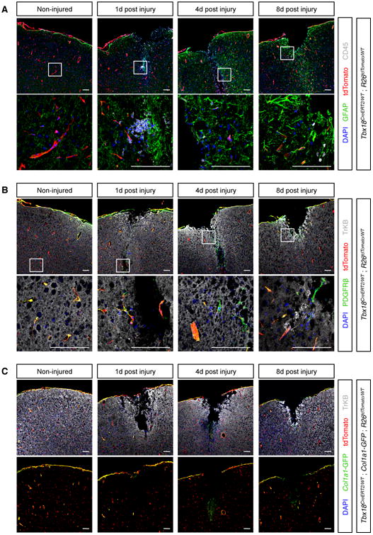
Pericytes are widely believed to function as mesenchymal stem cells (MSCs), multipotent tissue-resident progenitors with great potential for regenerative medicine. Cultured pericytes isolated from distinct tissues can differentiate into multiple cell types in vitro or following transplantation in vivo. However, the cell fate plasticity of endogenous pericytes in vivo remains unclear. Here, we show that the transcription factor Tbx18 selectively marks pericytes and vascular smooth muscle cells in multiple organs of adult mouse. Fluorescence-activated cell sorting (FACS)-purified Tbx18-expressing cells behaved as MSCs in vitro. However, lineage-tracing experiments using an inducible Tbx18-CreERT2 line revealed that pericytes and vascular smooth muscle cells maintained their identity in aging and diverse pathological settings and did not significantly contribute to other cell lineages. These results challenge the current view of endogenous pericytes as multipotent tissue-resident progenitors and suggest that the plasticity observed in vitro or following transplantation in vivo arises from artificial cell manipulations ex vivo.

Keywords: lineage tracing; mesenchymal stem cells; mural cells; pericytes.


Figures

Figure 1
Figure 1. Patterns of Tbxl8 expression in the adult mouse
To assess whether Tbxl8 was actively expressed in adult tissues, organs were harvested from 8-week-old Tbx18H2B:GFP/WT mice and processed for histological analyses with the nuclear dye DAPI and with the filamentous actin marker Phalloidin. Confocal microscopy revealed strong H2B:GFP signal (indicative of active Tbxl8 expression) in the membranous linings of: (A) the heart (epicardium), (C-E) the central nervous system (pia mater), and (O) the lungs (pleura). Expression by scattered interstitial cells was observed (A) within the cardiac ventricular walls, (B) in tibialis anterioris skeletal muscle, (C-E) in the central nervous system, (F) in the retina, (G, H) in interscapular brown and peri-gonadal white adipose depots, (I) in bone marrow, (J) in inguinal lymph nodes, and (K) in skin. Additionally, strong expression was observed (L) in the medial layer of the aorta, (M) in ureteric smooth muscle, and (N) in sinoatrial (SA) node pacemaker cells. No H2B:GFP signal could be detected (P) in the kidneys, nor (Q-T) the gastrointestinal tract or associated glands. Lum = lumen, Adv = adventitia. Bars = 200μm. See also Figure S1.
Figure 2
Figure 2. Interstitial Tbxl8-GFP+ cells were mural cells (pericytes and vascular smooth muscle)
(A) In all organs analyzed - heart, skeletal muscle of the tibialis anterioris (T.A.), cortex, retina, brown adipose tissue (B.A.T.) and white adipose tissue (W.A.T.) - interstitial Tbxl8-GFP+ cells (arrows) never expressed the endothelial marker CD31, but localized in the immediate vicinity of CD31+ endothelium and expressed the pan-pericyte marker PDGFRβ, emitting multiple cytoplasmic projections that enveloped adjacent blood vessels. Such location, morphology and cell-surface antigen profile placed interstitial Tbxl8-GFP+ cells as pericytes. Asterisks denote nuclei of endothelial cells. (B) In the vicinity of larger blood vessels, Tbxl8-GFP+ cells revealed a spindle shape and expressed the smooth muscle marker smooth muscle α actin (αSMA). (C,D) Importantly, in these organs, Tbx18 expression did not mark a subset of mural cells, but rather the totality of pericytes (PDGFRβ, CD146 double positive cells) and vascular smooth muscle (αSMA+ cells). In C and D data are represented as mean ± standard deviation. Bars = 30μm in (A) and 200μm in (B). See also Figures S2 and S3, and Table S1.
Figure 3
Figure 3. Pdgfrb-Cre was unsuitable for lineage tracing of pericytes
(A) PDGFRα (gray) and PDGFRβ (green) were expressed by multiple cell lineages during mid-gestational embryogenesis. Consequently, the constitutively active Pdgfrb-Cre extensively labeled multiple populations of embryonic progenitors (red channel), rendering it unsuitable for purposes of specific labeling of pericyte-derived lineages. Arrows indicate labeling of the aortic hemogenic endothelium by PDGFRβ and Pdgfrb-Cre in the aorta-gonad-mesonephros (AGM) region. (B) FACS analyses revealed that in adult animals Pdgfrb-Cre labeled the majority (approximately 90%) of all bone marrow cells, similar to the extent of labeling observed for the endothelial/hematopoietic Tie2-Cre. (C) Similarly, Pdgfrb-Cre labeled a majority of cells in multiple other tissues, including kidney, lungs, skeletal muscle and brain. Bars = 1000 μm in A and in brain montage, and 200 μm in all other panels. In B data are represented as mean ± standard deviation. See also Figure S4.
Figure 4
Figure 4. Pericytes and vascular smooth muscle cells maintained a mural cell phenotype during aging
(A) Strategy used to insert a CreERT2 cassette into the murine endogenous Tbxl8 locus. (B) Experimental design used for pulse-chase experiments aimed at determining the in vivo progenitor potential of pericytes during aging. (C, D) Intra-peritoneal administration of 1 mg of tamoxifen to adult (8-week-old) Tbxl8ERT2Cre/Wt;Rosa26tdTomato/Wt animals for 3 consecutive days labeled more than 90% of pericytes and more than 85% of vascular smooth muscle in brain, heart, brown adipose tissue (B.A.T.) and white adipose tissue (W.A.T.). (E-P) Short (8-week) and long (87-week) aging experiments revealed that tdTomato+ lineage traced cells (red) in brain (E-G), heart (H-J), B.A.T. (K-M) and W.A.T. (N-P) never expressed tissue-specific parenchymal markers (gray), and always retained expression of the mural cell marker PDGFRβ (green), indicating pericytes and vascular smooth muscle cells had maintained their mural cell identity throughout aging. In C and D data are represented as mean ± standard deviation. Bars = 50μm. See also Figure S5.
Figure 5
Figure 5. Mural cells were not adipogenic progenitors
(A) Experimental design for testing adipogenic potential of pericytes and vascular smooth muscle cells. (B) 6-week increase in body weight (expressed as percentage of starting weight) of animals fed a lean diet and animals fed a high-fat diet. (C) 6 weeks of high-fat diet resulted in hepatic steatosis. (D-F) Pdgfrb-Cre;Rosa26tdTomato/Wt lineage traced adipose depots isolated from regular diet fed animals were used as a positive control of adipocyte labeling by tdTomato. In these tissues, co-localization of the green (Perilipin) and red (tdTomato) signals produced an orange/yellow merge pattern. Boxed images are tdTomato-only panels to facilitate visualization of Pdgfrb-Cre-labeled cells. (G-L) In Tbx18ERT2Cre/Wt; Rosa26tdTomato/Wt animals, even after 6 weeks of high fat diet, lineage traced cells did not co-localize with the adipocyte marker Perilipin in any of the fat depots analyzed. G′-I′ are the tdTomato-only panels of the images shown in G-I, respectively. J-L are higher magnification images of the areas boxed in G-I, respectively. (M-O) In addition to not showing expression of Perilipin, Tbx18-CreERT2-lineage traced cells retained expression of the mural cell marker PDGFRβ, demonstrating that pericytes and vascular smooth muscle cells had not functioned as progenitors of new adipocytes. Bars = 50μm in C, J, K, L and 200μm in all other panels. In B data are represented as mean ± standard deviation. See also Figure S7.
Figure 6
Figure 6. Pericytes were not progenitors of fibroblasts or myocytes
(A) Experimental design for testing whether cardiac pericytes were progenitors of fibroblasts or cardiomyocytes post-TAC. (B-D) Trichrome staining of histological sections demonstrated progressive accumulation of collagen fibers in TAC animals. Boxed areas are shown in higher magnification. (E-G) Confocal microscopy imaging of sections adjacent to those displayed in (B-D) revealed lack of co-localization between Collal-GFPhigh fibroblasts and tdTomato+ pericytes and vascular smooth muscle in sham, but also TAC animals. E′-G′ are higher magnifications of areas boxed in E-G, respectively. (H) Confocal microscopy images showing absence of co-localization between tdTomato+ lineage traced cells and sarcomeric α-actinin+ cardiomyocytes. (I-I′) Non-injured tibialis anterioris muscle showing intact myofibers and non-overlapping populations of Collal-GFPhigh fibroblasts and tdTomato+ Tbxl8-CreERT2 lineage-traced mural cells. (J-J′) 1 day post-injury, dying myofibers were observed in the vicinity of the BaCl injection site. (K-K′) 3 day post-injury, dead myofibers had been replaced by an extensive scar composed of Collal-GFPhigh fibroblasts immune cells. Of note, the vast majority of Collal-GFPhigh fibroblasts were not lineage traced by Tbxl8-CreERT2, revealing they were not derived from pericytes or vascular smooth muscle. (L-L′) 7 days post-injury, most of the scar tissue had been replaced by new myofibers. Newly formed myofibers were not lineage-traced by Tbxl8-CreERT2, demonstrating that pericytes and vascular smooth muscle had not functioned as progenitors of new myocytes. Bars = 20μm in (E′-G′), (I″-L″), and 200μm in all other panels. See also Figures S6 and S7.
Figure 7
Figure 7. Pericytes of the brain were not neuronal or scar progenitors
(A) The brain responded to stab wounds by forming a glial scar mainly composed of GFAP+ reactive astrocytes (green) and CD45+ macrophages/microglia (gray). Importantly, such cells were not labeled by tdTomato, indicating they did not derive from pericytes or vascular smooth muscle. (B) After wounding, Tbxl8-CreERT2 lineage traced cells never co-localized with the neuronal marker TrKB and always expressed the pan-mural cell marker PDGFRβ, indicating they retained a pericyte or vascular smooth muscle cell phenotype even after an injury setting. (C) In non-injured cortical tissue, Collal-GFP expression was restricted to specific subsets of Tbxl8+ cells: pia mater and mural cells surrounding larger vessels. Although cortical stab wounds did not produce robust fibrosis, a small number of Collal-GFP+ fibroblasts could be detected 4-day post injury, at the tip of the wound. However, such Collal-GFP+ fibroblasts were not labeled by tdTomato, indicating they did not derive from pericytes or vascular smooth muscle. Bars = 100μm.

Comment in

References

    1. Alvarez-Dolado M, Pardal R, Garcia-Verdugo JM, Fike JR, Lee HO, Pfeffer K, Lois C, Morrison SJ, Alvarez-Buylla A. Fusion of bone-marrow-derived cells with Purkinje neurons, cardiomyocytes and hepatocytes. Nature. 2003;425:968–973. - PubMed
    1. Armulik A, Genove G, Betsholtz C. Pericytes: developmental, physiological, and pathological perspectives, problems, and promises. Developmental cell. 2011;21:193–215. - PubMed
    1. Ben-David U, Benvenisty N. The tumorigenicity of human embryonic and induced pluripotent stem cells. Nature reviews Cancer. 2011;11:268–277. - PubMed
    1. Bohnenpoll T, Bettenhausen E, Weiss AC, Foik AB, Trowe MO, Blank P, Airik R, Kispert A. Tbx18 expression demarcates multipotent precursor populations in the developing urogenital system but is exclusively required within the ureteric mesenchymal lineage to suppress a renal stromal fate. Dev Biol. 2013;380:25–36. - PubMed
    1. Buckingham ME, Meilhac SM. Tracing cells for tracking cell lineage and clonal behavior. Developmental cell. 2011;21:394–409. - PubMed

MeSH terms