. 2017 Jan 6:10:592.
doi: 10.3389/fnins.2016.00592. eCollection 2016.

Dynamic Expression of Serotonin Receptor 5-HT3A in Developing Sensory Innervation of the Lower Urinary Tract

Affiliations

Dynamic Expression of Serotonin Receptor 5-HT3A in Developing Sensory Innervation of the Lower Urinary Tract

K Elaine Ritter et al. Front Neurosci. .

Abstract

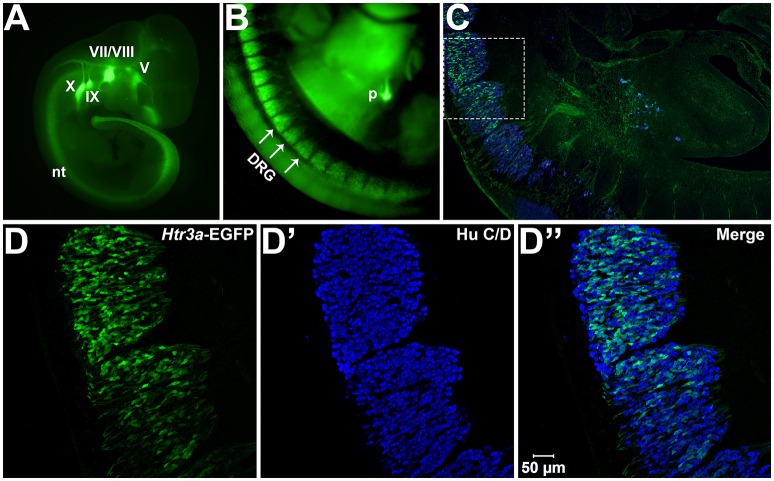
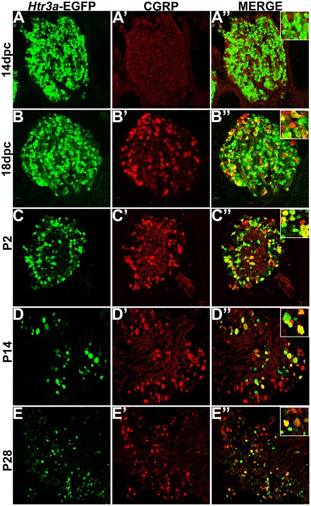
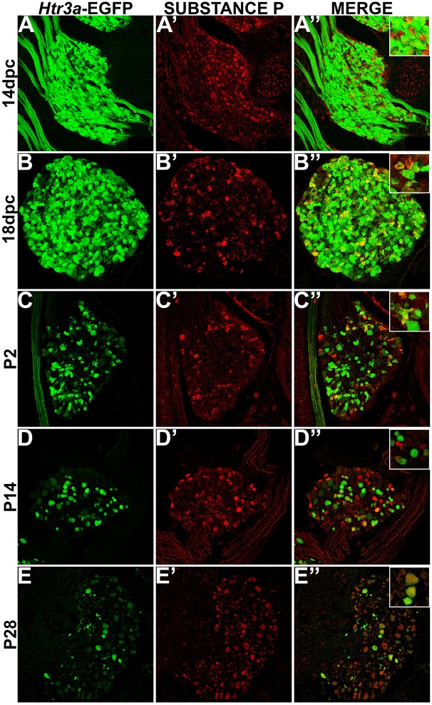
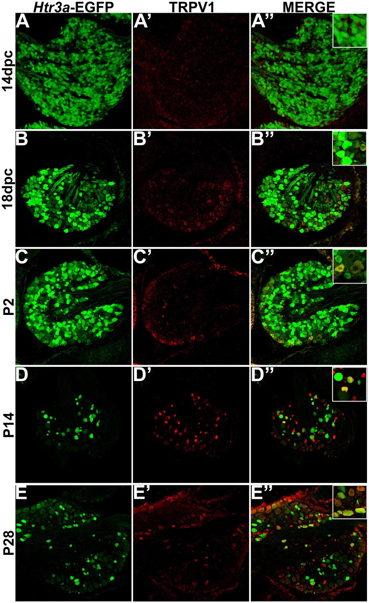
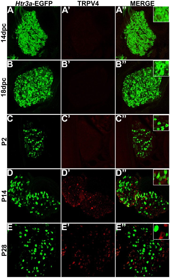
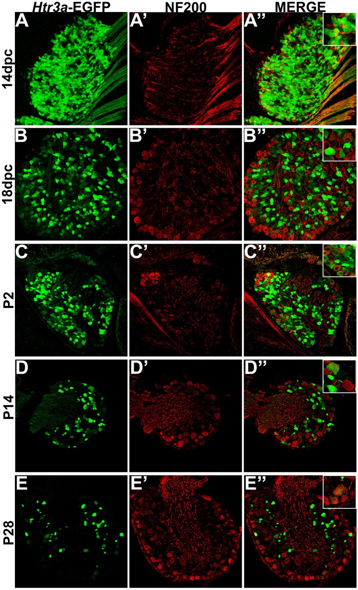
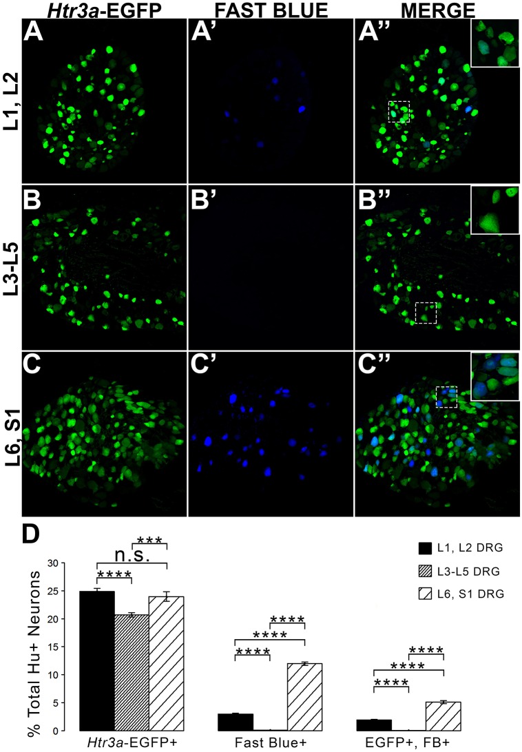
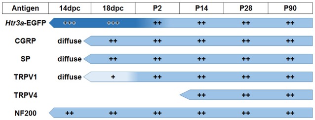
Sensory afferent signaling is required for normal function of the lower urinary tract (LUT). Despite the wide prevalence of bladder dysfunction and pelvic pain syndromes, few effective treatment options are available. Serotonin receptor 5-HT3A is a known mediator of visceral afferent signaling and has been implicated in bladder function. However, basic expression patterns for this gene and others among developing bladder sensory afferents that could be used to inform regenerative efforts aimed at treating deficiencies in pelvic innervation are lacking. To gain greater insight into the molecular characteristics of bladder sensory innervation, we conducted a thorough characterization of Htr3a expression in developing and adult bladder-projecting lumbosacral dorsal root ganglia (DRG) neurons. Using a transgenic Htr3a-EGFP reporter mouse line, we identified 5-HT3A expression at 10 days post coitus (dpc) in neural crest derivatives and in 12 dpc lumbosacral DRG. Using immunohistochemical co-localization we observed Htr3a-EGFP expression in developing lumbosacral DRG that partially coincides with neuropeptides CGRP and Substance P and capsaicin receptor TRPV1. A majority of Htr3a-EGFP+ DRG neurons also express a marker of myelinated Aδ neurons, NF200. There was no co-localization of 5-HT3A with the TRPV4 receptor. We employed retrograde tracing in adult Htr3a-EGFP mice to quantify the contribution of 5-HT3A+ DRG neurons to bladder afferent innervation. We found that 5-HT3A is expressed in a substantial proportion of retrograde traced DRG neurons in both rostral (L1, L2) and caudal (L6, S1) axial levels that supply bladder innervation. Most bladder-projecting Htr3a-EGFP+ neurons that co-express CGRP, Substance P, or TRPV1 are found in L1, L2 DRG, whereas Htr3a-EGFP+, NF200+ bladder-projecting neurons are from the L6, S1 axial levels. Our findings contribute much needed information regarding the development of LUT innervation and highlight the 5-HT3A serotonin receptor as a candidate for future studies of neurally mediated bladder control.

Keywords: autonomic nervous system; dorsal root ganglia; lower urinary tract; neural crest; serotonin.


Figures

Figure 1
Figure 1
Htr3a is expressed early in fetal development of sensory neurons. (A) Lateral whole-mount image of an Htr3a-EGFP transgenic fetus age 10 days post coitus (dpc). EGFP fluorescence is visible in the cranial ganglia (V, VII/VIII, IX, X) and neural tube (nt). (B) Lateral Whole-mount view of an Htr3a-EGFP 12 dpc mouse fetus at the trunk axial level between the forelimbs and hindlimbs. Htr3a-EGFP transgene expression is evident in the dorsal root ganglia (DRG) and pancreas (p). (C) Sagittal cryo-section of Htr3a-EGFP 12 dpc fetus, stained with Hu to label all neurons (blue). EGFP expression is observed in the lumbosacral DRG. (D–D”) 200x magnification of two sacral DRG seen in (C). Counter-staining with Hu C/D allows comparison of EGFP fluorescence to the total neuronal population.
Figure 2
Figure 2
Htr3a-EGFP and Calcitonin Gene Related Peptide (CGRP) are co-expressed in a subset of neurons through development and adulthood. Confocal images of cryosections from Htr3a-EGFP transgenic animals stained with anti-CGRP are shown. All DRG presented are from lumbosacral axial levels. (A–A”) Sagittal section of 14 dpc fetal DRG. (B–B”) Sagittal section of 18 dpc fetal DRG. (C–C”) Coronal section of P2 DRG. (D–D”) Cryosection of P14 male DRG. (E–E”) Cryosection of P28 male DRG. All zoom insets are 400X.
Figure 3
Figure 3
Some, but not all, Htr3a-EGFP neurons express Substance P neuropeptide. Confocal images of cryosections from Htr3a-EGFP transgenic animals stained with anti-Substance P are shown. All DRG presented are from lumbosacral axial levels. (A–A”) Sagittal section of 14 dpc fetal DRG. (B–B”) Sagittal section of 18 dpc fetal DRG. (C–C”) Coronal section of P2 DRG. (D–D”) Cryosection of P14 male DRG. (E–E”) Cryosection of P28 male DRG. All zoom insets are 400X.
Figure 4
Figure 4
The majority of adult TRPV1+ neurons also express Htr3a-EGFP. Confocal images of cryosections from Htr3a-EGFP transgenic animals stained with anti-TRPV1 are shown. All DRG presented are from lumbosacral axial levels. (A–A”) Sagittal section of 14 dpc fetal DRG. (B–B”) Sagittal section of 18 dpc fetal DRG. (C–C”) Coronal section of P2 DRG. (D–D”) Cryosection of P14 male DRG. (E–E”) Cryosection of P28 male DRG. All zoom insets are 400X.
Figure 5
Figure 5
Htr3a-EGFP does not co-localize with TRPV4 in lumbosacral DRG. Confocal images of cryosections from Htr3a-EGFP transgenic animals stained with anti-TRPV4 are shown. All DRG presented are from lumbosacral axial levels. (A–A”) Sagittal section of 14 dpc fetal DRG. (B–B”) Sagittal section of 18 dpc fetal DRG. (C–C”) Coronal section of P2 DRG. (D–D”) Cryosection of P14 male DRG. (E–E”) Cryosection of P28 male DRG. All zoom insets are 400X.
Figure 6
Figure 6
A subset of Htr3a-EGFP neurons express Neurofilament 200. Confocal images of cryosections from Htr3a-EGFP transgenic animals stained with anti-NF200 are shown. All DRG presented are from lumbosacral axial levels. (A–A”) Sagittal section of 14 dpc fetal DRG. (B–B”) Sagittal section of 18 dpc fetal DRG. (C–C”) Coronal section of P2 DRG. (D–D”) Cryosection of P14 male DRG. (E–E”) Cryosection of P28 male DRG. All zoom insets are 400X.
Figure 7
Figure 7
L1, L2 and L6, S1 axial levels harbor bladder-projecting neurons. Images of lumbosacral DRG harvested from Htr3a-EGFP transgenic male mice after retrograde tracing of bladder-projecting neurons are shown. (A–A”) L1, L2 axial level DRG sections. (B–B”) L3-L5 axial level DRG sections. (C–C”) L6, S1 axial level dorsal root ganglia sections. (D) Average percentages of Hu+ DRG neurons expressing the Htr3a-EGFP transgene, labeling with Fast Blue, and co-labeling with Htr3a-EGFP and Fast Blue at axial level groups L1, L2; L3-L5; and L6, S1. Error bars are standard error of the mean, n = 4 animals, 3–5 sections from 3 DRG quantified from each animal. ***p < 0.005, ****p < 0.001. Differences that are not significant (n.s.) are also indicated.
Figure 8
Figure 8
Distinct expression patterns of 5-HT3A receptor in subclasses of bladder-projecting afferents. Average percentages of bladder-projecting (Fast Blue+) neurons expressing the Htr3a-EGFP transgene and markers of sensory neuron subtypes in L1, L2 and L6, S1 DRG. Gray coloring represents L1, L2 proportions and black coloring represents L6, S1 proportions. Htr3a-EGFP average proportions in L1, L2 and L6, S1 DRG are the leftmost solid bars. Markers of sensory neuron subtypes CGRP, Substance P, TRPV1, NF200 (solid bars) and co-expression of Htr3a-EGFP and subtype markers (striped bars) are grouped by the marker used. Error bars are standard error of the mean, n = 4 animals, 3–5 sections from 3 DRG quantified from each animal. ***p < 0.005, ****p < 0.001. Differences that are not significant (n.s.) are also indicated.
Figure 9
Figure 9
Summary of developmental expression patterns in lumbosacral Htr3a-EGFP+ dorsal root ganglia neurons. At 14 and 18 dpc, Htr3a-EGFP fluorescence is uniformly intense in the majority of lumbosacral DRG neurons. Fluorescence intensity diversifies at postnatal day 2, with some cells still strongly expressing the transgene while others are moderate or dim. This pattern is maintained through postnatal development and adulthood. Neuropeptides CGRP and Substance P are both extremely weak and diffuse at 14 dpc but their expression level increases by 18 dpc. The expression patterns observed for these markers at 18 dpc is maintained through adulthood. TRPV1 expression is negligible at fetal stages but escalates after birth at P2 and maintains expression levels. TRPV4 is not detected until P2 but is maintained from that time point to adulthood. NF200, unlike the other markers, is expressed strongly in the majority of neurons at 14 dpc, and its expression levels are stable through all time points examined.

Similar articles

Cited by

References

    1. Arms L., Vizzard M. A. (2011). Neuropeptides in lower urinary tract function. Handb. Exp. Pharmacol. 202, 395–423. 10.1007/978-3-642-16499-6_19 - DOI - PMC - PubMed
    1. Bachy I., Franck M. C., Li L., Abdo H., Pattyn A., Ernfors P. (2011). The transcription factor Cux2 marks development of an A-delta sublineage of TrkA sensory neurons. Dev. Biol. 360, 77–86. 10.1016/j.ydbio.2011.09.007 - DOI - PubMed
    1. Baiou D., Santha P., Avelino A., Charrua A., Bacskai T., Matesz K., et al. . (2007). Neurochemical characterization of insulin receptor-expressing primary sensory neurons in wild-type and vanilloid type 1 transient receptor potential receptor knockout mice. J. Comp. Neurol. 503, 334–347. 10.1002/cne.21389 - DOI - PubMed
    1. Bhattacharya A., Dang H., Zhu Q. M., Schnegelsberg B., Rozengurt N., Cain G., et al. . (2004). Uropathic observations in mice expressing a constitutively active point mutation in the 5-HT3A receptor subunit. J. Neurosci. 24, 5537–5548. 10.1523/JNEUROSCI.5658-03.2004 - DOI - PMC - PubMed
    1. Birder L. A., Nakamura Y., Kiss S., Nealen M. L., Barrick S., Kanai A. J., et al. . (2002). Altered urinary bladder function in mice lacking the vanilloid receptor TRPV1. Nat. Neurosci. 5, 856–860. 10.1038/nn902 - DOI - PubMed

LinkOut - more resources