. 2017 Apr;38(4):539-550.
doi: 10.1038/aps.2016.146. Epub 2017 Jan 23.

Benzyl isothiocyanate induces protective autophagy in human lung cancer cells through an endoplasmic reticulum stress-mediated mechanism

Affiliations

Benzyl isothiocyanate induces protective autophagy in human lung cancer cells through an endoplasmic reticulum stress-mediated mechanism

Qi-Cheng Zhang et al. Acta Pharmacol Sin. 2017 Apr.

Abstract

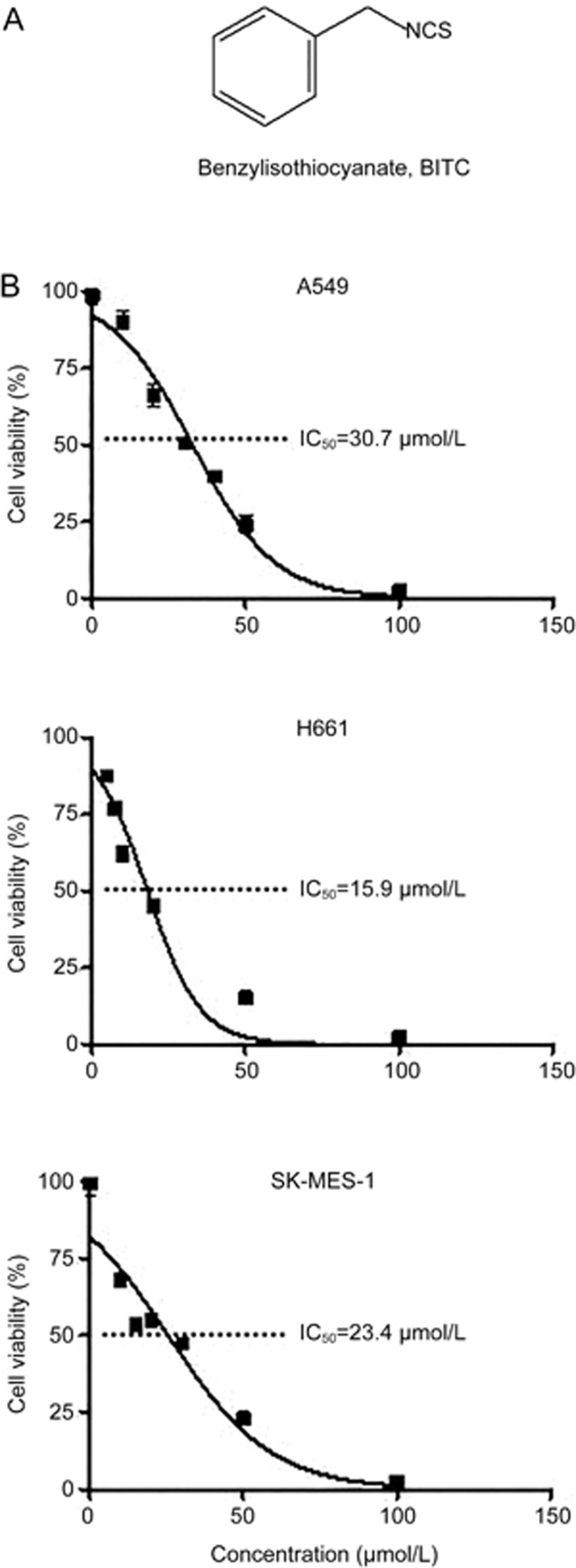
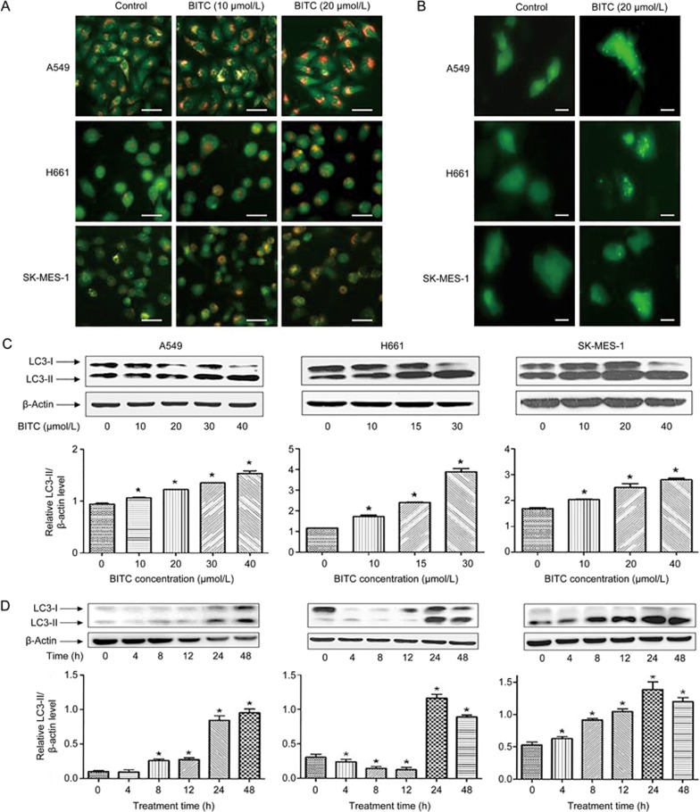
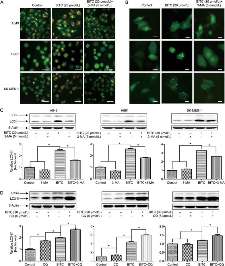
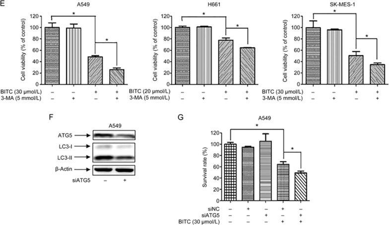
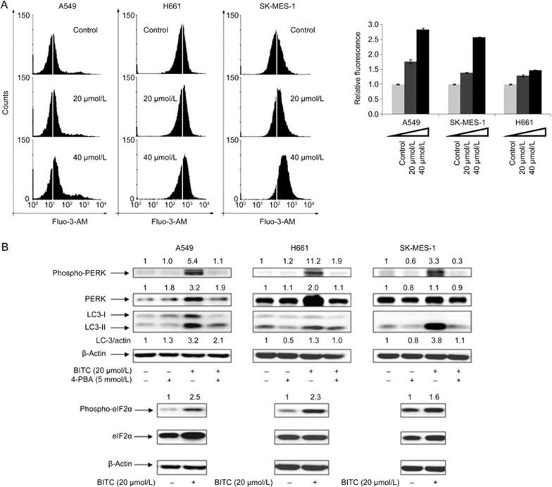
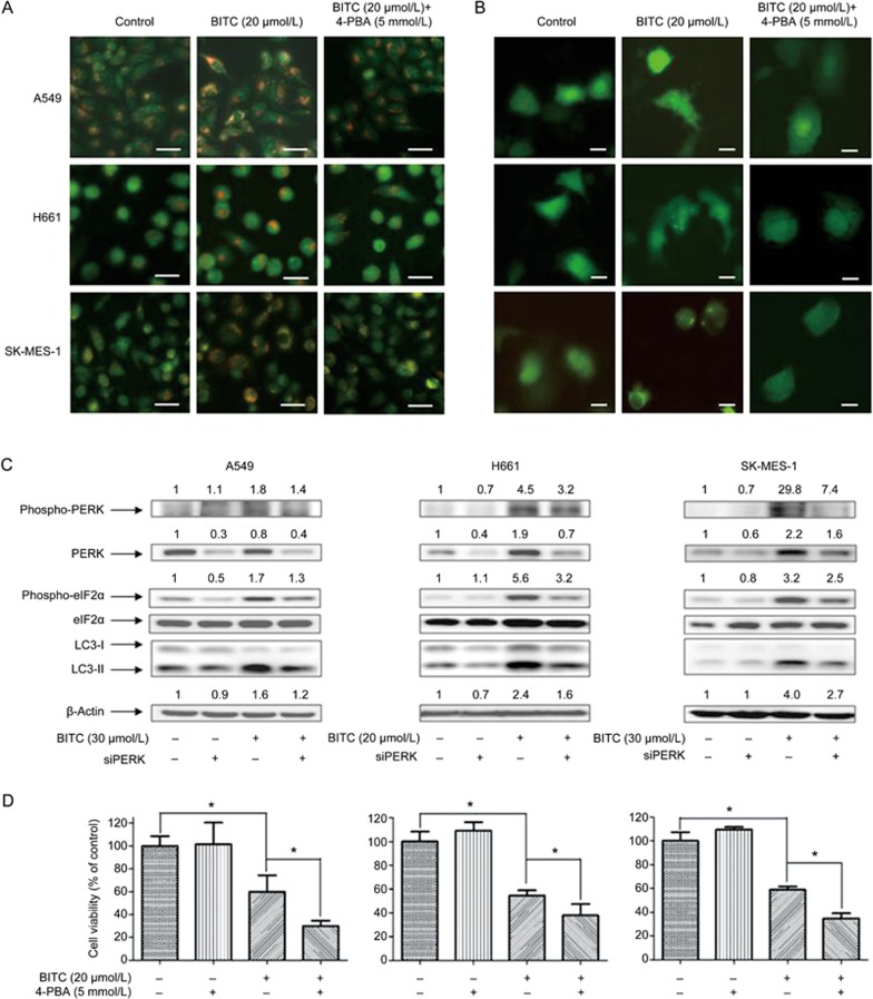
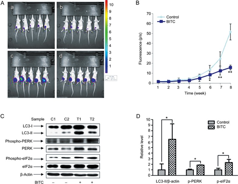
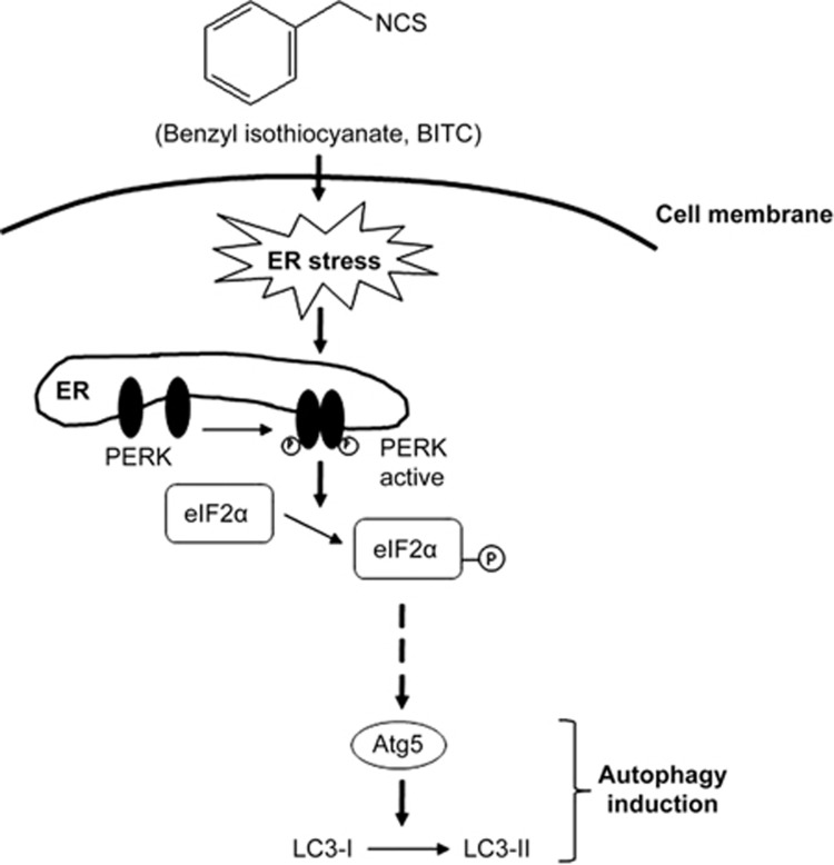
Isothiocyanates, such as allyl isothiocya¬nate (AITC), benzyl isothiocyanate (BITC), phenethyl isothio¬cyanate (PEITC) and sulforaphane (SFN), are natural compounds abundant in cruciferous vegetables, which have substantial chemopreventive activities against various human malignancies. However, the mechanisms underlying the inhibition of tumor cell growth by isothiocyanates are not fully understood. Since autophagy has dual functions in cancer, in the present study we investigated the effects of BITC on autophagy induction in human lung cancer cells in vitro and in vivo. BITC (1-100 μmol/L) dose-dependently inhibited the growth of 3 different human lung cancer cell lines A549 (adenocarcinoma), H661 (large cell carcinoma) and SK-MES-1 (squamous cell carcinoma) with IC50 values of 30.7±0.14, 15.9±0.22 and 23.4±0.11 μmol/L, respectively. BITC (10-40 μmol/L) induced autophagy in the lung cancer cells, evidenced by the formation of acidic vesicular organelles (AVOs), the accumulation of LC3-II, the punctate pattern of LC3, and the expression of Atg5. Pretreatment with the autophagy inhibitor 3-MA (5 mmol/L) significantly enhanced the BITC-caused growth inhibition in the lung cancer cells. Furthermore, BITC (20-40 μmol/L) activated ER stress, as shown by the increased cytosolic Ca2+ level and the phosphorylation of the ER stress marker proteins PERK and eIF2α in the lung cancer cells. Pretreatment with the ER stress inhibitor 4-PBA (5 mmol/L) attenuated the autophagy induction and potentiated the BITC-induced cell growth inhibition. In nude mice bearing A549 xenografts, administration of BITC (100 mg·kg-1·d-1, ip) for 8 weeks markedly suppressed the lung tumor growth, and significantly enhanced both autophagy and ER stress in the tumor tissues. Our results demonstrate that BITC inhibits human lung cancer cell growth in vitro and in vivo. In addition, BITC induces autophagy in the lung cancer cells, which protects the cancer cells against the inhibitory action of BITC; the autophagy induction is mediated by the ER stress response.


Figures

Figure 1
Figure 1
BITC inhibited human lung cancer cell growth. (A) Structure of benzyl isothiocyanate. (B) Cell viability of lung cancer cells. A549, H661, and SK-MES-1 cells were treated with 1–100 μmol/L BITC. The control group was treated with vehicle (DMSO). After 48 h, the cells were collected, and the viable cells were counted with the Cell Counting Kit-8. Values represent the mean±SD from three independent experiments.
Figure 2
Figure 2
BITC induced autophagy in human lung cancer cells. The induction of autophagy in cells by BITC treatment was assessed. (A) A549, H661, and SK-MES-1 cells were treated with 10 and 20 μmol/L BITC or vehicle for 24 h. The formation of acidic vesicles was detected by fluorescence microscopy using AO labeling. Scale bar=20 μm. (B) Lung cancer cells were transfected with pSELECT-GFP-LC3 and then treated with 20 μmol/L BITC for 24 h. The punctate pattern of LC3 was observed by fluorescence microscopy. Scale bar=30 μm. (C) Lung cancer cells were treated with 10–40 μmol/L BITC or vehicle for 24 h. The LC3 cleavage was assessed by immunoblotting. The relative band intensity of LC3-II was quantitated by densitometric scanning. (D) Lung cancer cells were treated with 20 μmol/L BITC or vehicle for 0–48 h. The LC3 cleavage was assessed by immunoblotting. The relative band intensity of LC3-II was quantitated by densitometric scanning. Values represent the mean±SD from three independent experiments. Columns, mean; bars, SD. *P<0.05.
Figure 3A–3D
Figure 3A–3D
Inhibition of autophagy enhanced the inhibitory effect of BITC on human lung cancer cells. (A) Lung cancer cells were pretreated with 5 mmol/L 3-MA for 2 h and then exposed to 20 μmol/L BITC or vehicle for 24 h. The formation of acidic vesicles was detected by fluorescence microscopy using AO labeling. Scale bar=20 μm. (B) Lung cancer cells were transfected with pSELECT-GFP-LC3. After 2 h of pretreatment with 5 mmol/L 3-MA, the cells were treated with 20 μmol/L BITC for 24 h. The punctate pattern of LC3 was observed by fluorescence microscopy. Scale bar=30 μm. (C) Lung cancer cells were pretreated with 5 mmol/L 3-MA for 2 h and then treated with 20 or 30 μmol/L BITC or vehicle for 24 h. The LC3 cleavage was assessed by immunoblotting. (D) Lung cancer cells were pretreated with 0 or 5 μmol/L CQ for 1 h and then treated with 20 or 30 μmol/L BITC or vehicle for 24 h. The LC3 cleavage was assessed by immunoblotting.
Figure 3E–3G
Figure 3E–3G
(E) Lung cancer cells were pretreated with 5 mmol/L 3-MA for 2 h and then exposed to 20 or 30 μmol/L BITC or vehicle for 24 h. The viable cells were counted. (F) The expression of ATG5 was knocked down using RNAi technology. The levels of ATG5 were analyzed by immunoblotting. (G) The expression of ATG5 was knocked down using RNAi, and the cells were then treated with 30 μmol/L BITC for 48 h. The viable cells were counted. Values represent the mean±SD from three independent experiments. Columns, mean; bars, SD. *P<0.05.
Figure 4
Figure 4
BITC activated ER stress in human lung cancer cells. The ER stress induced by BITC treatment was assessed. (A) Lung cancer cells were labeled with 5 μmol/L Fluo-3-AM for 30 min at 37 °C and then treated with 20–40 μmol/L BITC for 4 h. The level of intracellular Ca2+ was detected by measuring the intensity of fluorescence using flow cytometry. (B) Lung cancer cells were pretreated with 5 mmol/L 4-PBA for 2 h and then treated with 20 μmol/L BITC or vehicle for 24 h. The levels of p-PERK, PERK, p-eIF2α, eIF2α, LC3-I, and LC3-II were analyzed by immunoblotting. Values represent the mean±SD from three independent experiments. Columns, mean; bars, SD.
Figure 5
Figure 5
ER stress mediated the autophagy induction by BITC. (A) Lung cancer cells were pretreated with 5 mmol/L 4-PBA for 2 h and then exposed to 20 μmol/L BITC or vehicle for 24 h. The formation of acidic vesicles was detected by fluorescence microscopy using AO labeling. Scale bar=20 μm. (B) Lung cancer cells were transfected with pSELECT-GFP-LC3. After 2 h of pretreatment with 5 mmol/L 4-PBA, the cells were treated with 20 μmol/L BITC for 24 h. The punctate pattern of LC3 was observed by fluorescence microscopy. Scale bar=30 μm. (C) The cellular PERK expression was knocked down using RNA interference technology. The cells were then treated with 20–30 μmol/L BITC for 24 h. The levels of p-PERK, PERK, p-eIF2α, eIF2α, LC3-1, and LC3-II were analyzed by immunoblotting.
Figure 6
Figure 6
BITC inhibited tumor growth and induced autophagy in vivo. (A, B) A549-luc cells were subcutaneously injected into mice. The treatment was continued for 8 weeks. Tumors that formed from week 1 to week 8 were detected by an IVIS Imaging System. (a) and (b), control and BITC treatment groups at week 1. (c) and (d), control and BITC treatment groups at week 8. (C) Tumor tissues from two different mice in each group were used for immunoblot analysis. The relative band intensity was quantitated by densitometric scanning. C1 and C2, control group; T1 and T2, treatment group. Columns, mean; bars, SD. *P<0.05, **P<0.01 vs control.
Scheme 1
Scheme 1
A proposed model of ER stress-mediated induction of autophagy by BITC in lung cancer cells.

Similar articles

Cited by

References

    1. Ferlay J, Soerjomataram I, Dikshit R, Eser S, Mathers C, Rebelo M, et al. Cancer incidence and mortality worldwide: Sources, methods and major patterns in GLOBOCAN 2012. Int J Cancer 2015; 136: E359–86. - PubMed
    1. DeSantis CE, Lin CC, Mariotto AB, Siegel RL, Stein KD, Kramer JL, et al. Cancer treatment and survivorship statistics, 2014. CA Cancer J Clin 2014; 64: 252–71. - PubMed
    1. Mizushima N, Komatsu M. Autophagy: renovation of cells and tissues. Cell 2011; 147: 728–41. - PubMed
    1. Choi AM, Ryter SW, Levine B. Autophagy in human health and disease. N Engl J Med 2013; 368: 1845–6. - PubMed
    1. White E. Deconvoluting the context-dependent role for autophagy in cancer. Nat Rev Cancer 2012; 12: 401–10. - PMC - PubMed