

Cholesterol Synthetase DHCR24 Induced by Insulin Aggravates Cancer Invasion and Progesterone Resistance in Endometrial Carcinoma
- PMID: 28112250
- PMCID: PMC5256103
- DOI: 10.1038/srep41404
Cholesterol Synthetase DHCR24 Induced by Insulin Aggravates Cancer Invasion and Progesterone Resistance in Endometrial Carcinoma
Abstract
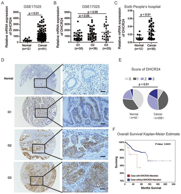
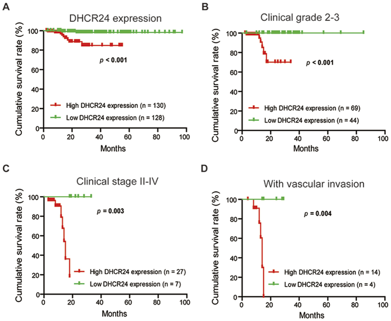
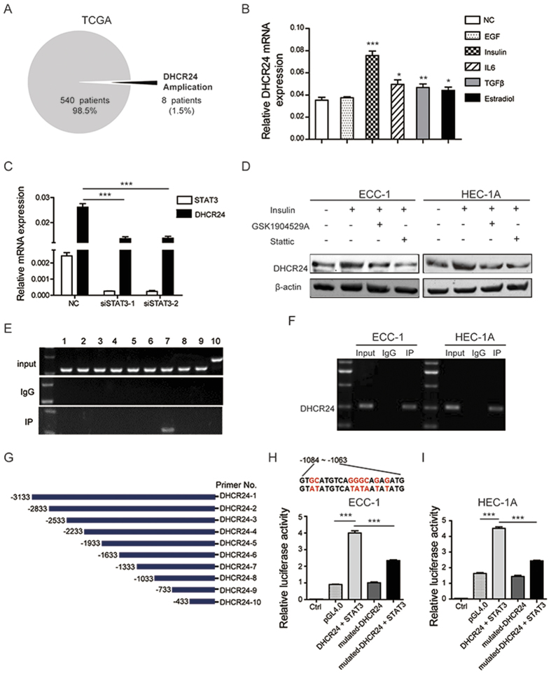
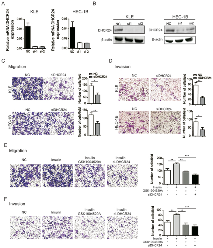
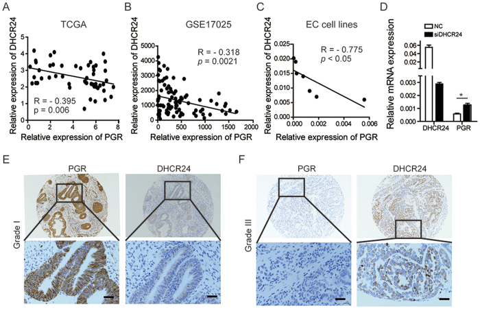
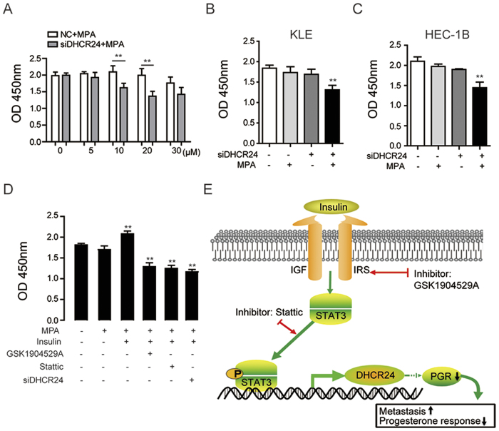
3β-Hydroxysteroid-Δ24 reductase (DHCR24), the final enzyme of the cholesterol biosynthetic pathway, has been associated with urogenital neoplasms. However, the function of DHCR24 in endometrial cancer (EC) remains largely elusive. Here, we analyzed the expression profile of DHCR24 and the progesterone receptor (PGR) in our tissue microarray of EC (n = 258), the existing EC database in GEO (Gene Expression Omnibus), and TCGA (The Cancer Genome Atlas). We found that DHCR24 was significantly elevated in patients with EC, and that the up-regulation of DHCR24 was associated with advanced clinical stage, histological grading, vascular invasion, lymphatic metastasis, and reduced overall survival. In addition, DHCR24 expression could be induced by insulin though STAT3, which directly binds to the promoter elements of DHCR24, as demonstrated by ChIP-PCR and luciferase assays. Furthermore, genetically silencing DHCR24 inhibited the metastatic ability of endometrial cancer cells and up-regulated PGR expression, which made cells more sensitive to progestin. Taken together, we have demonstrated for the first time the crucial role of the insulin/STAT3/DHCR24/PGR axis in the progression of EC by modulating the metastasis and progesterone response, which could serve as potential therapeutic targets for the treatment of EC with progesterone receptor loss.
Figures
Similar articles
-
CNR1 may reverse progesterone-resistance of endometrial cancer through the ERK pathway.Biochem Biophys Res Commun. 2021 Apr 9;548:148-154. doi: 10.1016/j.bbrc.2021.02.038. Epub 2021 Feb 25. Biochem Biophys Res Commun. 2021. PMID: 33640608
-
Knockdown of long non-coding HOTAIR enhances the sensitivity to progesterone in endometrial cancer by epigenetic regulation of progesterone receptor isoform B.Cancer Chemother Pharmacol. 2019 Feb;83(2):277-287. doi: 10.1007/s00280-018-3727-0. Epub 2018 Nov 15. Cancer Chemother Pharmacol. 2019. PMID: 30443761
-
Comprehensive bioinformatics analysis of acquired progesterone resistance in endometrial cancer cell line.J Transl Med. 2019 Feb 27;17(1):58. doi: 10.1186/s12967-019-1814-6. J Transl Med. 2019. PMID: 30813939 Free PMC article.
-
Desmosterol and DHCR24: unexpected new directions for a terminal step in cholesterol synthesis.Prog Lipid Res. 2013 Oct;52(4):666-80. doi: 10.1016/j.plipres.2013.09.002. Epub 2013 Oct 2. Prog Lipid Res. 2013. PMID: 24095826 Review.
-
The role of DHCR24 in the pathogenesis of AD: re-cognition of the relationship between cholesterol and AD pathogenesis.Acta Neuropathol Commun. 2022 Mar 16;10(1):35. doi: 10.1186/s40478-022-01338-3. Acta Neuropathol Commun. 2022. PMID: 35296367 Free PMC article. Review.
Cited by
-
Serine-arginine splicing factor 2 promotes oesophageal cancer progression by regulating alternative splicing of interferon regulatory factor 3.RNA Biol. 2023 Jan;20(1):359-367. doi: 10.1080/15476286.2023.2223939. RNA Biol. 2023. PMID: 37335045 Free PMC article.
-
OncoTrace-TOO: Interpretable Machine Learning Framework for Cancer Tissue-of-Origin Identification Using Transcriptomic Signatures.Cancer Rep (Hoboken). 2025 Aug;8(8):e70311. doi: 10.1002/cnr2.70311. Cancer Rep (Hoboken). 2025. PMID: 40784724 Free PMC article.
-
Aglianico Grape Seed Semi-Polar Extract Exerts Anticancer Effects by Modulating MDM2 Expression and Metabolic Pathways.Cells. 2023 Jan 4;12(2):210. doi: 10.3390/cells12020210. Cells. 2023. PMID: 36672146 Free PMC article.
-
Genkwadaphnin inhibits growth and invasion in hepatocellular carcinoma by blocking DHCR24-mediated cholesterol biosynthesis and lipid rafts formation.Br J Cancer. 2020 Nov;123(11):1673-1685. doi: 10.1038/s41416-020-01085-z. Epub 2020 Sep 22. Br J Cancer. 2020. PMID: 32958824 Free PMC article.
-
Oncogenic role of the SOX9-DHCR24-cholesterol biosynthesis axis in IGH-BCL2+ diffuse large B-cell lymphomas.Blood. 2022 Jan 6;139(1):73-86. doi: 10.1182/blood.2021012327. Blood. 2022. PMID: 34624089 Free PMC article.
References
Publication types
MeSH terms
Substances
Supplementary concepts
LinkOut - more resources
Full Text Sources
Other Literature Sources
Medical
Research Materials
Miscellaneous
