. 2017 Oct;232(10):2722-2730.
doi: 10.1002/jcp.25818. Epub 2017 Apr 25.

SUMO-modified insulin-like growth factor 1 receptor (IGF-1R) increases cell cycle progression and cell proliferation

Affiliations

SUMO-modified insulin-like growth factor 1 receptor (IGF-1R) increases cell cycle progression and cell proliferation

Yingbo Lin et al. J Cell Physiol. 2017 Oct.

Abstract

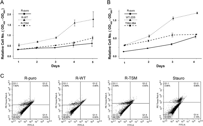
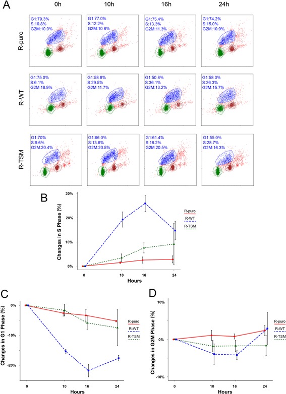
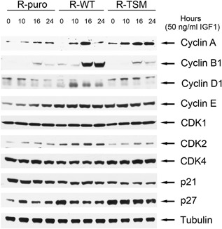
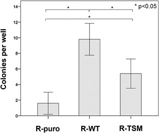
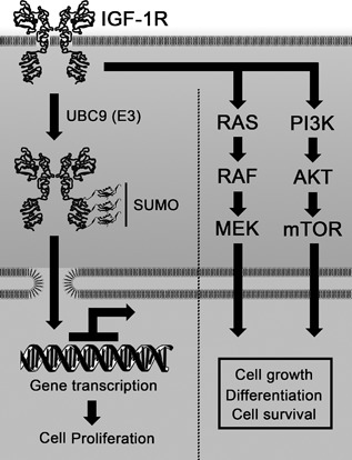
Increasing number of studies have shown nuclear localization of the insulin-like growth factor 1 receptor (nIGF-1R) in tumor cells and its links to adverse clinical outcome in various cancers. Any obvious cell physiological roles of nIGF-1R have, however, still not been disclosed. Previously, we reported that IGF-1R translocates to cell nucleus and modulates gene expression by binding to enhancers, provided that the receptor is SUMOylated. In this study, we constructed stable transfectants of wild type IGF1R (WT) and triple-SUMO-site-mutated IGF1R (TSM) using igf1r knockout mouse fibroblasts (R-). Cell clones (R-WT and R-TSM) expressing equal amounts of IGF-1R were selected for experiments. Phosphorylation of IGF-1R, Akt, and Erk upon IGF-1 stimulation was equal in R-WT and R-TSM. WT was confirmed to enter nuclei. TSM did also undergo nuclear translocation, although to a lesser extent. This may be explained by that TSM heterodimerizes with insulin receptor, which is known to translocate to cell nuclei. R-WT proliferated substantially faster than R-TSM, which did not differ significantly from the empty vector control. Upon IGF-1 stimulation G1-S-phase progression of R-WT increased from 12 to 38%, compared to 13 to 20% of R-TSM. The G1-S progression of R-WT correlated with increased expression of cyclin D1, A, and CDK2, as well as downregulation of p27. This suggests that SUMO-IGF-1R affects upstream mechanisms that control and coordinate expression of cell cycle regulators. Further studies to identify such SUMO-IGF-1R dependent mechanisms seem important.

Keywords: IGF-1R; SUMOylation; cancer; cell cycle; proliferation.


Figures

Figure 1
Figure 1
Characterization of R‐cells (igf1r−/−) stably expressing wild type (WT) or triple SUMO‐sites mutated (TSM) IGF1R. (A) Relative IFG1R mRNA transcription levels in selected transfected R‐cell clones were determined by qRT‐PCR. Two cell lines with equal expression of WT (WT‐2C4) and TSM (TSM‐2D4) IFG1R mRNA (marked with arrows) were chosen for further investigation and named R‐WT and R‐TSM, respectively. R‐puro was an empty vector control. (B) IGF‐1R and pre‐IGF1R protein expression in R‐puro, R‐WT, R‐TSM cells compared to the cancer cell lines MCF7 (breast cancer), H1299 (lung cancer), HCT116 (colon cancer) were determined by immunoblotting (IB) with anti‐IGF‐1Rβ. GAPDH was blotted as loading control. (C) SUMOylation of IGF‐1R in R‐puro, R‐WT, and R‐TSM cell lines were determined by immunoprecipitation (IP) of IGF‐1R and IB for SUMO1. The three predicted SUMO‐IGF‐1R bands are indicated by arrows. Re‐blot of IGF‐1Rβ as an input IP control. (D) IGF‐1R tyrosine kinase activity was assessed by receptor and substrate phosphorylation. IGF‐1R was IPed from lysates from R‐puro, R‐WT, and R‐TSM cells that had been subjected to 36‐h serum starvation with or without subsequent 10‐min IGF‐1 stimulation, and blotted with anti‐phospho‐tyrosine antibody. Re‐blot of IGF‐1R β served as input control. The lysates were also directly blotted for phosphorylated Erk (pErk) and phosphorylated Akt (pAkt). GAPDH was used as loading control. (E) R‐puro, R‐WT, and R‐TSM cells were fractionized and the nucleus portions were immunoblotted with anti‐IGF1Rβ. Histone 3 was blotted as loading control. (F) Association of IGF‐1R and InsR was investigated by co‐IP. IGF‐1R IPs from R‐puro, R‐WT, and R‐TSM cell lysates were blotted for InsRβ. Re‐blot of IGF‐1Rβ served as control of IP. (G) Co‐localizations between IGF‐1R and InsR were visualized by PLA (red dots) in R‐puro, R‐WT, and R‐TSM cells. IGF‐1R and cell nuclei were counterstained with Alexa Fluor® 488 (Green) and DAPI (Blue), respectively. Data are representative of 3–5 experiments
Figure 2
Figure 2
Comparison of cell proliferation and apoptotic cell death. (A) Equal numbers of R‐puro, R‐WT, or R‐TSM were seeded in 96‐well plates and cultured under basal condition. Proliferation of cells were monitored by XTT proliferation assay kit every 24 hr. Each time‐point represents the average (n = 5) proliferation, with whiskers representing a 95% confidence interval. (B) R‐puro, WT‐2D5, or TSM‐3B4 cells were investigated for proliferation exactly as described in A. (C) Apoptosis/cell death in R‐puro, R‐WT, or R‐TSM cells under basal condition was assessed using Annexin V (X‐axis)/PI (Y‐axis) protocol. A total of 1 μM (final concentration) staurosporine was used as positive control. Data are representative of three experiments
Figure 3
Figure 3
Comparison of ligand stimulated cell cycle progression. (A) R‐puro, R‐WT, and R‐TSM were synchronized by 36 hr serum starvation and then exposed to 50 ng/ml IGF‐1 for 0, 10, 16, and 24 hr. FACS analyses after double staining with 7‐amino actinomycin D (X‐axis) and BrdU (Y‐axis) are shown. (B–D) Percentage of changes of number of cells in S‐ (B), G1‐ (C), and G2M‐phase (D) upon IGF‐1 stimulation for 10, 16, and 24 hr as compared to unstimulated cells (0 hr). Experiments on R‐puro, R‐WT, and R‐TSM were performed as described in A. Results represent means of three independent experiments. Whiskers represent 95% confidence intervals
Figure 4
Figure 4
Comparison of expression of cell cycle proteins. R‐puro, R‐WT, and R‐TSM were synchronized by 36 hr serum starvation and then exposed to 50 ng/ml IGF‐1 for 0, 10, 16, and 24 hr. Protein expression of cyclin A, cyclin B1, cyclin D1, cyclin E, CDK1, CDK2, CDK4, p21, and p27 was detected by immunoblotting. Tubulin was blotted as loading control. Data are representative of >3 experiments
Figure 5
Figure 5
Comparison of colony formation. Soft agar colony formation assay on R‐puro, R‐WT, and R‐TSM was performed as described in Materials and Methods. Experiments were stopped after 14 days for counting of colonies. Each column represents the mean number of colonies formed per well (total 5‐wells per cell line and experiment). Whiskers represent 95% confidence intervals. p‐values are indicated. Data are representative of three experiments
Figure 6
Figure 6
Schematic model of cellular responses of IGF‐1R. Besides traditional signaling pathways, the SUMO‐modification of IGF‐1R may modulate cell proliferation through gene transactivation. The model represents a hypothesis based on present and previous results

References

    1. Aleksic, T. , Chitnis, M. M. , Perestenko, O. V. , Gao, S. , Thomas, P. H. , Turner, G. D. , … Macaulay, V. M. (2010). Type 1 insulin‐like growth factor receptor translocates to the nucleus of human tumor cells. Cancer Research, 70, 6412–6419. - PMC - PubMed
    1. Aslam, M. I. , Hettmer, S. , Abraham, J. , LaTocha, D. , Soundararajan, A. , Huang, E. T. , … Druker, B. J. (2013). Dynamic and nuclear expression of PDGFRα and IGF‐1R in alveolar Rhabdomyosarcoma. Molecular Cancer Research, 11, 1303–1313. - PMC - PubMed
    1. Asmane, I. , Watkin, E. , Alberti, L. , Duc, A. , Marec‐Berard, P. , Ray‐Coquard, I. , … Bergerat, J. P. (2012). Insulin‐like growth factor type 1 receptor (IGF‐1R) exclusive nuclear staining: A predictive biomarker for IGF‐1R monoclonal antibody (Ab) therapy in sarcomas. European Journal of Cancer, 48, 3027–3035. - PubMed
    1. Baserga, R. (2009). The insulin receptor substrate‐1: A biomarker for cancer? Experimental Cell Research, 315, 727–732. - PubMed
    1. Beckwith, H. & Yee, D. (2015). Minireview: Were the IGF Signaling Inhibitors All Bad? Molecular Endocrinology, 29, 1549–1557. - PMC - PubMed

MeSH terms