

Tinnitus distress is linked to enhanced resting-state functional connectivity from the limbic system to the auditory cortex
- PMID: 28112466
- PMCID: PMC6866871
- DOI: 10.1002/hbm.23525
Tinnitus distress is linked to enhanced resting-state functional connectivity from the limbic system to the auditory cortex
Abstract
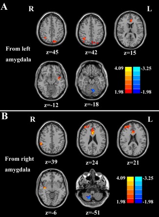
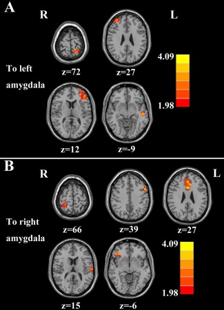
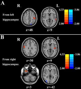
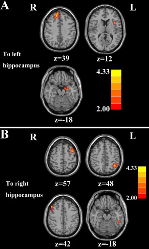
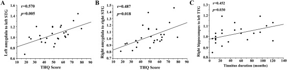
The phantom sound of tinnitus is believed to be triggered by aberrant neural activity in the central auditory pathway, but since this debilitating condition is often associated with emotional distress and anxiety, these comorbidities likely arise from maladaptive functional connections to limbic structures such as the amygdala and hippocampus. To test this hypothesis, resting-state functional magnetic resonance imaging (fMRI) was used to identify aberrant effective connectivity of the amygdala and hippocampus in tinnitus patients and to determine the relationship with tinnitus characteristics. Chronic tinnitus patients (n = 26) and age-, sex-, and education-matched healthy controls (n = 23) were included. Both groups were comparable for hearing level. Granger causality analysis utilizing the amygdala and hippocampus as seed regions were used to investigate the directional connectivity and the relationship with tinnitus duration or distress. Relative to healthy controls, tinnitus patients demonstrated abnormal directional connectivity of the amygdala and hippocampus, including primary and association auditory cortex, and other non-auditory areas. Importantly, scores on the Tinnitus Handicap Questionnaires were positively correlated with increased connectivity from the left amygdala to left superior temporal gyrus (r = 0.570, P = 0.005), and from the right amygdala to right superior temporal gyrus (r = 0.487, P = 0.018). Moreover, enhanced effective connectivity from the right hippocampus to left transverse temporal gyrus was correlated with tinnitus duration (r = 0.452, P = 0.030). The results showed that tinnitus distress strongly correlates with enhanced effective connectivity that is directed from the amygdala to the auditory cortex. The longer the phantom sensation, the more likely acute tinnitus becomes permanently encoded by memory traces in the hippocampus. Hum Brain Mapp 38:2384-2397, 2017. © 2017 Wiley Periodicals, Inc.
Keywords: effective connectivity; functional connectivity; limbic system; resting-state fMRI; tinnitus.
© 2017 Wiley Periodicals, Inc.
Figures
References
-
- Aldhafeeri FM, Mackenzie I, Kay T, Alghamdi J, Sluming V (2012): Neuroanatomical correlates of tinnitus revealed by cortical thickness analysis and diffusion tensor imaging. Neuroradiology 54:883–892. - PubMed
-
- Aron AR, Robbins TW, Poldrack RA (2014): Inhibition and the right inferior frontal cortex: One decade on. Trends Cogn Sci 18:177–185. - PubMed
-
- Bartels H, Staal MJ, Albers FW (2007): Tinnitus and neural plasticity of the brain. Otol Neurotol 28:178–184. - PubMed
-
- Biswal B, Zerrin Yetkin F, Haughton VM, Hyde JS (1995): Functional connectivity in the motor cortex of resting human brain using echo‐planar mri. Magn Reson Med 34:537–541. - PubMed
Publication types
MeSH terms
LinkOut - more resources
Full Text Sources
Other Literature Sources
Medical
