. 2017 Feb;35(2):154-163.
doi: 10.1038/nbt.3777. Epub 2017 Jan 23.

Combined small-molecule inhibition accelerates the derivation of functional cortical neurons from human pluripotent stem cells

Affiliations

Combined small-molecule inhibition accelerates the derivation of functional cortical neurons from human pluripotent stem cells

Yuchen Qi et al. Nat Biotechnol. 2017 Feb.

Abstract

Considerable progress has been made in converting human pluripotent stem cells (hPSCs) into functional neurons. However, the protracted timing of human neuron specification and functional maturation remains a key challenge that hampers the routine application of hPSC-derived lineages in disease modeling and regenerative medicine. Using a combinatorial small-molecule screen, we previously identified conditions to rapidly differentiate hPSCs into peripheral sensory neurons. Here we generalize the approach to central nervous system (CNS) fates by developing a small-molecule approach for accelerated induction of early-born cortical neurons. Combinatorial application of six pathway inhibitors induces post-mitotic cortical neurons with functional electrophysiological properties by day 16 of differentiation, in the absence of glial cell co-culture. The resulting neurons, transplanted at 8 d of differentiation into the postnatal mouse cortex, are functional and establish long-distance projections, as shown using iDISCO whole-brain imaging. Accelerated differentiation into cortical neuron fates should facilitate hPSC-based strategies for disease modeling and cell therapy in CNS disorders.


Conflict of interest statement

COMPETING FINANCIAL INTERESTS

The Memorial Sloan-Kettering Cancer Center has filed provisional patent application (US PRO 62/287821) on the methods described in the manuscript.

Figures
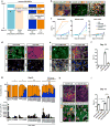
Figure 1
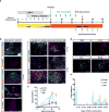
Figure 1. Rapid induction of cortical neurons from human pluripotent stem cells using a combinatorial small-molecule protocol
a) Illustration of pathway manipulations used to generate neural crest versus forebrain fates and neural precursor versus accelerated neuronal fates. SOX10+ neural crest precursor, PAX6+ CNS precursors and BRN3A/ISL1+ sensory neurons have been previously reported while the current study is focused on the rapid induction of cortical neurons from hPSCs. b) Validation of hESC-based reporter lines for PAX6::H2B-GFP, SOX10::GFP and SIX1::H2B-GFP by assessing co-labeling with matched protein markers. c) Time-course quantitative analysis of CNS (PAX6::H2B-GFP, left), NC (SOX10::GFP, middle) and placode (SIX1::H2B-GFP, right) induction under the various protocols. (LSB = dual SMAD inhibition; XAV = XAV939, tankyrase inhibitor (WNT-i); CHIR = CHIR99021, GSK3β-inhibition (WNT-activation). S = SU5402, FGFR inhibitor; D = DAPT, ɣ-secretase inhibitor (Notch-i). N = 3 independent batches of cell cultures per each condition and cell line. d) Immunocytochemistry for PAX6, SOX10 and TUJ1 during CNS versus NC based hPSC differentiation. e) CNS and NC-based hPSC differentiation in combination with S/D exposure to accelerate neuronal fate acquisition. f) Quantification of TUJ1 data from panels d and e at day 13 of differentiation by intracellular flow. Black dots represent values from independent experiments. From left to right, N = 3, 3, 5, 3. Statistics was done using unpaired t test with Welch’s correction (two-tailed). XAV vs. XAV+S/D: t=9.510 dF=2.160, P=0.0084. CHIR+S/D vs. XAV+S/D: t=13.40, df=4.718, P<0.0001. g) P, S single and combinatorial dose response analysis based on quantification of the percentages of NESTIN+ and TUJ1+ cells by intracellular flow cytometry (upper), and an estimate of the total neurons in culture per cm2 (for every hPSC plated at day 0 under P1S5D and P8S10D conditions, we obtained ~ 0.7 and 0.2 neurons respectively at day 13). P = PD0325901, ERK/MEK inhibitor. The digits following P, S, D represent the respective concentrations in μM. Each column represents N = 2 technical replicates per marker screened. h) Immunocytochemistry for PAX6/TUJ1 in P1S5D and P8S10D cultures at day 13. i) Quantification of TUJ1+ cells by intracellular flow cytometry at day 13. Black dots represent values from independent experiments. From left to right, N = 4, 5, 5, 4. Statistics was done using unpaired t test with Welch’s correction (two-tailed). XAV+P(1)/D vs. XAV+P(1)/S(5)/D: t=4.057, dF=6.045, P=0.0066. XAV+P(1)/S(5) vs. XAV+P(1)/S(5)/D: t=20.62, dF=4.880, P<0.0001. XAV+P(1)/S(5)/D vs. XAV+P(8)/S(10)/D: t=13.26, dF=6.397, P<0.0001. Scale bars: 100 μm (b), 200 μm (d, e, h). Error bars represent s. e. m. ** P<0.01, **** P<0.0001.
Figure 2
Figure 2. Temporal and phenotypic characterization of hPSC derived neurons by day 13 of differentiation
a) Differentiation scheme for KSR-based P1S5D and P8S10D neuronal induction protocol. hPSCs were plated one day prior to differentiation at 200,000/cm2 in conditioned hESC media supplemented with 10ng /ml FGF2 and 10 μM ROCK-Inhibitor Y-27632. Small molecules are added in the presence of dual SMAD inhibition (LDN193189, SB431542) and XAV939 treatment (LSB+X). Optimized timing for the application of PD0325901, SU5402 and DAPT (P/S/D) are shown. NB: neurobasal medium. BCA: BDNF, cAMP and ascorbic acid. b) Time-course analysis of neuronal induction efficiency by intracellular flow cytometry (N = 4 independent batches of cell cultures), unpaired t test with Welch’s correction (two-tailed) to compare mean difference between each group at day 13. LSBX vs. P1S5D: t=20.79 dF=3.042, P=0.0002. 3i vs. P1S5D: t=0.8277 dF=5.013, P=0.4455, N.S. P1S5D vs. P8S10D: t=12.79 dF=5.994, P<0.0001. c) Time-course qRT-PCR analysis at day 5, 8 11, 13 of differentiation. OCT4(POU5F1): Human pluripotency marker; PAX6: Dorsal cortical progenitor marker; FOXG1: Forebrain marker; DCX: Pan-neuron marker; TBR1: preplate, subplate and cortical Layer VI neuron marker; REELIN: cortical Layer I (Cajal-Retzius cell) neuron marker. FC: fold change. N = 3 independent batches of cell cultures. d) TBR1/TUJ1 expression by immunocytochemistry at day 13, with quantification of (e) the percentage of TBR1+ cells among total cells (t=3.151, dF=7.820, P=0.0140, two-tailed), or (f) among TUJ1+ neurons (t=1.094, dF=9.997, P=0.2994, two-tailed, N.S.). Black dots represent values from quantification of uniform random selection of six 150 μm × 150 μm areas from 3 independent batches of cell cultures. Statistics was done using unpaired t test with Welch’s correction. g) Alternative scheme of accelerated neuronal differentiation protocol using P/S/D in E6 medium. h) Validation of P/S/D protocol in E6 by immunocytochemistry of PAX6/TUJ1 and TBR1/TUJ1 expression at day 13. Scale bars: 50 μm, except for 100 μm in left and middle panels of (h). Error bars represent s. e. m. ** P<0.01, *** P<0.001, **** P<0.0001. N.S.: no significant.
Figure 3
Figure 3. Generation of neurons constituting multiple cortical layers upon long-term culture
a) Illustration of long-term culture protocols. In vitro differentiation before day 8 is the same as described in Fig.2a. For long-term culture, P1S5D and P8S10D cells were passaged at day 8 of differentiation at 150,000/cm2 and 300,000/cm2 respectively in NB/B27+BCA medium without adding inhibitors thereafter. Cells were fixed at various time points and processed for immunocytochemistry or RNA extraction and qRT-PCR analysis. b) Long-term maintenance of P1S5D and P8S10D cells produced neurons constituting distinct cortical layer fates: FOXP2 (layer V–VI), TLE4 (layer VI), CTIP2 (layer V), SATB2 (layer II–III, V), RGS4 (layer II–III, layer V), CUX2 (cortical progenitors and layer II–IV). c) Quantification of TBR1+, CTIP2+ and SATB2+ cells in total cell population using P1S5D differentiation. N = quantification of 6 randomly selected photo frames captured using a 20X objective from 2 independent batches of cell cultures. d) Representative image showing co-labeling of EdU with TBR1 and CTIP2 at day 40 of P1S5D differentiation. e) Quantification of the percentage of EdU positive among marker positive cells at day 40 of P1S5D differentiation. EdU was added to the cultures for 48 hrs at various time points of differentiation (as indicated on x-axis) and the cultures were fixed at day 40. Colored dots represent values of quantification results of individual photo frames from 2 independent batches of cell cultures. From left to right, N = 3,4,4; 0,0,0; 3,4,3; 3,3,4; 3,5,1; 1,3,4. Scale bars: 50 μm. Error bars represent s. e. m.
Figure 4
Figure 4. Accelerated induction yields hPSC-derived cortical neurons with mature electrophysiological properties in vitro
a) Illustration of six different treatment conditions for neuronal differentiation and maintenance. In vitro differentiation protocol up to day 8 is the same as described in Fig. 2a. P1S5D and P8S10D cells were then passaged at 150,000/cm2 and 300,000/cm2 respectively at day 8 in NB/B27+BCA. Both passaged P1S5D and P8S10D cells were further maintained in three conditions: without inhibitors (+none), with DAPT (+D), and with PD0315901 (1uM), SU5402 (5uM), DAPT, CHIR99021 (3uM) (+PSDC), making 6 different treatments of cells in total. b) Action potential firing traces at day 16 from cells representing the 6 differentiation conditions. c) Quantification of percentage of cells with indicated firing frequencies at day 16 of differentiation without current injection, and (d) with −10 pA current injection. Injecting −10 pA current triggered evoked firing and enabled an even larger proportion of cells to adopt high firing frequencies. From left to right, N = 15, 18, 11, 16, 17, 12 from 4 batches of independent cell cultures. PSDC: P(1 μM) S(5 μM) D+CHIR. e) Quantification of auto-firing electrophysiological properties at day 16 (without current injection). Black dots represent values of individual cells. From left to right, N = 15, 18, 11, 16, 17, 12 from 4 batches of independent cell cultures. f) Voltage-dependent sodium channel responses of P1S5D+none neurons at day 37 by whole-cell patch clamp, which could be blocked by Tetrodotoxin (TTX) that specifically blocks the sodium channel. Inset: protocol used for triggering sodium channel currents. g) Spontaneous excitatory postsynaptic currents (sEPSCs) recorded under P1S5D+none conditions at day 40 indicative of functional synapse formation. sEPSCs could be blocked by NBQX that selectively blocks AMPA receptors indicating excitatory synaptic currents. Error bars represent s. e. m.
Figure 5
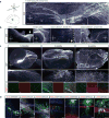
Figure 5. Extensive axonal projections and integration of hPSC-derived neuron using P1S5D induction grafted into the neonatal mouse brain as assessed by iDISCO-based whole mount brain imaging
a) Analysis of the grafted brain at 1–6 months post-grafting using whole brain immunohistochemistry and imaging by light-sheet microscopy. Dorsal view of the graft core and its cortical projections at 1.5 months. b) Projections (100 μm thick) showing the graft core morphology and the major projection regions (frontal cortex and corpus callosum). Dotted line: midline, arrow: aberrant longitudinal projections in the corpus callosum. c) iDISCO based imaging of half brains showing the morphology of the graft projections at 1.5, 3 and 6 months. Top panels: views of the half brain showing the graft cores and their major cortical projections. Central panels: projections (100 μm thick) showing the details of the fiber morphology from the boxed regions, and their increased branching over time. Lower panel: projections (100 μm thick) showing hSynaptophysin co-labeled with GFP in the hippocampus. The hSyn signal was absent at 1.5 months, extremely faint at 3 months, but very high at 6 months. d) iDISCO based immunohistochemical analysis of graft derived GFP+ fiber projections at 1.5 and 6 months post grafting (left, middle panels). Right panel: human specific synaptophysin expression in host hippocampus. e) Immunohistochemical analysis for markers of cortical identity in graft derived neurons identified by human specific cytoplasm marker (SC121) or GFP expression. ~ 60% of the grafted cells expressed TBR1, ~ 50% expressed CTIP2 (more than half of all CTIP2+ cells co-expressed TBR1), and ~ 30% expressed SATB2. N = 5 animals for 1, 1.5 and 3 months analyzed, and 2 animals for 6 months analyzed. Scale bars: 500 μm (a,b,c), 50 μm (c, bottom panel and d).
Figure 6
Figure 6. Summary of rapid cortical neuron induction paradigm
Dual SMAD inhibition by LSB inhibits trophectoderm, mesendoderm, and non-neural ectoderm cell fates promoting CNS fates. XAV939 promotes anterior CNS identity while SU5402/PD0325901 accelerate exit from pluripotency toward neuroectodermal fates. A highly transient anterior neuroectodermal precursor state is driven toward post-mitotic cortical fates in the presence of DAPT and SU5402/PD0325901. Immature cortical neurons can acquire functional maturity in vitro by day 16 of differentiation and day 8 neurons, grafted into neonatal mouse host brain, show widespread axonal projections and integration in cortex.

References

    1. Lee G, et al. Modelling pathogenesis and treatment of familial dysautonomia using patient-specific iPSCs. Nature. 2009;461:402–406. - PMC - PubMed
    1. Mica Y, Lee G, Chambers SM, Tomishima MJ, Studer L. Modeling neural crest induction, melanocyte specification, and disease-related pigmentation defects in hESCs and patient-specific iPSCs. Cell reports. 2013;3:1140–1152. - PMC - PubMed
    1. Menendez L, Yatskievych TA, Antin PB, Dalton S. Wnt signaling and a Smad pathway blockade direct the differentiation of human pluripotent stem cells to multipotent neural crest cells. Proceedings of the National Academy of Sciences of the United States of America. 2011;108:19240–19245. - PMC - PubMed
    1. Maroof AM, et al. Directed differentiation and functional maturation of cortical interneurons from human embryonic stem cells. Cell stem cell. 2013;12:559–572. - PMC - PubMed
    1. Sun L, et al. Design, synthesis, and evaluations of substituted 3-[(3- or 4-carboxyethylpyrrol-2-yl)methylidenyl]indolin-2-ones as inhibitors of VEGF, FGF, and PDGF receptor tyrosine kinases. Journal of medicinal chemistry. 1999;42:5120–5130. - PubMed

METHODS ONLY REFERENCES

    1. Hoya-Arias R, Tomishima M, Perna F, Voza F, Nimer SD. L3MBTL1 deficiency directs the differentiation of human embryonic stem cells toward trophectoderm. Stem Cells Dev. 2011;20:1889–1900. - PMC - PubMed
    1. Sanjana NE, et al. A transcription activator-like effector toolbox for genome engineering. Nat Protoc. 2012;7:171–192. - PMC - PubMed
    1. Kaech S, Banker G. Culturing hippocampal neurons. Nat Protoc. 2006;1:2406–2415. - PubMed

Publication types

MeSH terms

Substances