. 2017 Jan 23;12(1):e0170716.
doi: 10.1371/journal.pone.0170716. eCollection 2017.

Polyporus squamosus Lectin 1a (PSL1a) Exhibits Cytotoxicity in Mammalian Cells by Disruption of Focal Adhesions, Inhibition of Protein Synthesis and Induction of Apoptosis

Affiliations

Polyporus squamosus Lectin 1a (PSL1a) Exhibits Cytotoxicity in Mammalian Cells by Disruption of Focal Adhesions, Inhibition of Protein Synthesis and Induction of Apoptosis

Dipankar Manna et al. PLoS One. .

Abstract

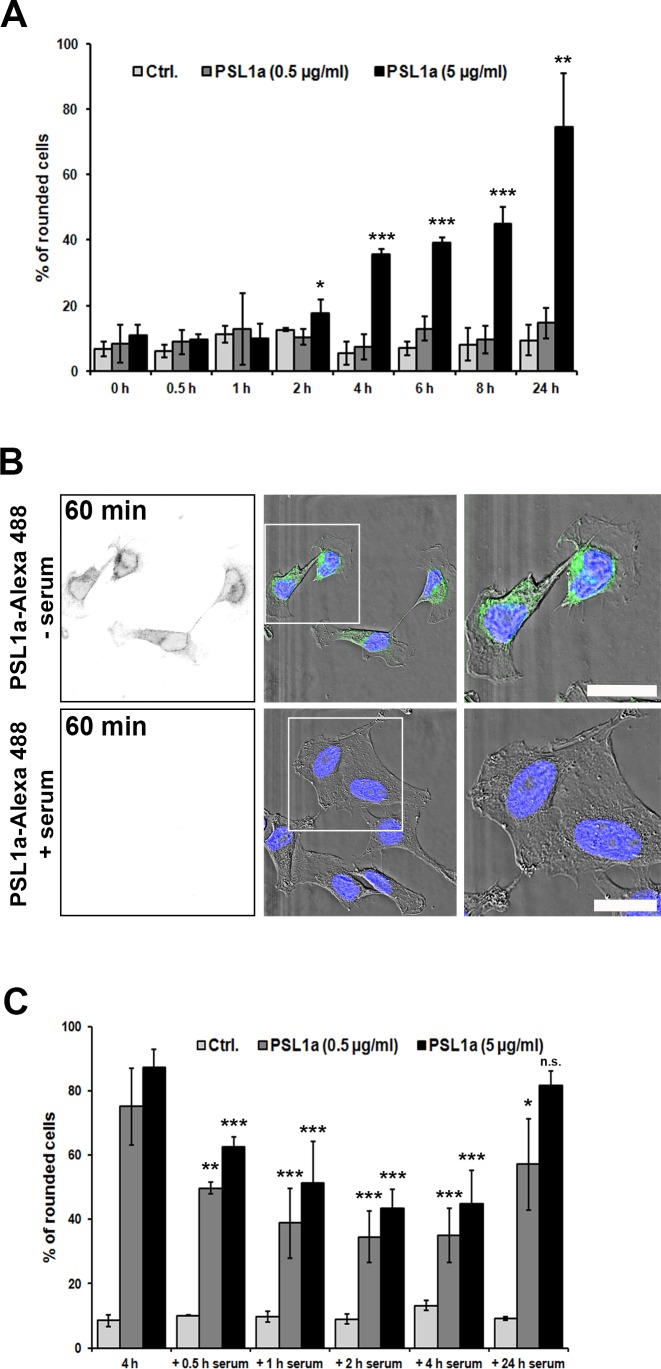
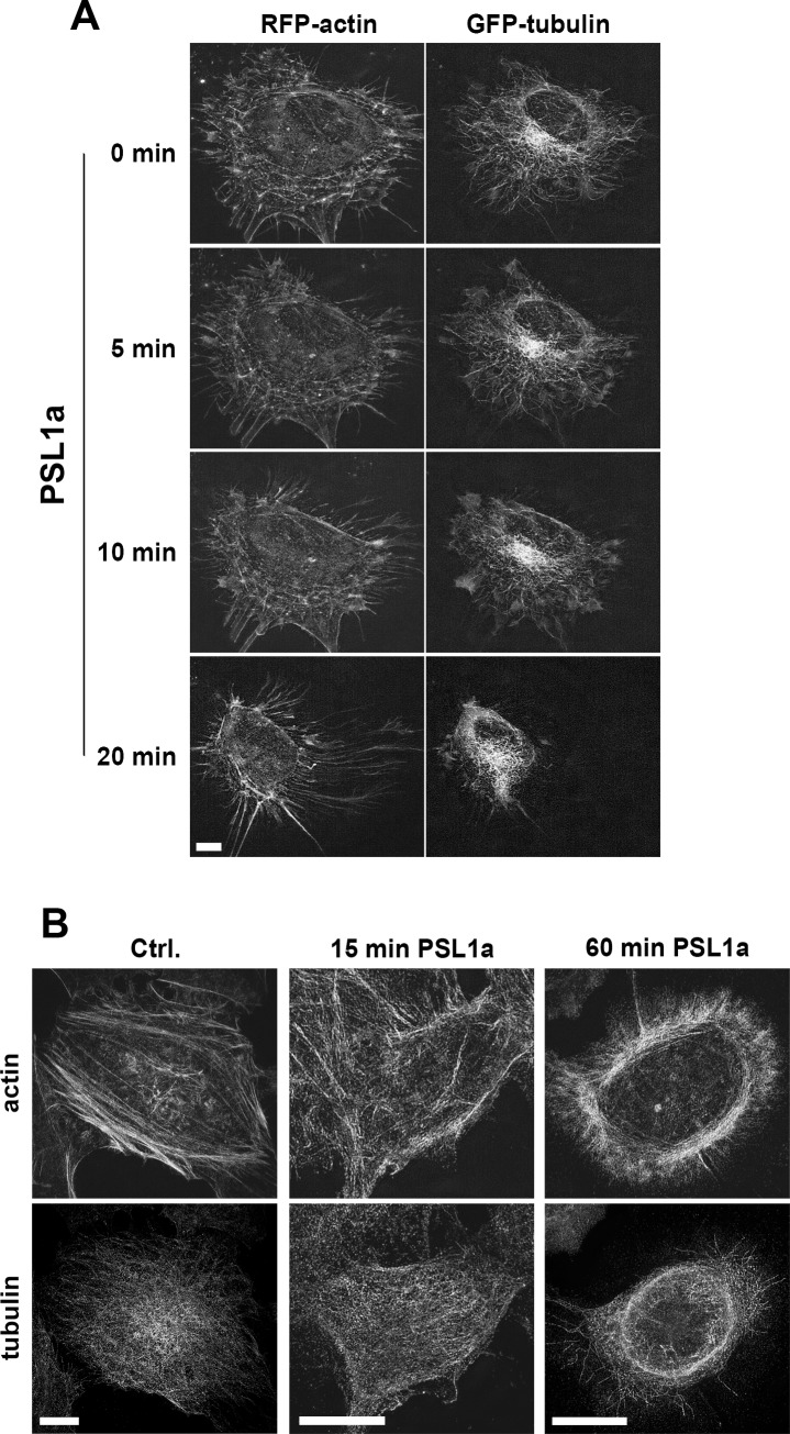
PSL1a is a lectin from the mushroom Polyporus squamosus that binds to sialylated glycans and glycoconjugates with high specificity and selectivity. In addition to its N-terminal carbohydrate-binding domain, PSL1a possesses a Ca2+-dependent proteolytic activity in the C-terminal domain. In the present study, we demonstrate that PSL1a has cytotoxic effects on mammalian cancer cells, and we show that the cytotoxicity is dependent on the cysteine protease activity. PSL1a treatment leads to cell rounding and detachment from the substratum, concomitant with disruption of vinculin complexes in focal adhesions. We also demonstrate that PSL1a inhibits protein synthesis and induces apoptosis in HeLa cells, in a time- and concentration-dependent manner.


Conflict of interest statement

The authors have declared that no competing interests exist.

Figures

Fig 1
Fig 1. PSL1a treatment leads to cell rounding.
(A) HeLa, HEp-2, SKBR-3 and PC3 cells were treated with 5 μg/ml of PSL1a in serum-free medium and incubated for 4 h at 37°C. PSL1a treatment leads to cell rounding in all tested cell lines. Scale bar: 200 μm. (B) HeLa cells were treated with 0.5 μg/ml and 5 μg/ml of PSL1a in serum-free medium at 37°C for 4 h in total. The cell morphology was analyzed by light microscopy at given time points and the numbers of rounded cells were quantified. Results are shown as mean values +/- SEM from at least three independent experiments; 180–300 cells per condition, per time point in each experiment (*P<0.05; **P<0.01; ***P<0.001 compared to untreated control). (C) RPE cells were treated for 4 h with 0.5 μg/ml and 5 μg/ml of PSL1a in serum-free medium at 37°C. Only slight morphological changes but no cell rounding could be observed even upon treatment with 5 μg/ml of PSL1a. Scale bar: 200 μm. (D) Quantification of rounded RPE cells after treatment with PSL1a, as described in (C). No significant cell rounding was observed. Results are shown as mean values +/- SEM from at least three independent experiments (210–300 cells per condition, per time point in each experiment).
Fig 2
Fig 2. PSL1a induced cell rounding is partially inhibited by the presence of serum.
(A) HeLa cells were treated with 0.5 μg/ml or 5 μg/ml of PSL1a in the presence of serum for 24 h in total. After the indicated time points the number of rounded cells was quantified. The presence of serum completely inhibits the effect of PSL1a at the lower concentration. At higher concentrations the effect of PSL1a is strongly reduced and the cellular response is delayed (compare Fig 1B and Fig 2A). Results are shown as mean values +/- SEM from at least three independent experiments (90–180 cells per condition, per time point in each experiment, *P<0.05; **P<0.01; ***P<0.001 compared to untreated control). (B) HeLa cells were treated with 1 μg/ml of Alexa-488 labeled PSL1a in the presence or absence of 10% serum for 60 min at 37°C. Subsequently, cells were washed, fixed and analyzed by fluorescence microscopy. After 60 min of incubation without serum PSL1a binds to cells (fluorescence signal in the left panel) and induces morphological changes (upper panel). In contrast, no binding of PSL1a is observed in the presence of serum (lower panel). Scale bars: 20 μm. (C) HeLa cells were incubated with 0.5 μg/ml or 5 μg/ml of PSL1a in serum-free medium. After 4 h of PSL1a treatment, the medium was supplemented with serum to a final concentration of 10% and the cells were incubated for additional 24 h. At the indicated time points the number of rounded cells was quantified. The cells gradually recovered from the rounded morphology within 2 h after addition of serum. After longer incubation (24 h) the protective effect of serum is abolished. The results are shown as mean values +/- SEM from at least three independent experiments (120–200 cells per condition, per time point in each experiment *P<0.05; **P<0.01; ***P<0.001 compared to cells treated for 4 h without serum).
Fig 3
Fig 3. PSL1a is internalized and accumulates in intracellular vesicles.
HeLa cells were treated with Alexa-488 labeled PSL1a (0.5 μg/ml) in serum-free medium and analyzed by live-cell imaging. PSL1a rapidly binds to the plasma membrane, and 60 min after addition PSL1a appears in intracellular vesicles and accumulates over time. Scale bar: 10 μm.
Fig 4
Fig 4. PSL1a treatment does not lead to disruption of actin filaments or the tubulin network.
(A) HeLa cells were transiently transfected with mRFP-actin and β-tubulin-GFP. Fluorescence live cell imaging was performed to analyze the effects on filament dynamics after treatment with 0.5 μg/ml PSL1a in serum-free medium. PSL1a treatment leads to significant morphological changes, but does not induce disruption or fragmentation of actin or tubulin filaments. Scale bar: 7 μm. (B) HeLa cells were treated for 15 or 60 minutes with PSL1a, subsequently fixed and stained for F-actin and β-tubulin and analyzed by 3D SIM. PSL1a treatment induces cell rounding, but cells still contain a filamentous actin and tubulin network. Scale bars: 10 μm.
Fig 5
Fig 5. Vinculin positive focal adhesion points disappear upon treatment with PSL1a.
(A) Confocal analysis of HeLa cells treated with 1 μg/ml PSL1a in serum-free medium for 30 min, fixed and stained for vinculin. PSL1a treatment leads to a strong reduction in the number and intensity of intracellular vinculin patches. Scale bar: 20μm. The quantification of vinculin intensity per cell upon treatment with PSL1a (1 μg/ml, 15–60 min) shows a significant reduction after 30 min. All data represent mean values +/- SEM from at least three independent experiments (90–120 cells per condition, per time point in each experiment, * P<0.05; *** P<0.001 compared to untreated control). (B) HeLa cells were transfected with RFP-vinculin and dynamic changes were analyzed by live cell imaging (see S1 File). PSL1a treatment leads to disappearance of vinculin patches (marked area) in a time-dependent manner. Scale bars: 10 μm.
Fig 6
Fig 6. PSL1a treatment inhibits protein and DNA synthesis in mammalian cells.
(A) HeLa, Hep-2, SKBR-3, PC3, and RPE cells were incubated with varying concentrations (5 ng/ml to 5 μg/ml) of PSL1a in serum-free medium for 4 h at 37°C. The level of protein synthesis was determined as described in the Experimental procedures. In contrast to all other tested cell lines, protein synthesis was not affected in non-cancer RPE cells. (B) HeLa cells were incubated with varying concentrations (5 ng/ml to 5 μg/ml) of PSL1a for 4 h at 37°C. The level of DNA synthesis, in parallel to the level of protein synthesis, was determined as described in Experimental procedures. All data represent mean values +/- SEM from at least three independent experiments (* P<0.05; ** P<0.01; *** P<0.001 compared to untreated control). (C) HeLa cells were treated with 5 μg/ml of PSL1a in serum-free medium at 37°C and the level of protein synthesis was measured at the indicated time-points. All data represent mean values +/- SEM from three independent experiments (* P<0.05; ** P<0.01 compared to untreated control).
Fig 7
Fig 7. Impact on protein synthesis upon treatment with PSL1a (WT) and PSL1a (C208A) mutant.
(A) HeLa cells were incubated with the indicated concentrations of PSL1a (WT) or PSL1a (C208A) mutant in the presence or absence of E-64 (10 μM) and incubated for 4 h at 37°C. The level of protein synthesis was determined as described in the Experimental procedures. The data represent mean values +/- SEM from at least three independent experiments (*** P< 0.001). (B) HeLa cells were incubated with 5 μg/ml of PSL1a (WT) or PSL1a (C208A) in serum-free medium at 37°C. Microscopic images were captured after 18 h of incubation. Treatment with PSL1a (WT) leads to morphological changes and a reduced cell number, whereas PSL1a (C208) mutant does not induce cytotoxic effects. Scale bar: 200 μm.
Fig 8
Fig 8. PSL1a induces apoptosis, but not autophagy.
(A) HeLa cells were treated with 5 μg/ml of WT PSL1a or PSL1a (C208A) mutant in complete medium for 6 h in the presence or absence of 50 nM concanamycin A (Conc A). Lysates were prepared for immunoblotting and probed with the indicated antibodies. The blots shown are representative of three independent experiments. (B) HeLa cells were grown in full medium or starved in EBSS for 6 hours in the presence or absence of 50 nM Conc A. Lysates were prepared for immunoblotting and probed with the indicated antibodies. (C) HeLa cells were treated with 5 μg/ml of WT PSL1a or PSL1a (C208A) mutant in complete medium for 24 h at 37°C and the levels of PARP, caspase-3 and actin were detected by immunoblotting. The cleavage fragments of PARP and caspase-3 are indicated. Lysate of cells treated for 3 h with 1 μM staurosporine was used as a positive control (+ Staur.). (D) HeLa cells were treated with PSL1a at 0.5 or 5 μg/ml in serum-free medium. After 8 h incubation cells were fixed, stained for cleaved PARP by a specific antibody and analyzed by confocal microscopy. In contrast to untreated control cells, PSL1a treatment leads to cell rounding and induction of apoptosis as indicated by the appearance of cells positive for cleaved PARP. Scale Bar: 20 μm.

Similar articles

Cited by

References

    1. Goldstein IJ, Hughes R. C., Monsigny M., Osawa T., and Sharon N. What should be called a lectin? Nature. 1980;66:285.
    1. Sharon N, Lis H. Lectins as cell recognition molecules. Science. 1989;246(4927):227–34. - PubMed
    1. Dam TK, Brewer CF. Multivalent lectin-carbohydrate interactions energetics and mechanisms of binding. Adv Carbohydr Chem Biochem. 2010;63:139–64. 10.1016/S0065-2318(10)63005-3 - DOI - PubMed
    1. Dam TK, Roy R, Das SK, Oscarson S, Brewer CF. Binding of multivalent carbohydrates to concanavalin A and Dioclea grandiflora lectin. Thermodynamic analysis of the "multivalency effect". J Biol Chem. 2000;275(19):14223–30. - PubMed
    1. Lakhtin V, Lakhtin M, Alyoshkin V. Lectins of living organisms. The overview. Anaerobe. 2011;17(6):452–5. 10.1016/j.anaerobe.2011.06.004 - DOI - PubMed

LinkOut - more resources