. 2017 Jan 23;12(1):e0170770.
doi: 10.1371/journal.pone.0170770. eCollection 2017.

Contribution of the Pseudomonas fluorescens MFE01 Type VI Secretion System to Biofilm Formation

Affiliations

Contribution of the Pseudomonas fluorescens MFE01 Type VI Secretion System to Biofilm Formation

Mathias Gallique et al. PLoS One. .

Abstract

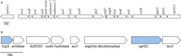
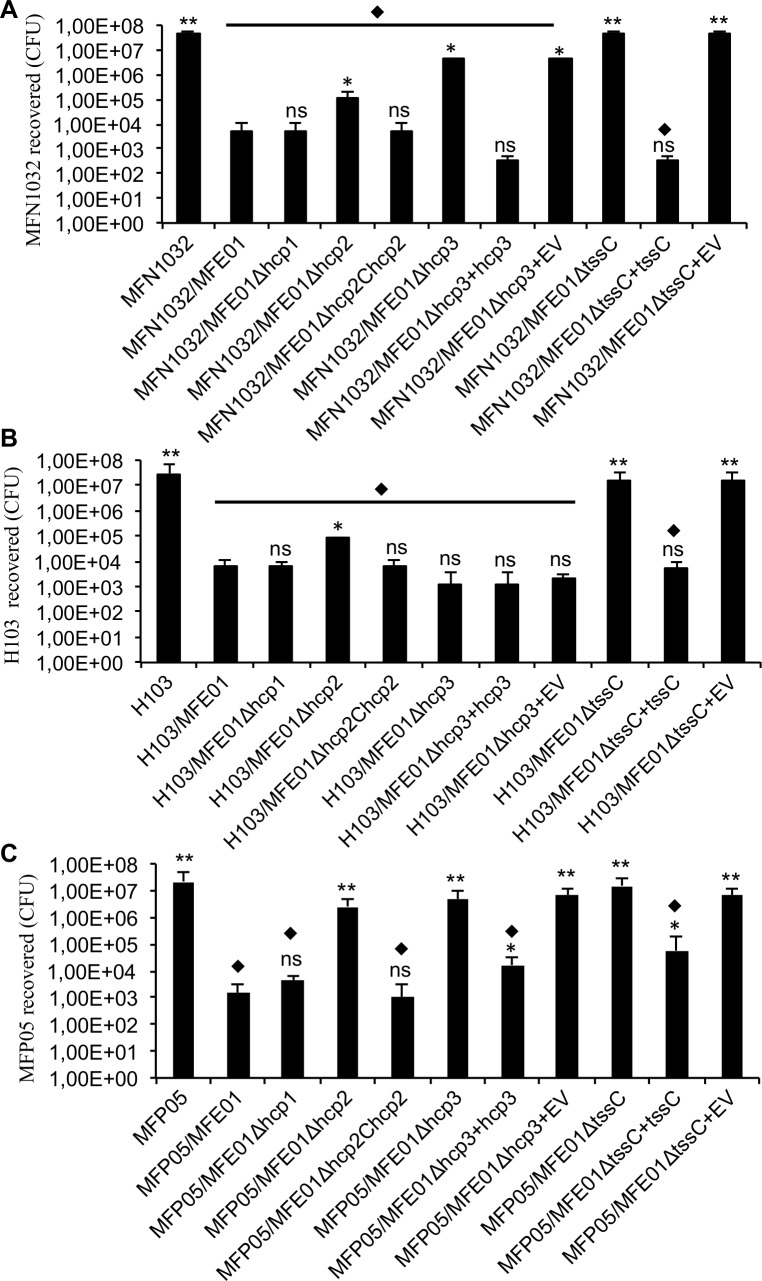
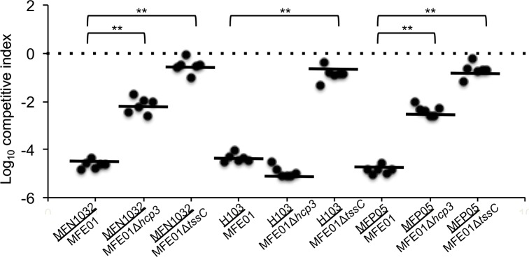
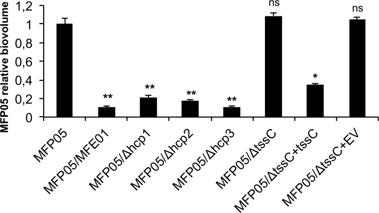
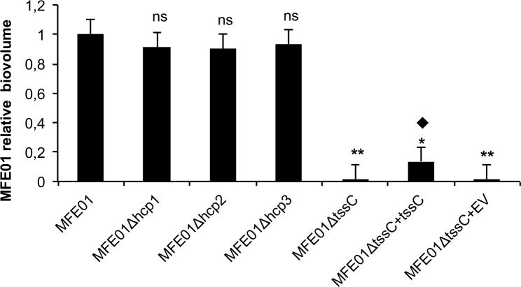
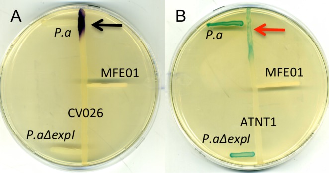
Type VI secretion systems (T6SSs) are widespread in Gram-negative bacteria, including Pseudomonas. These macromolecular machineries inject toxins directly into prokaryotic or eukaryotic prey cells. Hcp proteins are structural components of the extracellular part of this machinery. We recently reported that MFE01, an avirulent strain of Pseudomonas fluorescens, possesses at least two hcp genes, hcp1 and hcp2, encoding proteins playing important roles in interbacterial interactions. Indeed, P. fluorescens MFE01 can immobilise and kill diverse bacteria of various origins through the action of the Hcp1 or Hcp2 proteins of the T6SS. We show here that another Hcp protein, Hcp3, is involved in killing prey cells during co-culture on solid medium. Even after the mutation of hcp1, hcp2, or hcp3, MFE01 impaired biofilm formation by MFP05, a P. fluorescens strain isolated from human skin. These mutations did not reduce P. fluorescens MFE01 biofilm formation, but the three Hcp proteins were required for the completion of biofilm maturation. Moreover, a mutant with a disruption of one of the unique core component genes, MFE01ΔtssC, was unable to produce its own biofilm or inhibit MFP05 biofilm formation. Finally, MFE01 did not produce detectable N-acyl-homoserine lactones for quorum sensing, a phenomenon reported for many other P. fluorescens strains. Our results suggest a role for the T6SS in communication between bacterial cells, in this strain, under biofilm conditions.


Conflict of interest statement

The authors have declared that no competing interests exist.

Figures

Fig 1
Fig 1. Genomic organisation of the T6SS core component locus and the hcp3 locus in MFE01.
A. Genomic organisation of the T6SS core component locus in MFE01. Genes are represented as arrows, indicating the direction of transcription. The sequences of the T6SS core component genes have been deposited in GenBank under the following accession numbers: vgrGc1: KX941475, tssA: KX941476, tssB: KX941477, tssC: KX941478, tssE: KX941479, paar-motif: KX941480, unkown1: KX907122, unknown2: KX941481, tssF: KX941482, tssG: KX941483, tssH: KX941484, sfa2: KX941485, tagU: KX941486, tagH: KX941487, tssJ: KX941488, tssK: KX941489, tssL: KX941490, tssM: KX941491, pppA: KX941492, ppkA: KX941493, vgrGc2: KX941494. B. Genomic organisation of the hcp3 locus. Genes are represented as arrows, indicating the direction of transcription. The sequences of the hcp3 locus genes have been deposited in GenBank under the following accession numbers: hcp3: KX941495, vgrG3: KX941496, arginine-decarboxylase: KX941497, sui1: KX941498, nudix-hydrolase: KX941499, duf2333: KX941500, amidase: KX941501, tec3: KX941502.
Fig 2
Fig 2. Killing activity of MFE01 and mutant strains against P. fluorescens MFN1032, P. aeruginosa H103, and P. fluorescens MFP05 during contact on solid media.
Quantitative co-culture assays. A. Prey cells (MFN1032 carrying pSMC2.1 gfp) were cultured alone or mixed with P. fluorescens MFE01 or various MFE01 T6SS mutants in a 1:5 ratio. After incubation for 4 h at 28°C, the MFN1032 colonies were counted (n = 6, the error bars represent the standard error of the mean). ** indicates a significant difference in the number of MFN1032 cfu (p-value < 0.01) relative to MFN1032 co-cultured with MFE01; * indicates a significant difference in the number of MFN1032 cfu (p-value < 0.05) relative to MFN1032 co-cultured with MFE01; ns indicates no significant difference. ⧫ indicates a significant difference in the number of MFN1032 cfu (p-value < 0.05) relative to the MFN1032-alone control assay. EV means empty pPSV35 (plasmid control). B. Prey cells (P. aeruginosa H103 carrying pSMC2.1 gfp) were cultured alone or mixed with P. fluorescens MFE01 or various MFE01 T6SS mutants in a 1:5 ratio. After 4 h at 28°C, H103 colonies were counted (n = 6, the error bars represent the standard error of the mean). ** indicates a significant difference in the number of H103 cfu (p-value < 0.01) relative to H103 co-cultured with MFE01; * indicates a significant difference in the number of H103 cfu (p-value < 0.05) relative to H103 co-cultured with MFE01; ns indicates no significant difference. ⧫ indicates a significant difference in the number of H103 cfu (p-value < 0.05) relative to the H103-alone control assay. EV means empty pPSV35 (plasmid control). C. Prey cells (MFP05 carrying pSMC2.1 gfp) were cultured alone or mixed with P. fluorescens MFE01 or various MFE01 T6SS mutants in a 1:5 ratio. EV indicates empty vector for the pPSV35 control. After 4 h at 28°C, MFP05 colonies were counted (n = 6, the error bars represent the standard error of the mean). ** indicates a significant difference in the number of MFP05 cfu (p-value < 0.01) relative to MFE05 co-cultured with MFE01; * indicates a significant difference in the number of MFE05 cfu (p-value < 0.05) relative to MFE05 co-cultured with MFE01; ns indicates no significant difference. ⧫ indicates a significant difference in the number of MFE05 cfu (p-value < 0.05) relative to the MFE05-alone control assay.
Fig 3
Fig 3. Competitive index of MFE01, MFE01ΔtssC and MFE01Δhcp3.
MFE01, MFE01ΔtssC or MFE01Δhcp3 (attackers) were mixed with MFP05, MFN1032 or H103 (preys). The mixture was incubated on nutrient agar for 4 h, and survival were enumerated by plating survivors on appropriate selective plates. The competitive index is calculated using the equation: (input attacker/input prey)/(output attacker/output prey). Horizontal bars indicate the arithmetic mean of log-transformed data. ** indicates statistical significance (Wilcoxon signed rank test, p-value < 0.05), n = 6.
Fig 4
Fig 4. Effect of MFE01 and T6SS mutants on MFP05 biofilm formation.
Biofilms were grown on a glass surface, for 48 h at 28°C, under a flow of LB medium. Biovolumes of fluorescent bacteria were determined by COMSTAT analysis after confocal laser scanning microscopy observation. P. fluorescens MFP05 bearing pSMC2.1 gfp, encoding green fluorescent protein, was co-cultured alone or with MFE01 or derivatives, in a 1:5 ratio. Each histogram represents the biovolume of fluorescent MFP05, relative to that of fluorescent MFP05 when MFP05-gfp is cultivated alone. Comparisons were made with the control MFP05-gfp; ** p-value < 0.01; * p-value < 0.05; ns = non-significant; n = 6 (the error bars represent the standard error of the mean).
Fig 5
Fig 5. Effects of hcp and tssC gene mutations on biofilm biovolume in P. fluorescens strain MFE01.
Biofilms were grown on a glass surface, for 48 h at 28°C, under a flow of LB medium. Bacteria were visualised with the Syto 9® green fluorescent nucleic acid stain. Biovolumes were determined by COMSTAT analysis, after confocal laser scanning microscopy observation. The values shown are biofilm biovolumes relative to that of wild-type MFE01. The data presented are the mean values for at least five independent experiments and the error bars represent the standard error of the mean. Statistical analyses were performed with non-parametric Mann-Whitney tests (two-tailed): ns indicates no significant difference in biovolume (p-value > 0.05) relative to the MFE01 biofilm, * and ** indicates significant difference in biovolume relative to the MFE01 biofilm: *p-value < 0.05, ** p-value < 0.01. ⧫ indicates a significant difference in biovolume (p-value < 0.05) relative to the MFE01ΔtssC biofilm. EV means empty pPSV35 (plasmid control).
Fig 6
Fig 6. Effect of hcp and tssC gene mutations on the maturation of P. fluorescens MFE01 biofilms.
Biofilms were grown on a glass surface for 48 h at 28°C, under a flow of LB medium. Bacteria were visualised with the Syto 9® green fluorescent nucleic acid stain. A 3D shadow representation and a side-view projection are shown at the top and bottom, respectively, for each strain. Images show representative data from at least five independent biofilm assays. Bars, 10 μm.
Fig 7
Fig 7. Detection of AHLs with biosensor strains.
P.a indicates Pectobacterium atrosepticum 6276 strain, which produces C8- NAHLs, used as positive control. P.aΔexp1 means Pectobacterium atrosepticum 6276 mutant strain, which does not produce AHLs, used as negative control. A. Detection of short-chain AHLs with Chromobacterium violaceum CV026 (CV026). CV026 indicates C.violaceum CV026 strain, which produces vioalacein in contact with C4-C8 NAHLs, used as biosensor strain. Black arrow indicates violacein production by C.violaceum CV026 strain. LB plates were incubated for 72 h at 37°C (n = 3). B. Detection of long-chain AHLs with Agrobacterium tumefaciens NTI (ANTI1). ANT1 means Agrobacterium tumefaciens NT1 strain, which produces β-galactosidase in contact with C6-C12 NAHLs, used as biosensor strain. Red arrow indicates β-galactosidase production by Agrobacterium tumefaciens strain. LB plates containing X-Gal (40 μg/mL) were incubated for 72 h at 28°C (n = 3).

References

    1. Coulthurst SJ. The Type VI secretion system—a widespread and versatile cell targeting system. Res Microbiol. 2013;164: 640–654. 10.1016/j.resmic.2013.03.017 - DOI - PubMed
    1. Hood RD, Singh P, Hsu F, Guvener T, Carl MA, Trinidad RRS, et al. A type VI secretion system of Pseudomonas aeruginosa targets a toxin to bacteria. Cell Host Microbe. 2010;7: 25–37. 10.1016/j.chom.2009.12.007 - DOI - PMC - PubMed
    1. Zoued A, Brunet YR, Durand E, Aschtgen M-S, Logger L, Douzi B, et al. Architecture and assembly of the Type VI secretion system. Biochim Biophys Acta. 2014;1843: 1664–1673. 10.1016/j.bbamcr.2014.03.018 - DOI - PubMed
    1. Alcoforado Diniz J, Coulthurst SJ. Intraspecies Competition in Serratia marcescens Is Mediated by Type VI-Secreted Rhs Effectors and a Conserved Effector-Associated Accessory Protein. J Bacteriol. 2015;197: 2350–2360. 10.1128/JB.00199-15 - DOI - PMC - PubMed
    1. Hachani A, Wood TE, Filloux A. Type VI secretion and anti-host effectors. Curr Opin Microbiol. 2016;29: 81–93. 10.1016/j.mib.2015.11.006 - DOI - PubMed

LinkOut - more resources