. 2017 Jan 23;8(1):10.
doi: 10.1186/s13287-016-0459-0.

TGF-β1-induced differentiation of SHED into functional smooth muscle cells

Affiliations

TGF-β1-induced differentiation of SHED into functional smooth muscle cells

Jian Guang Xu et al. Stem Cell Res Ther. .

Abstract

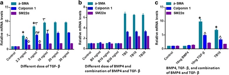
Background: Adequate vascularization is crucial for supplying nutrition and discharging metabolic waste in freshly transplanted tissue-engineered constructs. Obtaining the appropriate building blocks for vascular tissue engineering (i.e. endothelial and mural cells) is a challenging task for tissue neovascularization. Hence, we investigated whether stem cells from human exfoliated deciduous teeth (SHED) could be induced to differentiate into functional vascular smooth muscle cells (vSMCs).

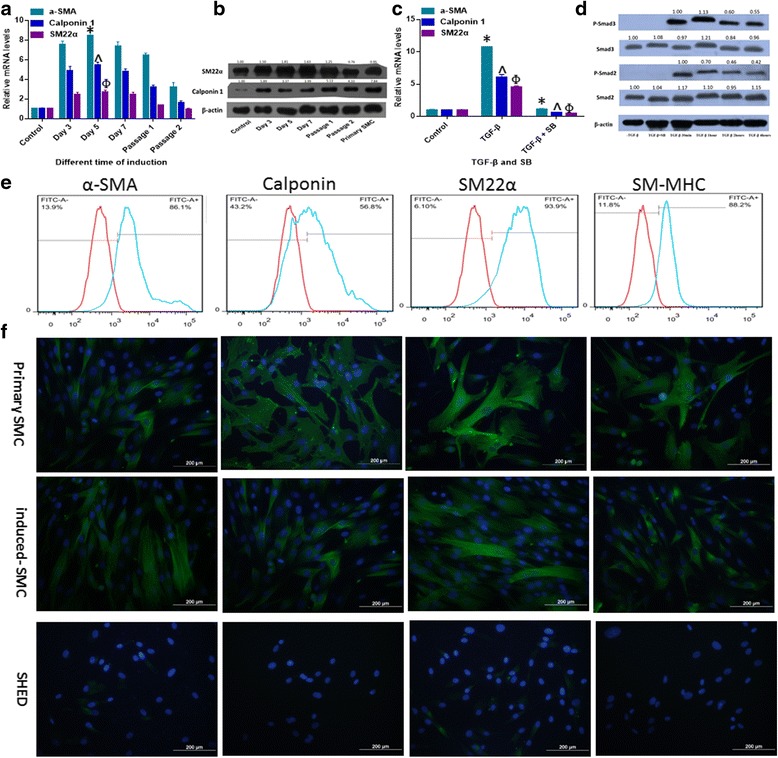
Methods: We utilized two cytokines of the TGF-β family, transforming growth factor beta 1 (TGF-β1) and bone morphogenetic protein 4 (BMP4), to induce SHED differentiation into SMCs. Quantitative real-time polymerase chain reaction (RT-qPCR) was used to assess mRNA expression, and protein expression was analyzed using flow cytometry, western blot and immunostaining. Additionally, to examine whether these SHED-derived SMCs possess the same function as primary SMCs, in vitro Matrigel angiogenesis assay, fibrin gel bead assay, and functional contraction study were used here.

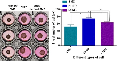
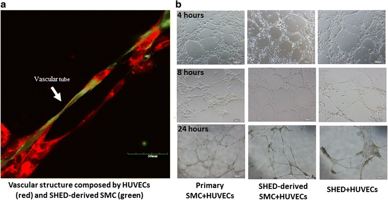
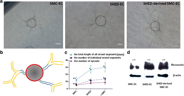
Results: By analyzing the expression of specific markers of SMCs (α-SMA, SM22α, Calponin, and SM-MHC), we confirmed that TGF-β1, and not BMP4, could induce SHED differentiation into SMCs. The differentiation efficiency was relatively high (α-SMA+ 86.1%, SM22α+ 93.9%, Calponin+ 56.8%, and SM-MHC+ 88.2%) as assessed by flow cytometry. In vitro Matrigel angiogenesis assay showed that the vascular structures generated by SHED-derived SMCs and human umbilical vein endothelial cells (HUVECs) were comparable to primary SMCs and HUVECs in terms of vessel stability. Fibrin gel bead assay showed that SHED-derived SMCs had a stronger capacity for promoting vessel formation compared with primary SMCs. Further analyses of protein expression in fibrin gel showed that cultures containing SHED-derived SMCs exhibited higher expression levels of Fibronectin than the primary SMCs group. Additionally, it was also confirmed that SHED-derived SMCs exhibited functional contractility. When SB-431542, a specific inhibitor of ALK5 was administered, TGF-β1 stimulation could not induce SHED into SMCs, indicating that the differentiation of SHED into SMCs is somehow related to the TGF-β1-ALK5 signaling pathway.

Conclusions: SHED could be successfully induced into functional SMCs for vascular tissue engineering, and this course could be regulated through the ALK5 signaling pathway. Hence, SHED appear to be a promising candidate cell type for vascular tissue engineering.

Keywords: Angiogenesis; Dental pulp stem cells; Smooth muscle cells; Stemness; Tissue engineering.


Figures

Fig. 1
Fig. 1
SMC-specific marker gene expression profile of SHED-derived SMCs induced with different concentrations and combinations of TGF-β1 and BMP4. Expression levels of SMC-specific marker genes relative to GAPDH with (a) different doses of TGF-β1 induction. There was significant upregulation of α-SMA and Calponin 1 gene expression levels as the TGF-β1 concentration is increased from 2.5 ng/ml to 10 ng/ml (p < 0.05). There were however no significant differences between the 10 ng/ml, 20 ng/ml and 30 ng/ml TGF-β1 treatment groups (p > 0.05). SM22α gene expression was a little different, by which 5 ng/ml TGF-β1 had a same effect with 10 ng/ml, 20 ng/ml and 30 ng/ml. b Different doses of BMP4 induction and with different combinations of TGF-β (10 ng/ml) and BMP4 (5, 10, 30 ng/ml). There were no significant differences amongst the different doses of BMP4 (p > 0.05), and also no significant differences amongst the different combinations of TGF-β and BMP4 (p > 0.05). c 10 ng/ml TGF-β1, 10 ng/ml BMP4 and the combination of 10 ng/ml TGF-β1 with BMP4. The effect of TGF-β1 was weakened when combined with BMP4 (p < 0.05). * ^ Φ: p < 0.05. B means BMP4; TB means TGF-β and BMP4. All experiments were performed three times (N = 3). BMP4 bone morphogenetic protein 4, TGF-β1 transforming growth factor beta 1, α-SMA alpha-smooth muscle actin
Fig. 2
Fig. 2
Characterization of SHED-derived SMCs. a Expression levels of SMC-specific marker gene expression relative to GAPDH at different induction time points. The expression levels of all SMC-specific marker genes peaked on day 5 (p < 0.05, compared with other time points). At passage 2, the gene expression levels of α-SMA and Calponin 1 declined but were still significantly higher than the control group (p < 0.05); while the gene expression level of SM22α declined to the same level as the control group (p > 0.05). * ^ Φ: p < 0.05 compared to all other time points. b The protein expression levels of SM22α and Calponin 1 were analyzed by western blot utilizing β-actin as the internal marker. Numbers depict the band density normalized against untreated controls and β-actin. c SB-431542 suppressed TGF-β1-mediated SMC differentiation. * ^ Φ: p < 0.05 between TGF-β1 group and SB-431542 + TGF-β1-treated group. d Smad2/3 became phosphorylated within 30 minutes of exposure to TGF-β1, and SB-431542 inhibited this phosphorylation. Numbers depict the band density normalized against untreated controls and β-actin. e SHED-derived SMCs were also analyzed for expression of SMC-specific markers (α-SMA, Calponin, SM22α and SM-MHC) with flow cytometry (α-SMA+ 86.1%, SM22α + 93.9%, Calponin + 56.8%, and SM-MHC+ 88.2% respectively) (the isotype control in red) and (f) immunofluorescence staining (nuclei in blue). All experiments were performed three times (N = 3). SM-MHC smooth muscle-myosin heavy chain, SHED stem cells from human exfoliated deciduous teeth, α-SMA alpha-smooth muscle actin
Fig. 3
Fig. 3
Collagen gel contraction assay. Gel surface areas were measured and further analyzed using the Image J software. *: p < 0.05, ^: p > 0.05. HUVECs human umbilical vein endothelial cells, SHED stem cells from human exfoliated deciduous teeth
Fig. 4
Fig. 4
Vascular tube formation by SHED-derived SMCs and ECs on Matrigel. a Immunofluorescence image of vascular structure (EC in red; SHED-derived SMCs in green). (b) Phase-contrast images (×10) of vascular structures from 4 hours to 24 hours after seeding ECs and SHED-derived SMCs on Matrigel. HUVECs human umbilical vein endothelial cells, SHED stem cells from human exfoliated deciduous teeth, SMCs smooth muscle cells
Fig. 5
Fig. 5
SHED-derived SMCs promote EC vessel formation. a Photomicrographs of EC-coated microbeads cultured in fibrin gels with SMC, SHED and SHED-induced SMC on the gel surface. b The schematic representation of calculating tube formation from each bead: (i) the number of vessels which sprout from the bead directly (in blue); (ii) the total number of individual vessel segments (including blue and yellow); and (iii) the total length of all the vessel segments. c Quantification of the vessel structures in the fibrin gel bead assay in the presence of SMC, SHED and SHED-induced SMC.*: p < 0.05 versus SMCs group, ^: p < 0.05 between SHED and SHED-derived SMCs. d Western blot showing fibronectin expression within each experimental group. All experiments were performed three times (N = 3). ECs endothelial cells, SHED stem cells from human exfoliated deciduous teeth, SMCs smooth muscle cells

Similar articles

Cited by

References

    1. Adams RH, Alitalo K. Molecular regulation of angiogenesis and lymphangiogenesis. Nat Rev Mol Cell Biol. 2007;8:464–78. doi: 10.1038/nrm2183. - DOI - PubMed
    1. Carmeliet P. Mechanisms of angiogenesis and arteriogenesis. Nat Med. 2000;6:389–95. doi: 10.1038/74651. - DOI - PubMed
    1. Gökçinar-Yagci B, Uçkan-Çetinkaya D, Çelebi-Saltik B. Pericytes: properties, functions and applications in tissue engineering. Stem Cell Rev. 2015;11:549–59. doi: 10.1007/s12015-015-9590-z. - DOI - PubMed
    1. Kim S, von Recum H. Endothelial stem cells and precursors for tissue engineering: cell source, differentiation, selection, and application. Tissue Eng Part B Rev. 2008;14:133–47. doi: 10.1089/teb.2007.0304. - DOI - PubMed
    1. Patsch C, Challet-Meylan L, Thoma EC, Urich E, Heckel T, O'Sullivan JF. Generation of vascular endothelial and smooth muscle cells from human pluripotent stem cells. Nat Cell Biol. 2015;17:994–1003. doi: 10.1038/ncb3205. - DOI - PMC - PubMed

Publication types

MeSH terms