. 2017 Jan 23;8(1):7.
doi: 10.1186/s13287-016-0464-3.

Astaxanthin attenuates total body irradiation-induced hematopoietic system injury in mice via inhibition of oxidative stress and apoptosis

Affiliations

Astaxanthin attenuates total body irradiation-induced hematopoietic system injury in mice via inhibition of oxidative stress and apoptosis

Xiao-Lei Xue et al. Stem Cell Res Ther. .

Abstract

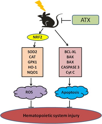
Background: The hematopoietic system is especially sensitive to total body irradiation (TBI), and myelosuppression is one of the major effects of TBI. Astaxanthin (ATX) is a powerful natural anti-oxidant with low toxicity. In this study, the effect of ATX on hematopoietic system injury after TBI was investigated.

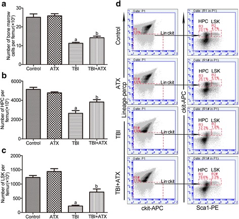
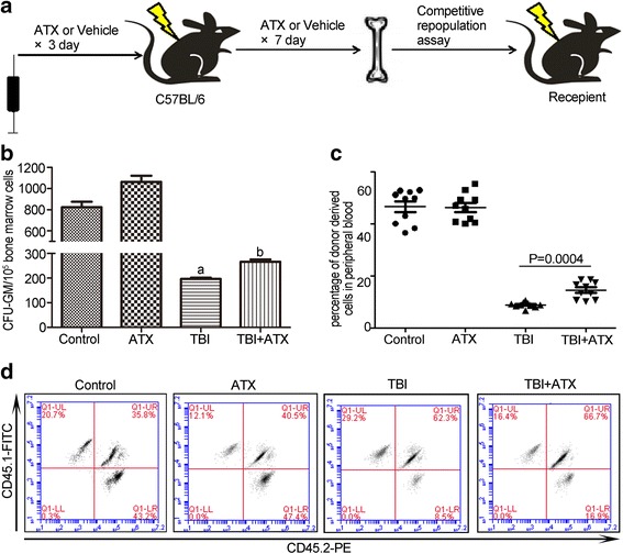
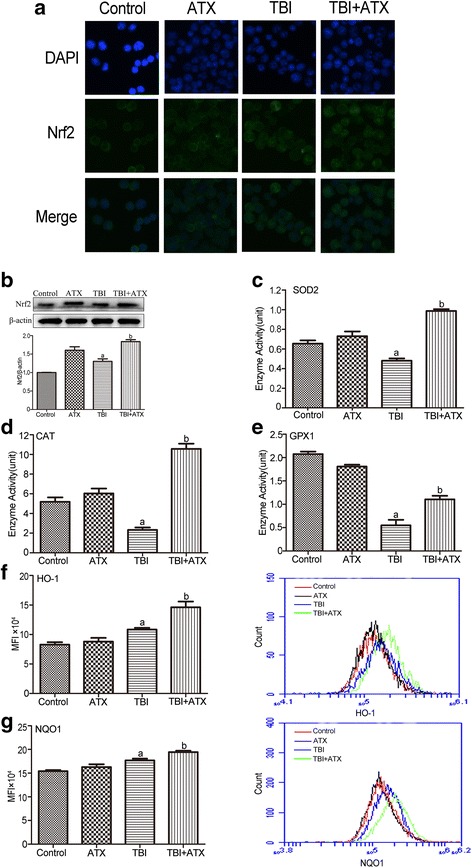
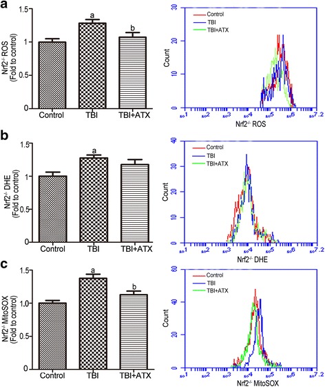
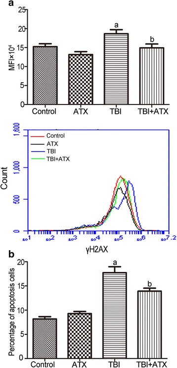
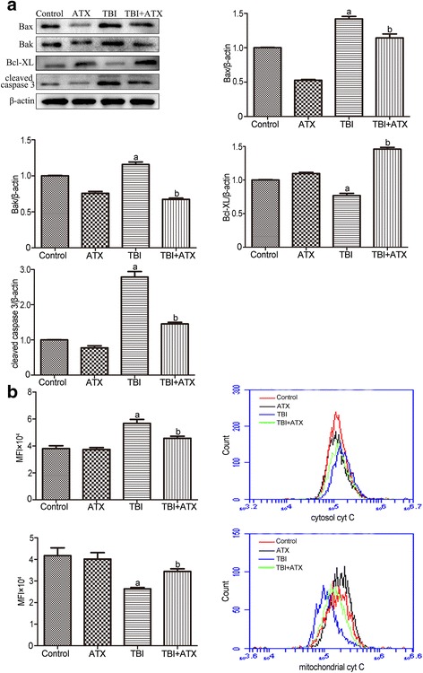
Methods: Flow cytometry was used to detect the proportion of hematopoietic progenitor cells (HPCs) and hematopoietic stem cells (HSCs), the level of intracellular reactive oxygen species (ROS), expression of cytochrome C, cell apoptosis, and NRF2-related proteins. Immunofluorescence staining was used to detect Nrf2 translocation. Western blot analysis was used to evaluate the expression of apoptotic-related proteins. Enzymatic activities assay kits were used to analyze SOD2, CAT, and GPX1 activities.

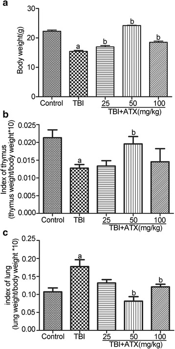
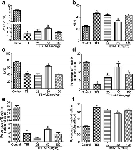
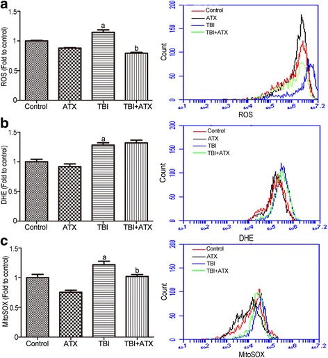
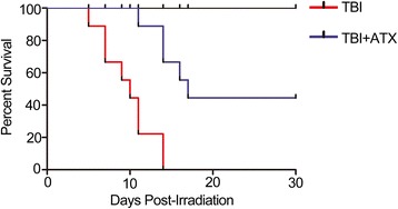
Results: Compared with the TBI group, ATX can improve radiation-induced skewed differentiation of peripheral blood cells and accelerate hematopoietic self-renewal and regeneration. The radio-protective effect of ATX is probably attributable to the scavenging of ROS and the reduction of cell apoptosis. These changes were associated with increased activation of Nrf2 and downstream anti-oxidative proteins, and regulation of apoptotic-related proteins.

Conclusions: This study suggests that ATX could be used as a potent therapeutic agent to protect the hematopoietic system against TBI-induced bone marrow suppression.

Keywords: Astaxanthin; Cell apoptosis; Hematopoietic stem cells; Ionizing radiation; Reactive oxygen species.


Figures

Fig. 1
Fig. 1
Astaxanthin (ATX) rescues the body weight loss and the organ index changes caused by total body irradiation (TBI) in mice. Mice were randomly divided into five groups and were administered either DMSO or different concentrations of ATX by gavage for 3 days before exposure to 4 Gy TBI and then continuously for 7 days after irradiation. Control mice were sham-irradiated. a Bar graph showing the average body weight of each group. b Bar graph showing the thymus index. c Bar graph showing the lung index. The data are presented as the mean ± SEM (N = 5). a P < 0.05 vs control; b P < 0.05 vs TBI
Fig. 2
Fig. 2
Astaxanthin (ATX) attenuates total body irradiation (TBI)-induced multilineage differentiation disorders and maintains hematopoietic homeostasis. Mice were sham-irradiated as a control or irradiated with 4 Gy TBI after receiving DMSO or various concentrations of ATX in a manner similar to that illustrated in Fig. 1. a Bar graph showing the numbers of white blood cells (WBC). b Bar graph showing the percentage of neutrophilic granulocytes (NE%) and (c) lymphocytes (LY%). The percentage of T cells (d), B cells (e), and myeloid cells (f) in peripheral blood detected by flow cytometry. The data are presented as the mean ± SEM (N = 5). a P < 0.05 vs control; b P < 0.05 vs TBI
Fig. 3
Fig. 3
Astaxanthin (ATX) treatment attenuates total body irradiation (TBI)-induced bone marrow cell reduction in vivo. Mice were randomly divided into four groups and were administered DMSO or 50 mg/kg ATX by gavage for 3 days before exposure to 4 Gy TBI and then continuously for 7 days after irradiation. Control/ATX mice were sham-irradiated. All mice were sacrificed at 12 days after exposure to TBI. The numbers and frequencies of hematopoietic progenitor cells (HPCs) and LSK cells in bone marrow were then analyzed by flow cytometry. a Bar graph showing the numbers of bone marrow cells per femur. b The numbers of HPCs (LineageSca1c-kit+ bone marrow cells) per femur. c The numbers of LSKs (LineageSca1+c-kit+ bone marrow cells) per femur. d Representative FACS plots show the percentages of HPC and LSK. The data are presented as the mean ± SEM (N = 5). a P < 0.05 vs control; b P < 0.05 vs TBI
Fig. 4
Fig. 4
Astaxanthin (ATX) reduces total body irradiation (TBI)-induced suppression of BM cell clonogenic function and induces HSC reconstitution in vivo. a Schematic diagram shows the recipients were transplanted with BM cells from the various treated groups. b Bar graph showing the number of CFU-GM per 105 BM cells. c The scatter plot shows the percentage of donor-derived cells in peripheral blood cells 2 months after transplantation. d Representative results of donor cell engraftment. The data are presented as the mean ± SEM (N = 6 in b and N = 10 in c). a P < 0.05 vs control; b P < 0.05 vs TBI
Fig. 5
Fig. 5
Astaxanthin (ATX) treatment inhibits total body irradiation (TBI)-induced reactive oxygen species (ROS) generation in BM c-kit-positive cells. a The levels of ROS detected by the DCFDA MFI and the representative analysis of ROS levels by flow cytometry. b The levels of ROS detected by the DHE MFI and the representative analysis of DHE levels by flow cytometry. c The levels of ROS in mitochondria detected by the MitoSOX MFI and the representative analysis of MitoSOX levels by flow cytometry. The data are presented as the mean ± SEM (N = 5). a P < 0.05 vs control; b P < 0.05 vs TBI
Fig. 6
Fig. 6
Astaxanthin (ATX) scavenges total body irradiation (TBI)-induced excessive ROS by upregulating NRF2 and its downstream anti-oxidant proteins. a The NRF2 protein level in c-kit-positive cells was determined using the immunofluorescence method. b Expression of NRF2 protein was analyzed by Western blotting. The enzyme activities of c SOD2, d CAT, and e GPX1 were analyzed by respective assay kit. The levels of intracellular f HO-1 and g NQO1 expression was analyzed by flow cytometry. The data are presented as the mean ± SEM (N = 3 in be and N = 5 in f, g). a P < 0.05 vs control; b P < 0.05 vs TBI. MFI mean fluorescence intensity
Fig. 7
Fig. 7
Astaxanthin (ATX) alleviates total body irradiation (TBI)-induced Nrf2 −/− reactive oxygen species (ROS) generation in c-kit-positive cells. a The levels of ROS detected by the DCF MFI and the representative analysis of ROS levels by flow cytometry. b The levels of ROS detected by the DHE MFI and the representative analysis of DHE levels by flow cytometry. c The levels of ROS in mitochondria detected by the MitoSOX MFI and the representative analysis of MitoSOX levels by flow cytometry. The data are presented as the mean ± SEM (N = 5). a P < 0.05 vs control; b P < 0.05 vs TBI
Fig. 8
Fig. 8
Astaxanthin (ATX) reduces total body irradiation (TBI)-induced DNA DSBs and apoptosis in c-kit-positive cells. a Representative analysis of γH2AX expression in c-kit-positive cells by flow cytometry. b Bar graph showing the percentage of apoptosis in c-kit-positive cells. The data are presented as the mean ± SEM (N = 5). a P < 0.05 vs control; b P < 0.05 vs TBI. MFI mean fluorescence intensity
Fig. 9
Fig. 9
Astaxanthin (ATX) protects the hematopoietic system from total body irradiation (TBI) by regulating the apoptotic pathway. a The protein expression of BAX, BAK, BCL-XL, and cleaved CASPASE 3 in c-kit-positive cells was analyzed by Western blotting. b Representative analysis of cytochrome C (cyt C) release by flow cytometry. The data are presented as the mean ± SEM (N = 3 in a and N = 5 in b). a P < 0.05 vs control; b P < 0.05 vs TBI. MFI mean fluorescence intensity
Fig. 10
Fig. 10
Astaxanthin (ATX) administration increases survival. Mice were treated with DMSO or ATX (50 mg/kg) by gavage for 3 days before exposure to a lethal dose (7.2 Gy) of total body irradiation (TBI) and then continuously for 7 days after TBI. Kaplan–Meier analysis of mouse survival after exposure to the lethal dose of TBI (N = 19 mice/group)
Fig. 11
Fig. 11
A Summary on the radioprotective mechanism of astaxanthin (ATX). ROS reactive oxygen species

References

    1. Shao L, Luo Y, Zhou D. Hematopoietic stem cell injury induced by ionizing radiation. Antioxid Redox Signal. 2014;20:1447–62. doi: 10.1089/ars.2013.5635. - DOI - PMC - PubMed
    1. Coleman CN, Stone HB, Moulder JE, Pellmar TC. Medicine. Modulation of radiation injury. Science. 2004;304:693–94. doi: 10.1126/science.1095956. - DOI - PubMed
    1. Acharya SS, Fendler W, Watson J, Hamilton A, Pan Y, Gaudiano E, et al. Serum microRNAs are early indicators of survival after radiation-induced hematopoietic injury. Sci Transl Med. 2015;7:287ra69. doi: 10.1126/scitranslmed.aaa6593. - DOI - PMC - PubMed
    1. Wang Y, Schulte BA, LaRue AC, Ogawa M, Zhou D. Total body irradiation selectively induces murine hematopoietic stem cell senescence. Blood. 2006;107:358–66. doi: 10.1182/blood-2005-04-1418. - DOI - PMC - PubMed
    1. Meng A, Wang Y, Brown SA, Van Zant G, Zhou D. Ionizing radiation and busulfan inhibit murine bone marrow cell hematopoietic function via apoptosis-dependent and -independent mechanisms. Exp Hematol. 2003;31:1348–56. doi: 10.1016/j.exphem.2003.08.014. - DOI - PubMed

Publication types

MeSH terms