

Potential application of klotho in human chronic kidney disease
- PMID: 28115282
- PMCID: PMC5474175
- DOI: 10.1016/j.bone.2017.01.017
Potential application of klotho in human chronic kidney disease
Abstract
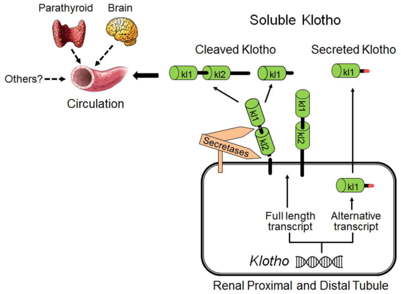
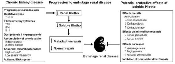
The extracellular domain of transmembrane alpha-Klotho (αKlotho, hereinafter simply called Klotho) is cleaved by secretases and released into the circulation as soluble Klotho. Soluble Klotho in the circulation starts to decline early in chronic kidney disease (CKD) stage 2 and urinary Klotho possibly even earlier in CKD stage 1. Therefore soluble Klotho could serve as an early and sensitive marker of kidney function decline. Moreover, preclinical animal data support Klotho deficiency is not just merely a biomarker, but a pathogenic factor for CKD progression and extrarenal CKD complications including cardiovascular disease and disturbed mineral metabolism. Prevention of Klotho decline, re-activation of endogenous Klotho production or supplementation of exogenous Klotho are all associated with attenuation of renal fibrosis, retardation of CKD progression, improvement of mineral metabolism, amelioration of cardiomyopathy, and alleviation of vascular calcification in CKD. Therefore Klotho is not only a diagnostic and/or prognostic marker for CKD, but the treatment of Klotho deficiency may be a promising strategy to prevent, retard, and decrease the burden of comorbidity in CKD.
Keywords: AKI; CKD; Klotho; Phosphate; Uremic cardiomyopathy; Vascular calcification; Vitamin D.
Copyright © 2017 Elsevier Inc. All rights reserved.
Figures
References
-
- Kuro-o M, Matsumura Y, Aizawa H, Kawaguchi H, Suga T, Utsugi T, Ohyama Y, Kurabayashi M, Kaname T, Kume E, Iwasaki H, Iida A, Shiraki-Iida T, Nishikawa S, Nagai R, Nabeshima YI. Mutation of the mouse klotho gene leads to a syndrome resembling ageing. Nature. 1997;390(6655):45–51. - PubMed
-
- Ito S, Kinoshita S, Shiraishi N, Nakagawa S, Sekine S, Fujimori T, Nabeshima YI. Molecular cloning and expression analyses of mouse betaklotho, which encodes a novel Klotho family protein. Mech Dev. 2000;98(1–2):115–9. - PubMed
-
- Ito S, Fujimori T, Hayashizaki Y, Nabeshima Y. Identification of a novel mouse membrane-bound family 1 glycosidase-like protein, which carries an atypical active site structure. Biochim Biophys Acta. 2002;1576(3):341–5. - PubMed
-
- Kato Y, Arakawa E, Kinoshita S, Shirai A, Furuya A, Yamano K, Nakamura K, Iida A, Anazawa H, Koh N, Iwano A, Imura A, Fujimori T, Kuro-o M, Hanai N, Takeshige K, Nabeshima Y. Establishment of the anti-Klotho monoclonal antibodies and detection of Klotho protein in kidneys. Biochem Biophys Res Commun. 2000;267(2):597–602. - PubMed
Publication types
MeSH terms
Substances
Grants and funding
LinkOut - more resources
Full Text Sources
Other Literature Sources
Medical
