. 2017 Jan 23;6(1):e003456.
doi: 10.1161/JAHA.116.003456.

Females Are Protected From Iron-Overload Cardiomyopathy Independent of Iron Metabolism: Key Role of Oxidative Stress

Affiliations

Females Are Protected From Iron-Overload Cardiomyopathy Independent of Iron Metabolism: Key Role of Oxidative Stress

Subhash K Das et al. J Am Heart Assoc. .

Abstract

Background: Sex-related differences in cardiac function and iron metabolism exist in humans and experimental animals. Male patients and preclinical animal models are more susceptible to cardiomyopathies and heart failure. However, whether similar differences are seen in iron-overload cardiomyopathy is poorly understood.

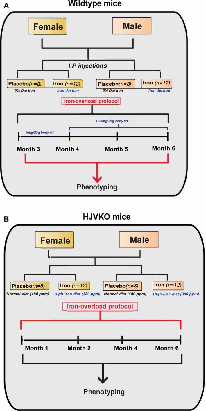
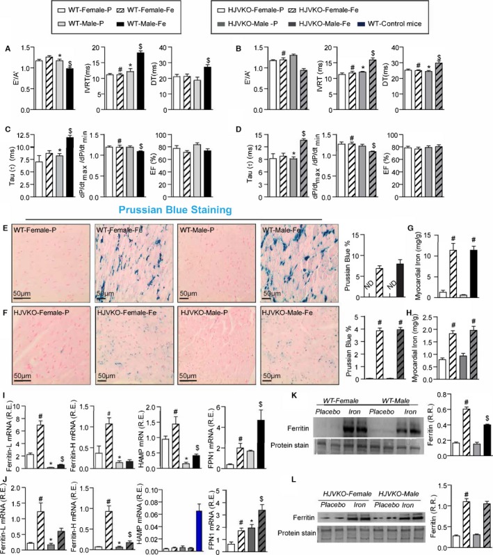
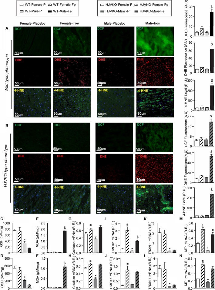
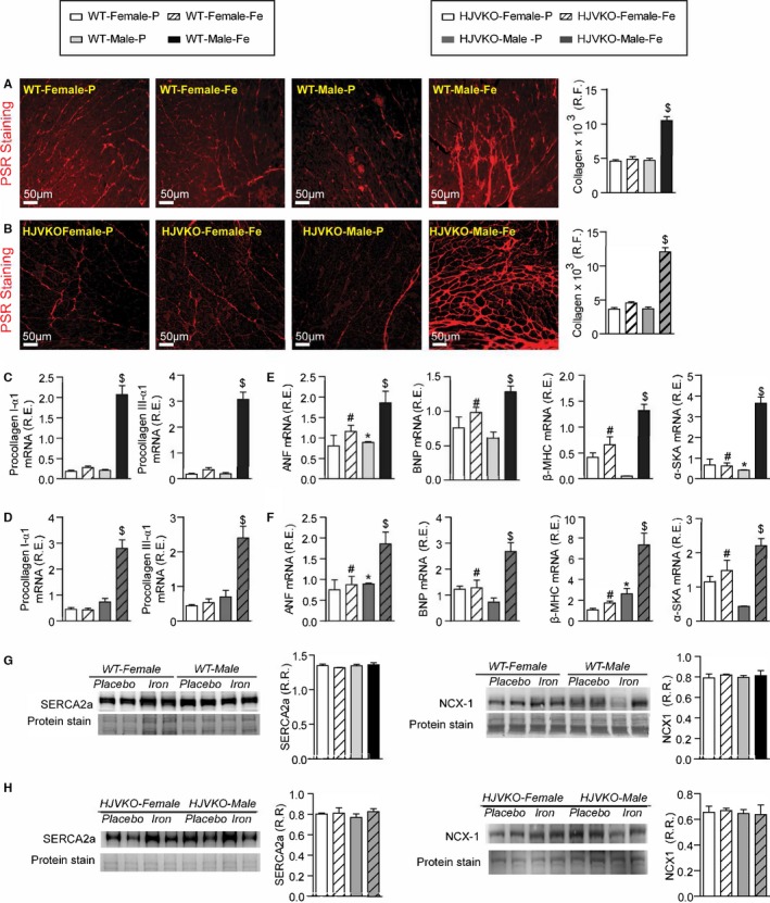
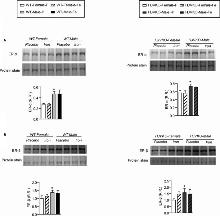
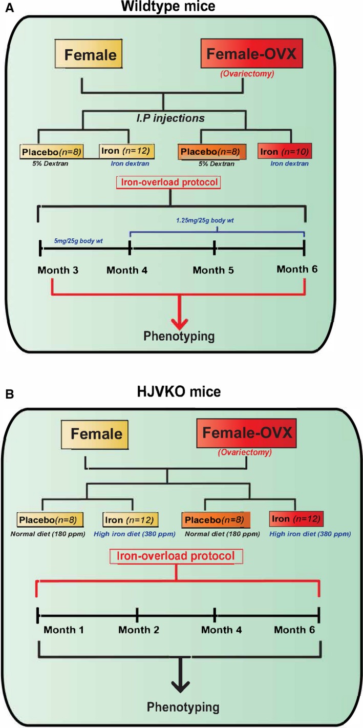
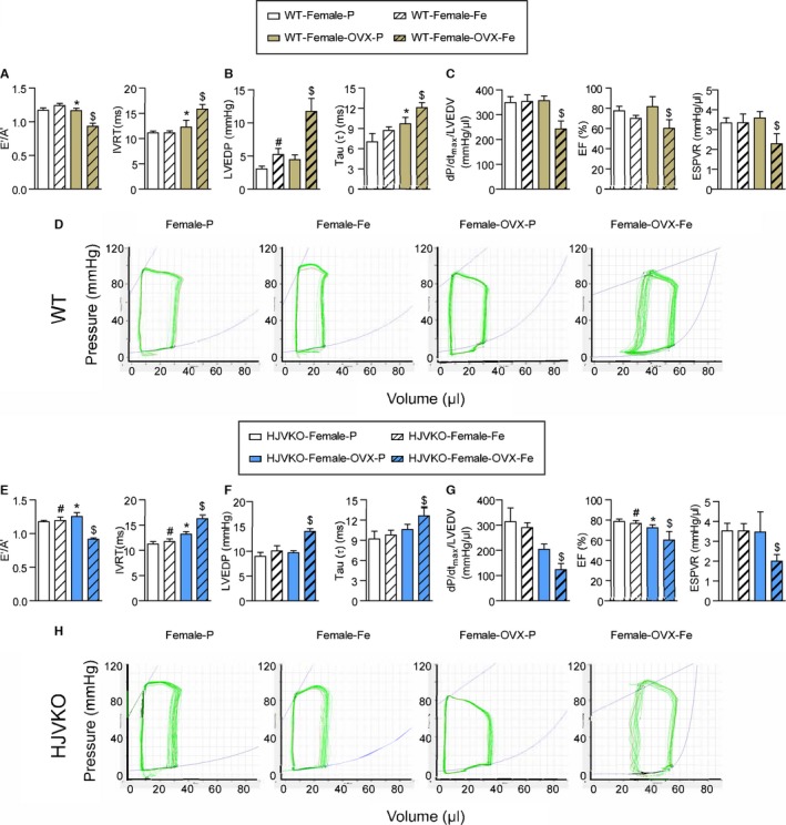
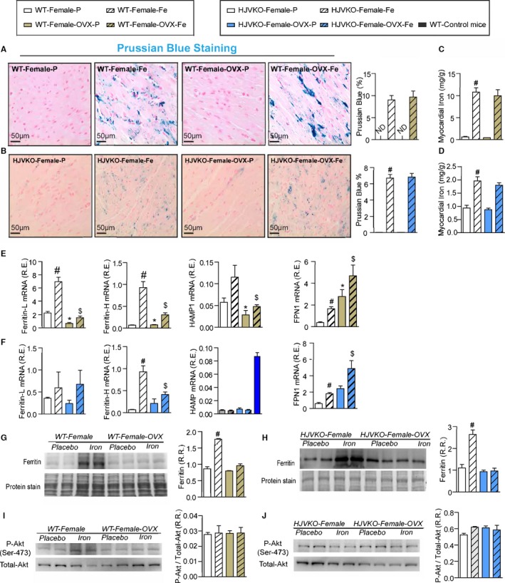
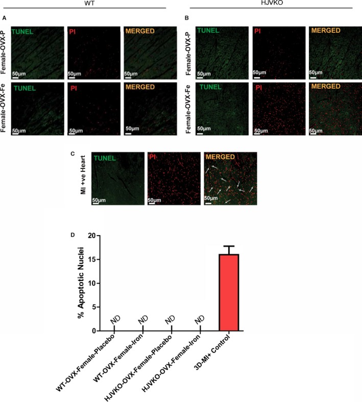
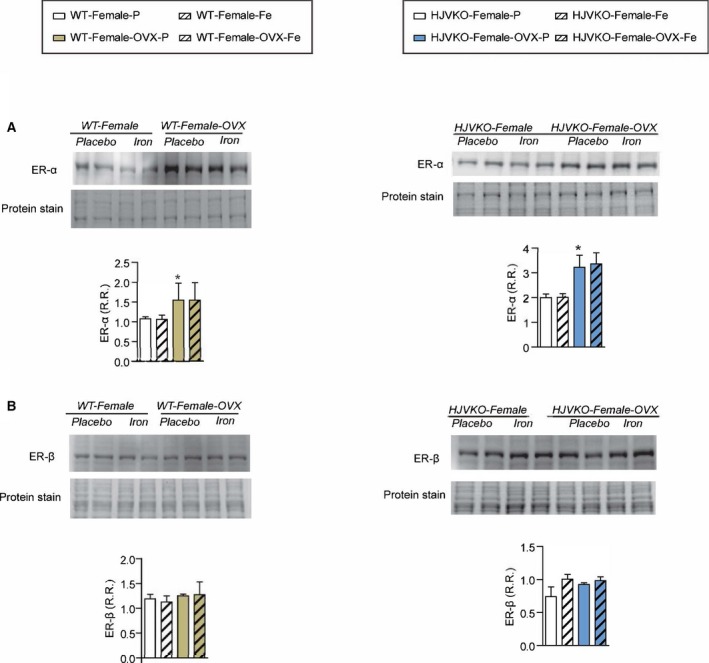
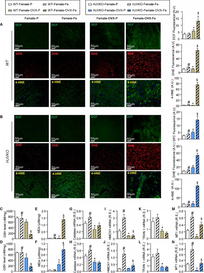
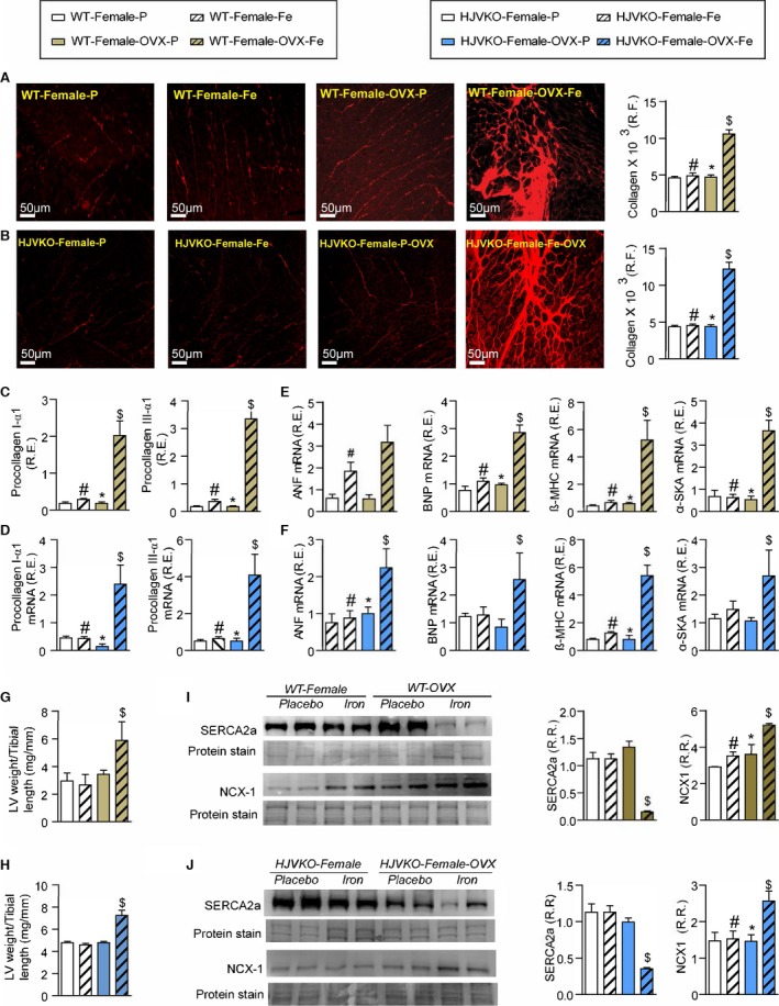
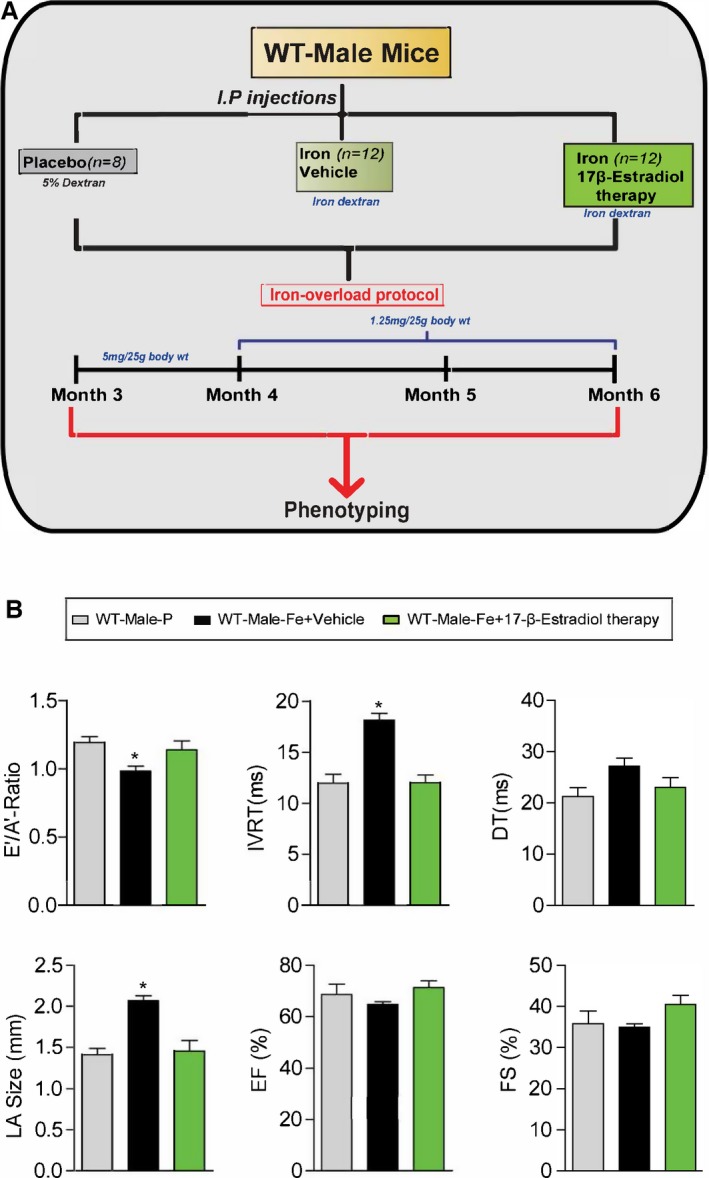
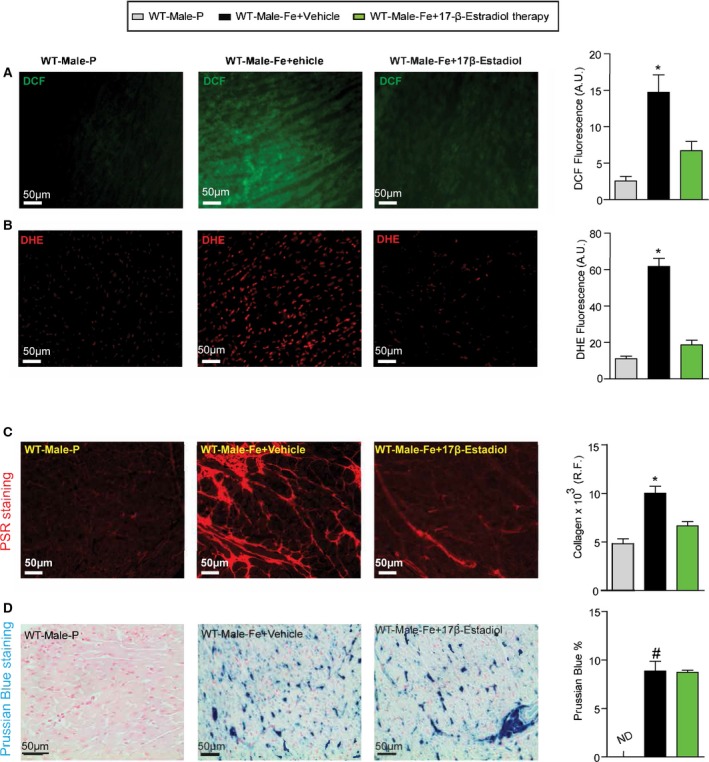
Methods and results: Male and female wild-type and hemojuvelin-null mice were injected and fed with a high-iron diet, respectively, to develop secondary iron overload and genetic hemochromatosis. Female mice were completely protected from iron-overload cardiomyopathy, whereas iron overload resulted in marked diastolic dysfunction in male iron-overloaded mice based on echocardiographic and invasive pressure-volume analyses. Female mice demonstrated a marked suppression of iron-mediated oxidative stress and a lack of myocardial fibrosis despite an equivalent degree of myocardial iron deposition. Ovariectomized female mice with iron overload exhibited essential pathophysiological features of iron-overload cardiomyopathy showing distinct diastolic and systolic dysfunction, severe myocardial fibrosis, increased myocardial oxidative stress, and increased expression of cardiac disease markers. Ovariectomy prevented iron-induced upregulation of ferritin, decreased myocardial SERCA2a levels, and increased NCX1 levels. 17β-Estradiol therapy rescued the iron-overload cardiomyopathy in male wild-type mice. The responses in wild-type and hemojuvelin-null female mice were remarkably similar, highlighting a conserved mechanism of sex-dependent protection from iron-overload-mediated cardiac injury.

Conclusions: Male and female mice respond differently to iron-overload-mediated effects on heart structure and function, and females are markedly protected from iron-overload cardiomyopathy. Ovariectomy in female mice exacerbated iron-induced myocardial injury and precipitated severe cardiac dysfunction during iron-overload conditions, whereas 17β-estradiol therapy was protective in male iron-overloaded mice.

Keywords: 17‐β‐estradiol; heart failure; hemojuvelin; iron overload; myocardial fibrosis; ovariectomy; oxidative stress; sex.


Figures

Figure 1
Figure 1
Experimental protocol to study the effects of sex on iron‐overload cardiomyopathy in WT (A) and HJV knockout (B) mice. HJVKO indicates hemojuvelin; WT, wild‐type.
Figure 2
Figure 2
Marked sex differences in iron‐overload cardiomyopathy. Noninvasive echocardiographic assessment of heart function by tissue Doppler imaging and transmitral filling pattern and invasive hemodynamic assessment using pressure‐volume loops in WT (A and B) and HJVKO mice (C and D) showing preserved cardiac function in female mice and heart failure with preserved ejection fraction in male mice in response to iron overload. Representative Prussian blue staining images (E and F) and quantification of myocardial tissue iron levels (G and H) in WT and HJVKO mice showing equivalent cardiac iron deposition in male and female mice in response to iron overload. Taqman real‐time PCR analysis of ferritin light (L) and heavy (H) chain, hepcidin (HAMP) and ferroportin (FPN1) myocardial mRNA expression in WT (I) and HJVKO (J) mice. Western blot analysis for total ferritin in WT (K) and HJVKO (L) hearts showing a smaller increase in ferritin levels in WT male mice in response to iron overload. The blue bar indicates the data from WT controls. A indicates tissue Doppler due to atrial contraction; DT, deceleration time; E, early tissue Doppler velocity; EF, ejection fraction; IVRT, isovolumetric relaxation time; ND, not detected; R.R., relative ratio; n=8 to 12 for functional studies; n=8 for gene expression analysis; n=4 for Western blot analysis. HJVKO indicates hemojuvelin‐null; WT, wild‐type. *P<0.05 for effect of sex; # P<0.05 for effect of iron; $ P<0.05 for interaction.
Figure 3
Figure 3
Female mice are protected against iron‐overload‐induced oxidative stress. Representative dichlorodihydrofluorescein (DCF) (green), dihydroethidium (DHE) fluorescence (red), 4‐hydroxynonenal (4‐HNE) immunofluorescence (green), and quantification showing a relative lack of iron‐induced myocardial oxidative stress in female WT (A) and HJVKO (B) mice, whereas iron overload resulted in increased oxidative stress in male mice. Biochemical analysis of myocardial reduced glutathione (GSH) (C and D) and lipid peroxidation product, malondialdehyde (MDA) (E and F) levels in WT (C and E) and HJVKO hearts (D and F) showing increased oxidative stress in male mice in contrast to unchanged oxidative stress in female mice in response to iron overload. Myocardial gene expression analysis using Taqman real‐time PCR showing sex‐specific and iron‐overloaded related alteration in mRNA expression for catalase (G and H), heme oxygenase 1 (HMOX1) (I and J), thioredoxin 1 (TRXN1) (K and L), and metallothionein 1 (MT1) (M and N) in WT and HJVKO mice, respectively; n=4 for histology; n=8 for gene expression and biochemical analyses. HJVKO indicates hemojuvelin‐null; WT, wild‐type. *P<0.05 for effect of sex; # P<0.05 for effect of iron; $ P<0.05 for interaction.
Figure 4
Figure 4
Female mice are protected against iron‐overload‐induced myocardial fibrosis and heart disease. Representative picro‐sirius red (PSR) staining and quantification of myocardial fibrosis (A and B), and Taqman real‐time PCR expression analysis of procollagen type Iα1 and procollagen type IIIα1 mRNA (C and D) in male and female WT and HJVKO mice showing a clear protection against iron‐overload‐induced myocardial fibrosis in female mice. Expression analysis of cardiac disease markers, atrial natriuretic factor (ANF), brain natriuretic peptide (BNP), and β‐myosin heavy chain (β‐MHC) and α‐skeletal actin (α‐SkA) in WT (E) and HJVKO mice (F), showing a potential intrinsic cardioprotective effect against iron‐overload cardiomyopathy in female mice. Western blot analyses in male and female WT and HJVKO mice showing no change in myocardial sarco/endoplasmic reticulum Ca2+ ATPase 2a (SERCA2a) and sodium‐calcium exchanger 1 (NCX1) levels in WT (G) and HJVKO (H) mice in response to iron overload. R.E. indicates relative expression; R.F., relative fraction; R.R., relative ratio; n=4 for histology and Western blot analyses; n=8 for gene expression analysis. HJVKO indicates hemojuvelin‐null; WT, wild‐type. *P<0.05 for effect of sex; # P<0.05 for effect of iron; $ P<0.05 for interaction.
Figure 5
Figure 5
Lack of myocardial apoptosis in chronic iron‐overloaded mice based on terminal deoxynucleotidyl transferase dUTP nick end labeling (TUNEL) staining in WT (A) and HJVKO (B) female hearts (n=3 each). Two sections from each heart were stained and imaged and 5 fields examined from each section. Positive TUNEL staining (white arrows) is shown from 3‐day postmyocardial infarction murine hearts using the LAD ligation technique (C). LAD indicates left anterior descending; HJVKO, hemojuvelin‐null; WT, wild‐type.
Figure 6
Figure 6
Western blot analysis of myocardial estrogen receptor‐α (ER‐α) (A) and estrogen receptor‐β (ER‐β) (B) levels showing increased ER‐α in male WT hearts with no changes observed in response to iron‐overload. n=6 per group. HJVKO indicates hemojuvelin‐null; WT, wild‐type. *P<0.05 for effect of sex.
Figure 7
Figure 7
Experimental protocol to study the effects of ovariectomy (OVX) on iron‐overload cardiomyopathy in WT (A) and HJV knockout (B) mice. HJV indicates hemojuvelin; WT, wild‐type.
Figure 8
Figure 8
Histological analysis of ovaries and plasma 17β‐estradiol levels in WT mice and HJVKO mice. Hematoxylin and eosin staining of surgically removed ovaries from a WT (A) and HJVKO (C) mouse and plasma 17β‐estradiol levels in WT (B) and HJVKO (D) mice illustrating the effectiveness of ovariectomy (OVX). Scale bars represent 250 μm (left) and 50 μm (right); n=8 per group. HJVKO indicates hemojuvelin‐null; WT, wild‐type. *P<0.05 for effect of OVX.
Figure 9
Figure 9
Ovariectomy (OVX) precipitates diastolic and systolic dysfunction in response to myocardial iron overload. Noninvasive echocardiographic assessment of heart function illustrated by tissue Doppler (E/A) and transmitral filling (IVRT) (A), invasive hemodynamics illustrated by LVEDP and LV pressure exponential decay constant (τ) (B) showing diastolic dysfunction in OVX WT females with iron overload. Invasive hemodynamic assessment based on dP/dtmax/LVEDV, EF, and ESPVR (C) showing systolic dysfunction in OVX WT female iron‐overloaded mice. Representative pressure‐volume loops illustrating diastolic and systolic dysfunction in OVX WT female iron‐overloaded mice (D). Noninvasive echocardiographic assessment of heart function illustrated by tissue Doppler (E/A) and transmitral filling (IVRT) (E), invasive hemodynamics illustrated by LVEDP and LV pressure exponential decay constant (τ) (F) showing diastolic dysfunction in OVX HJVKO females with iron overload. Invasive hemodynamic assessment based on dP/dtmax/LVEDV, EF, and ESPVR (G) showing systolic dysfunction in OVX HJVKO female iron‐overloaded mice. Representative pressure‐volume loops illustrating diastolic and systolic dysfunction in OVX WT female iron‐overloaded mice (H). A indicates tissue Doppler due to atrial contraction; E, early tissue Doppler velocity; EF, ejection fraction; ESPVR, end‐systolic pressure‐volume relationship; HJVKO, hemojuvelin‐null; IVRT, isovolumetric relaxation time; LVEDP, LV end‐diastolic pressure; LVEDV, LV end‐diastolic volume; WT, wild‐type; n=8 for placebo and n=10 for iron‐overload groups. *P<0.05 for effect of OVX; # P<0.05 for effect of iron; $ P<0.05 for interaction.
Figure 10
Figure 10
Ovariectomy (OVX) does not modulate myocardial iron deposition but suppresses the upregulation of ferritin expression. Prussian blue staining (A and B) and quantification of myocardial tissue iron levels (C and D) in WT (A and C) and HJVKO (B and D) female hearts and in response to OVX, confirming the presence of myocardial iron deposition without a differential response to OVX. Taqman real‐time PCR analysis of ferritin light (L) and heavy (H) chain, hepcidin (HAMP) and ferroportin (FPN1) myocardial mRNA expression in female WT (E) and HJVKO (F) mice in response to OVX and iron overload. Western blot analysis of myocardial ferritin levels in WT (G) and HJVKO (H) females in response to iron overload showing a complete lack of an increase in myocardial ferritin level in OVX mice. Western blot analysis of phospho‐Akt/total‐Akt in WT (I) and HJVKO (J) female hearts showing no change in myocardial phospho‐Akt/PKB(Ser‐473) level in response to OVX and iron overload. ND indicates not detected; R.E., relative expression; R.R., relative ratio; n=8 for iron quantification and gene expression analysis; n=4 for histology and Western blot analysis. HJVKO indicates hemojuvelin‐null; WT, wild‐type. *P<0.05 for effect of ovariectomy; # P<0.05 for effect of iron; $ P<0.05 for interaction.
Figure 11
Figure 11
Lack of myocardial apoptosis in chronic iron‐overloaded mice based on terminal deoxynucleotidyl transferase dUTP nick end labeling (TUNEL) staining in WT (A) and HJVKO (B) female hearts (n=3 each). Two sections from each heart were stained and imaged, and 5 fields were examined from each section. Positive TUNEL staining is shown (white arrows) from 3‐day post–myocardial infarction murine hearts using the LAD ligation technique (C). Quantification of TUNEL images did not show TUNEL‐positive nuclei in WT and HJVKO female hearts. Positive control showed markedly increased TUNEL‐positive nuclei (D). LAD indicates left anterior descending; HJVKO, hemojuvelin‐null; MI, myocardial infarction; ND, not detectable; OVX, ovariectomy; PI: propidium iodide; WT, wild‐type.
Figure 12
Figure 12
Western blot analysis of myocardial estrogen receptor‐α (ER‐α) (A) and estrogen receptor‐β (ER‐β) (B) levels showing increased ER‐α in male WT hearts with no changes observed in response to iron overload; n=6 per group; *P<0.05 for effect of ovariectomy. HJVKO indicates hemojuvelin‐null; OVX, ovariectomy; R.R., relative ratio; WT, wild‐type.
Figure 13
Figure 13
Iron‐overload‐induced myocardial oxidative stress is potentiated in ovariectomized (OVX) female mice. Increased iron‐induced myocardial oxidative stress in OVX females with iron overload detecting reactive oxygen species (ROS) by dichlorodihydrofluorescein (DCF) fluorescence (green), dihydroethidium (DHE) fluorescence (red), 4‐hydroxyneonal (4‐HNE) immunofluorescence (green), and quantification in wild‐type (WT) (A) and hemojuvelin‐null (HJVKO) (B) females in response to OVX and iron overload. Biochemical analysis of myocardial reduced glutathione (GSH) (C and D) and lipid peroxidation product malondialdehyde (MDA) (E and F) levels in female WT (C and E) and HJVKO hearts (D and F), clearly illustrating increased myocardial oxidative injury in OVX female iron‐overloaded hearts. Gene expression analysis in hearts showing a marked downregulation in key antioxidant enzymes catalase (G and H) and heme oxygenase 1 (HMOX1) (I and J) and antioxidant molecules thioredoxin 1 (TRXN1) (K and L), and metallothionein 1 (MT1) (M and N) in WT and HJVKO mice, respectively, following OVX and a lack of upregulation of their expression following iron overload; n=4 for histology analysis; n=8 for gene expression and biochemical analysis. R.E. indicates relative expression; *P<0.05 for effect of ovariectomy; # P<0.05 for effect of iron; $ P<0.05 for interaction.
Figure 14
Figure 14
Exacerbation of pathological myocardial remodeling in iron‐overloaded ovariectomized (OVX) female mice. Picro‐sirius red (PSR) staining and quantification of myocardial fibrosis (A and B) and gene expression of procollagen type Iα1 and procollagen type IIIα1 (C and D) in wild‐type (WT) and hemojuvelin‐null (HJVKO) females clearly demonstrating that OVX potentiates iron‐overload‐mediated myocardial fibrosis. Expression of disease markers, atrial natriuretic factor (ANF), brain natriuretic peptide (BNP), β‐myosin heavy chain (β‐MHC), and α‐skeletal actin (α‐SkA) in WT (E) and HJVKO (F) mice illustrating pathological myocardial remodeling in iron‐overloaded OVX female hearts. Morphometric assessment of hypertrophy showing increased LV weights in iron‐overloaded OVX female WT (G) and HJVKO (H) hearts. Western blot analysis and quantification clearly showed significant downregulation in myocardial sarco/endoplasmic reticulum Ca2+ ATPase 2a (SERCA2a) and sodium‐calcium exchanger 1 (NCX1) levels in female WT (I) and HJVKO (J) iron‐overloaded hearts following OVX. R.E. indicates relative expression; R.F., relative fraction; R.R., relative ratio; n=8 for gene expression analysis; n=4 for histology and Western blot analyses. *P<0.05 for effect of OVX; # P<0.05 for effect of iron; $ P<0.05 for interaction.
Figure 15
Figure 15
Experimental protocol for exogenous 17β‐estradiol therapy (0.04 mg/kg per day) in male iron‐overloaded WT mice (A) and functional assessment showing a complete rescue of iron‐overload‐induced diastolic dysfunction in response to 17β‐estradiol therapy (B). A indicates tissue Doppler due to atrial contraction; DT, deceleration time; E, early tissue Doppler velocity; EF, ejection fraction; FS, fractional shortening; IVRT, isovolumetric relaxation time; LA, left atrial; WT, wild‐type; n=8 to 12 for functional studies. *P<0.05 compared to all the groups.
Figure 16
Figure 16
Reduction in myocardial oxidative stress and fibrosis in male iron‐overloaded mice in response to 17β‐estradiol therapy. Representative dichlorodihydrofluorescein (DCF) fluorescence (green) (A) and dihydroethidium (DHE) fluorescence (red) (B) and quantification showing a marked suppression of myocardial oxidative stress in male WT iron‐overloaded mice in response to 17β‐estradiol therapy, with a similar change seen in myocardial fibrosis as determined by picro‐sirius red (PSR) staining (C) without suppression of myocardial iron deposition as illustrated by Prussian blue staining (D); n=4 for histology analysis. R.F. indicates relative fraction; WT, wild‐type. *P<0.05 compared with all the groups; # P<0.05 compared with the placebo group.

Comment in

  • Iron and Sex Cross Paths in the Heart.
    Shapiro JS, Chang HC, Ardehali H. Shapiro JS, et al. J Am Heart Assoc. 2017 Jan 23;6(1):e005459. doi: 10.1161/JAHA.116.005459. J Am Heart Assoc. 2017. PMID: 28115313 Free PMC article. No abstract available.

References

    1. Sawicki KT, Chang HC, Ardehali H. Role of heme in cardiovascular physiology and disease. J Am Heart Assoc. 2015;4:e001138 doi: . - DOI - PMC - PubMed
    1. Conrad ME, Umbreit JN. Disorders of iron metabolism. N Engl J Med. 2000;342:1293–1294. - PubMed
    1. Pietrangelo A. Hereditary hemochromatosis—a new look at an old disease. N Engl J Med. 2004;350:2383–2397. - PubMed
    1. Hentze MW, Muckenthaler MU, Andrews NC. Balancing acts: molecular control of mammalian iron metabolism. Cell. 2004;117:285–297. - PubMed
    1. Olivieri NF. The beta‐thalassemias. N Engl J Med. 1999;341:99–109. - PubMed

MeSH terms

Grants and funding

LinkOut - more resources