. 2017 Jun 1;19(6):796-807.
doi: 10.1093/neuonc/now287.

Immunosuppressive tumor-infiltrating myeloid cells mediate adaptive immune resistance via a PD-1/PD-L1 mechanism in glioblastoma

Affiliations

Immunosuppressive tumor-infiltrating myeloid cells mediate adaptive immune resistance via a PD-1/PD-L1 mechanism in glioblastoma

Joseph P Antonios et al. Neuro Oncol. .

Abstract

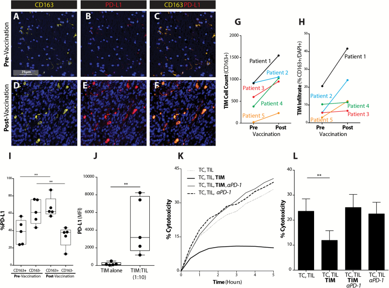
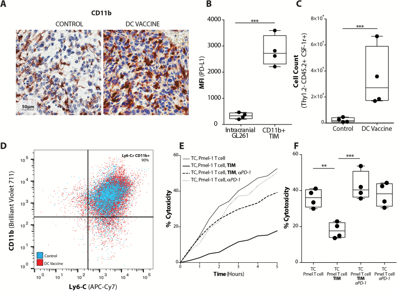
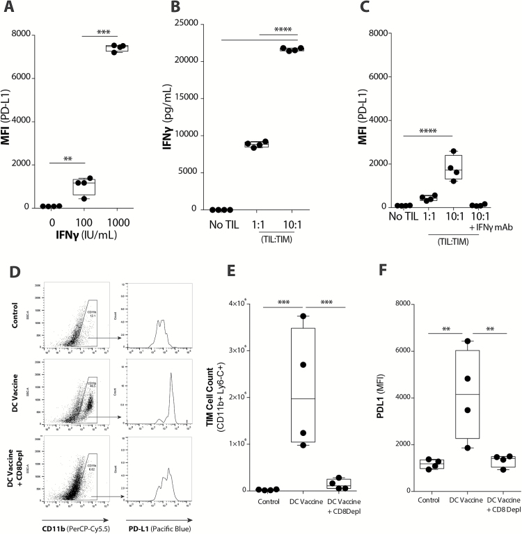
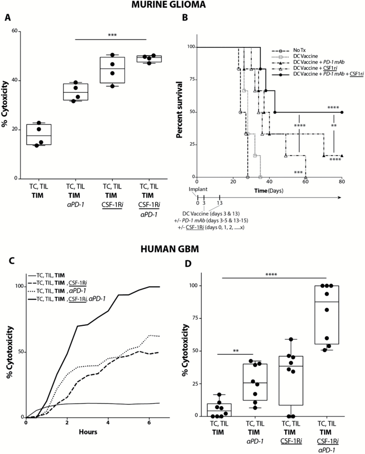
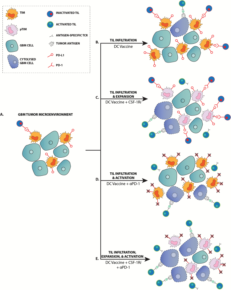
Background: Adaptive immune resistance in the tumor microenvironment appears to attenuate the immunotherapeutic targeting of glioblastoma (GBM). In this study, we identified a tumor-infiltrating myeloid cell (TIM) population that expands in response to dendritic cell (DC) vaccine treatment. The aim of this study was to understand how this programmed death ligand 1 (PD-L1)-expressing population restricts activation and tumor-cytolytic function of vaccine-induced tumor-infiltrating lymphocytes (TILs).

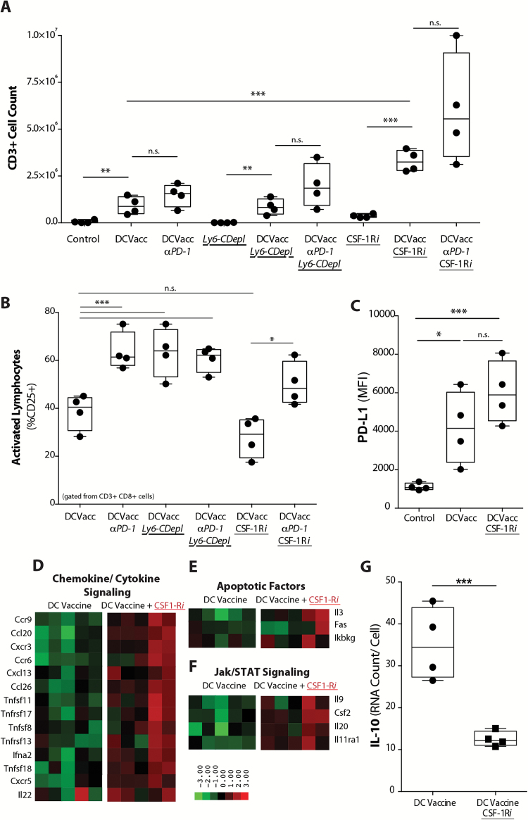
Methods: To test this hypothesis in our in vivo preclinical model, we treated mice bearing intracranial gliomas with DC vaccination ± murine anti-PD-1 monoclonal antibody (mAb) blockade or a colony stimulating factor 1 receptor inhibitor (CSF-1Ri) (PLX3397) and measured overall survival. We then harvested and characterized the PD-L1+ TIM population and its role in TIL activation and tumor cytolysis in vitro.

Results: Our data indicated that the majority of PD-L1 expression in the GBM environment is contributed by TIMs rather than by tumor cells themselves. While PD-1 blockade partially reversed the TIL dysfunction, targeting TIMs directly with CSF-1Ri altered TIM expression of key chemotactic factors associated with promoting increased TIL infiltration after vaccination. Neither PD-1 mAb nor CSF-1Ri had a demonstrable therapeutic benefit alone, but when combined with DC vaccination, a significant survival benefit was observed. When the tripartite regimen was given (DC vaccine, PD-1 mAb, PLX3397), long-term survival was noted together with an increase in the number of TILs and TIL activation.

Conclusion: Together, these studies elucidate the role that TIMs play in mediating adaptive immune resistance in the GBM microenvironment and provide evidence that they can be manipulated pharmacologically with agents that are clinically available. Development of immune resistance in response to active vaccination in GBM can be reversed with dual administration of CSF-1Ri and PD-1 mAb.

Keywords: CSF-1R; PD-1; cancer; checkpoint inhibitors; dendritic cell vaccine; glioblastoma; immunotherapy.


Figures

Fig. 1
Fig. 1
GBM TIMs expand to inhibit vaccine-induced T-cell mediated tumor cytolysis via PD-1/PD-L1 regulatory pathway. (A, D) CD163, DAPI, (B, E) PD-L1, DAPI, and (C, F) CD163, PD-L1, and DAPI co-staining are shown across pre- and post-DC vaccination samples from a GBM patient at 40× magnification (scale bar represents 75µm). (G) CD163+ cell count across pre– and post–DC vaccine treatment patient samples was quantified (n = 5). (H) Percent of CD163+ cells of total number of cells (DAPI+) was quantified (n = 5). (I) The percent of CD163+ cells dually expressing PD-L1 was quantified before and after vaccination (n = 5) (**P < .01). (J) PD-L1 expression on CD11b+ TIMs in the absence or presence of CD3+ TILs from freshly resected GBM shown (**P < .01) (n = 5). (K) Representative plot demonstrating TIL cytolysis of tumor cells (TC) over time in the absence or presence of TIMs or PD-1 mAb shown for freshly resected GBM. (L) GBM tumor cell cytolysis at selected time point of 4 hours in the absence or presence of TIMs or PD-1 mAb shown (n = 11/group) (**P < .01).
Fig. 2
Fig. 2
Murine glioma TIMs expand in response to vaccination to inhibit T cell-mediated tumor cytolysis. (A) CD11b IHC staining of nontreatment control and DC vaccinated tumor-bearing mice (scale bar represents 50µm). (B) Mean fluorescence intensity for PD-L1 expression on FACS intracranial GL261 gliomas (CD11b− CD3− cells) and TIMs (CD11b+ CD3−); (n = 4/group) (***P < .001). Flow cytometric characterization of (C) the absolute number of Thy1.2−, CD45.2+, CSF-1R+ cells (n = 4/group) (***P < .001), and (D) representative scatter plot of percent CD11b+, Ly6-C+ TIMs from tumors of control and DC vaccinated mice. (E) Representative xCELLigence plot of Pmel-1 T cell cytolysis of GL261-hgp100 cells (TC) over time in the absence or presence of TIMs or PD-1 mAb. (F) Quantitation of Pmel-1 T-cell induced tumor cell cytolysis at 4 hours in the absence or presence of TIMs or PD-1 mAb (n = 4) (**P < .01, ***P < .001).
Fig. 3
Fig. 3
TIMs upregulate PD-L1 in response to vaccine-induced TIL population. (A) Upregulation of PD-L1 on TIMs in the presence of increasing IFNɣ concentrations was quantified with flow cytometry (n = 4/group) (***P < .001). (B) IFNɣ levels present in TIL-TIM co-cultures in vitro was quantified using a Luminex assay (n = 4/group) (****P < .0001). (C) Upregulation of PD-L1 on TIMs in the presence of increasing concentrations of TILs or blockade of IFNɣ (IFNɣ mAb) was quantified with flow cytometry (n = 4/group) (****P < .0001). (D) Representative plots demonstrate PD-L1 expression on the CD11b+ TIM population across treatment groups. (E) CD11b+ Ly6-C+ TIM cell count and (F) PD-L1 expression in non-treatment control, DC vaccinated, and DC vaccinated + CD8 mAb depletion tumor-bearing mice (n = 4/group) (**P < .01, ***P < .001). Statistical analyses were performed using Student’s t-test (A–F).
Fig. 4
Fig. 4
TIM-mediated PD-1/PD-L1 immunoregulatory mechanism reduces T cell infiltration and activation in tumor. (A) Absolute TIL count (CD3+) and (B) TIL activation (%CD8+ CD3+ CD25+) in tumors from mice treated with DC vaccine and PD-1 mAb, along with Ly6-C depleting mAb (Ly6-C Depl) or CSF-1Ri (n = 4/ group) (*P < .05, **P < .01, ***P < .001, ****P < .0001). (C) PD-L1 expression across treatment groups (n = 5/group) (**P < .01, ***P < .001). Unbiased heatmap ranking and quantification of (D) chemokine/cytokine signaling, (E) apoptotic factors, and (F) Jak/STAT pathway signaling factors on CD11b+ TIMs from tumor-bearing mice receiving DC vaccination alone or with adjuvant CSF-1Ri treatment (n = 5/group). (G) Quantification of IL-10 expression by CD11b+ TIMs from tumor-bearing mice treated with DC vaccine or DC vaccine + CSF-1Ri is shown. (n = 4/group) (***P < .001).
Fig. 5
Fig. 5
Combination treatment with PD-1 mAb and CSF-1Ri maximally enhances vaccination-induced immune responses in both murine glioma and ex vivo human GBM. (A) Murine GL261-hgp100 glioma cell (TC) cytolysis at 4 hours following co-culture of TC, Pmel-1 T cells, and TIMs FACS-subjected from intracranial tumor-bearing mice treated with DC vaccination. PD-1 mAb or CSF-1Ri were added to Pmel-1 T cells or TIM ex vivo, respectively (n = 4/ group) (***P < .001). (B) Mice were randomized into control (tumor-bearing, no treatment), DC vaccine, DC vaccine + PD-1 mAb, DC vaccine + CSF-1Ri, and DC vaccine + PD-1 mAb + CSF-1Ri treatment groups. Graph depicts comparison of survival using method of Kaplan–Meier (n = 6/group) (**P < .01, ***P < .001, ****P < .0001) (P values indicate statistical difference from no treatment control unless otherwise indicated). (C) Human GBM tumor cell (TC) cytolysis over time following co-culture of TC, TIL, and TIM for representative GBM patient. PD-1 mAb or CSF-1Ri were added to autologous TIL or TIM ex vivo, respectively. (D) Compilation of human GBM tumor cell (TC) cytolysis at 4 hours following co-culture of TC, TIL, and TIM. PD-1 mAb or CSF-1Ri were added to TIL and TIM ex vivo, respectively (n = 8 patient samples/group) (**P < .01, ****P < .0001).
Fig. 6
Fig. 6
PD-1 mAb blockade and CSF-1R inhibitors enhance vaccine-induced immune responses by distinct mechanisms. (A) Malignant gliomas lack a significant antitumor immune response in the nontreatment setting. (B) DC vaccination elicits an infiltrating TIL response, but is largely ineffective due to inactivation of TILs via the PD-1/PD-L1 signaling mechanism. (C) Treatment with CSF-1Ri alters the TIM population to become pro-inflammatory TIM (pTIM) and results in the expansion of the TIL population, increasing the potential for TIL-tumor cell interaction and tumor cytolysis. (D) While PD-1 mAb treatment does not increase the TIL population over what is generated by DC vaccination, it does promote activation of TILs and subsequent tumor cytolysis. (E) PD-1 mAb and CSF-1Ri together promote the expansion and activation of the DC vaccine-generated TIL population such that there is a maximal tumor cytolysis.

References

    1. Liau LM, Prins RM, Kiertscher SM, et al. Dendritic cell vaccination in glioblastoma patients induces systemic and intracranial T-cell responses modulated by the local central nervous system tumor microenvironment. Clin Cancer Res. 2005;11(15):5515–5525. - PubMed
    1. Prins RM, Wang X, Soto H, et al. Comparison of glioma-associated antigen peptide-loaded versus autologous tumor lysate-loaded dendritic cell vaccination in malignant glioma patients. J Immunother. 2013;36(2):152–157. - PMC - PubMed
    1. Heimberger AB, Hussain SF, Aldape K, et al. Tumor-specific peptide vaccination in newly-diagnosed patients with GBM. J Clin Oncol. 2006;24(18):107s.
    1. Kanaly CW, Ding D, Heimberger AB, et al. Clinical applications of a peptide-based vaccine for glioblastoma. Neurosurg Clin N Am. 2010;21(1):95–109. - PMC - PubMed
    1. Prins RM, Soto H, Konkankit V, et al. Gene expression profile correlates with T-cell infiltration and relative survival in glioblastoma patients vaccinated with dendritic cell immunotherapy. Clin Cancer Res. 2011;17(6):1603–1615. - PMC - PubMed

MeSH terms