. 2017 Feb 7;114(6):1401-1406.
doi: 10.1073/pnas.1607541114. Epub 2017 Jan 23.

Superresolution imaging reveals activity-dependent plasticity of axon morphology linked to changes in action potential conduction velocity

Affiliations

Superresolution imaging reveals activity-dependent plasticity of axon morphology linked to changes in action potential conduction velocity

Ronan Chéreau et al. Proc Natl Acad Sci U S A. .

Abstract

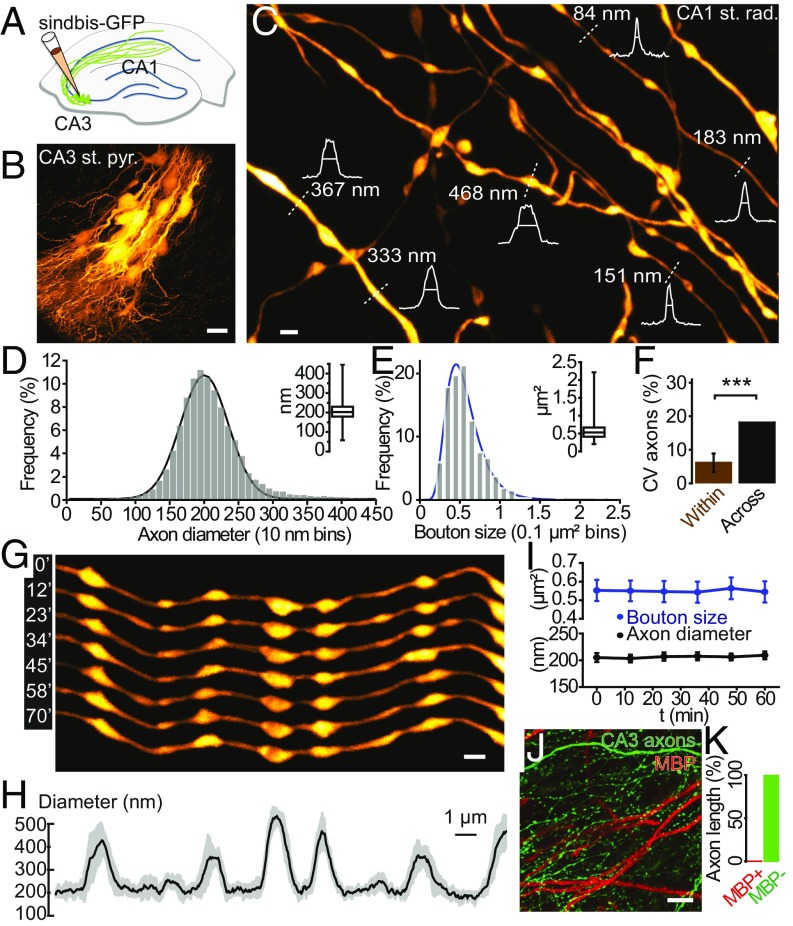
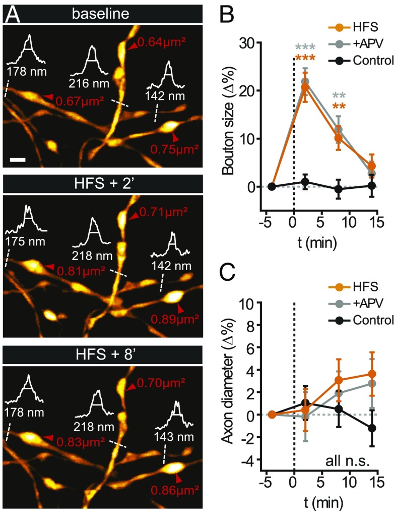
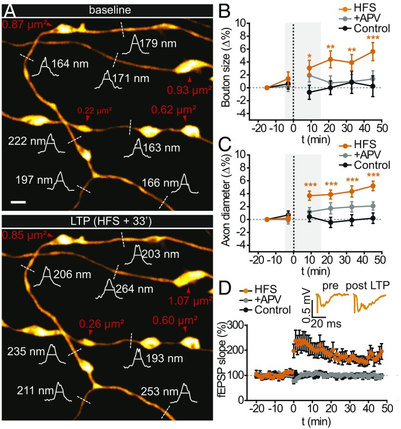
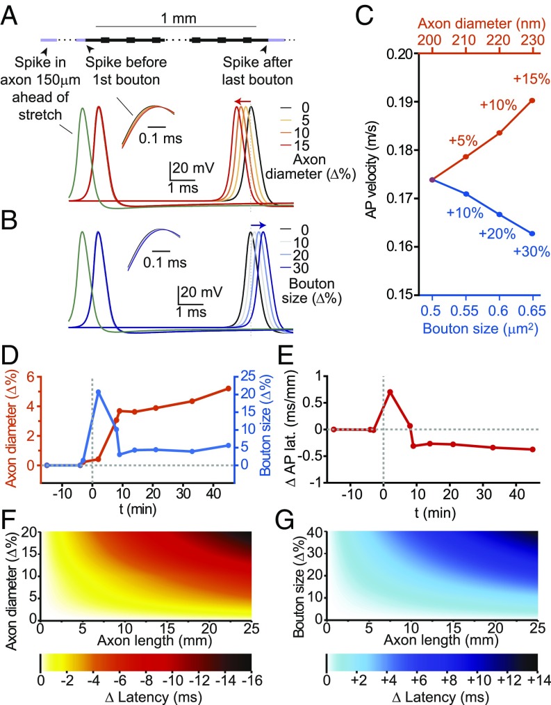
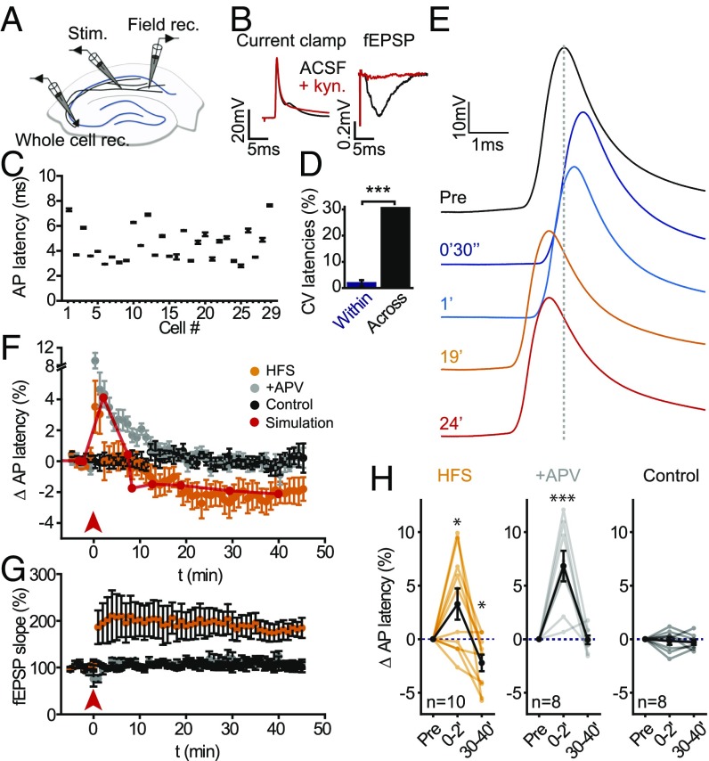
Axons convey information to nearby and distant cells, and the time it takes for action potentials (APs) to reach their targets governs the timing of information transfer in neural circuits. In the unmyelinated axons of hippocampus, the conduction speed of APs depends crucially on axon diameters, which vary widely. However, it is not known whether axon diameters are dynamic and regulated by activity-dependent mechanisms. Using time-lapse superresolution microscopy in brain slices, we report that axons grow wider after high-frequency AP firing: synaptic boutons undergo a rapid enlargement, which is mostly transient, whereas axon shafts show a more delayed and progressive increase in diameter. Simulations of AP propagation incorporating these morphological dynamics predicted bidirectional effects on AP conduction speed. The predictions were confirmed by electrophysiological experiments, revealing a phase of slowed down AP conduction, which is linked to the transient enlargement of the synaptic boutons, followed by a sustained increase in conduction speed that accompanies the axon shaft widening induced by high-frequency AP firing. Taken together, our study outlines a morphological plasticity mechanism for dynamically fine-tuning AP conduction velocity, which potentially has wide implications for the temporal transfer of information in the brain.

Keywords: STED microscopy; action potential conduction velocity; axons; plasticity; synaptic boutons.


Conflict of interest statement

The authors declare no conflict of interest.

Figures

Fig. 1.
Fig. 1.
Heterogeneity and dynamics of CA3 axons. (A) Labeling strategy. (B) CA3 area ∼36 h after virus injection, showing a cluster of GFP-positive pyramidal neurons. (C) STED image of axons in the stratum radiatum in the CA1 area. Intensity profiles (white) and FWHM measurements (straight lines) of axon and bouton diameters were taken at the locations indicated by the white dotted lines for illustrative purposes. (D) Distribution of axon diameters. Curved line is a Gaussian fit. Inset shows median value and interquartile and 0.01–99.9% range. (E) Distribution of synaptic bouton sizes (area). Inset shows median value and interquartile and range. (F) Coefficient of variations of 52 axon segment diameters within and across individual measurements. (G) Example of an axon segment imaged over more than 1 h. (H) Temporal profile of axon diameter, showing average diameter (black) and SD over time (gray). (I) Time-lapse data of morphological parameters over 1 h (geometric mean with 95% confidence interval), showing no alterations induced by the imaging. (J and K) Myelin basic protein immunostaining (red) showing no colocalization of the myelin with GFP-labeled CA3 axons (green) in CA1 stratum radiatum. Representative image (J) and quantification showing that more than 99.5% of total axon length is unmyelinated (K). [Scale bars, 20 µm (B), 1 µm (C and G), 10 µm (J).]
Fig. 2.
Fig. 2.
STED imaging of short-term dynamics of axonal morphology. (A) Synaptic boutons rapidly enlarged relative to pre-HFS baseline levels (red arrowheads), whereas axon diameters remained unchanged initially (white curve, intensity profiles; dashed lines, FWHM). (B and C) Quantification of early morphological changes. (B) Synaptic boutons enlarged rapidly and mostly transiently after HFS (orange), whereas this effect was not blocked by APV (gray), and boutons remained stable during control conditions (black). (C) From the same axon segments, no significant changes in axon diameter were observed in this time frame. (Scale bar, 1 µm.)
Fig. 3.
Fig. 3.
STED imaging of long-term dynamics of synaptic boutons and axon diameters. (A) Synaptic boutons (red arrowheads) remained slightly enlarged, and axon diameters (white curve, intensity profiles; dashed lines, FWHM) grew wider relative to pre-HFS baseline levels. (B and C) Quantification of late morphological changes. (B) Normalized synaptic bouton sizes for HFS, control, and APV conditions. (C) Normalized axon diameters for these conditions. The gray boxes in B and C correspond to the time window of the short-term experiments in Fig. 2. (D) Normalized time course of the fEPSP slope during LTP induction. Inset shows representative fEPSP traces. LTP was blocked by APV, and the recordings were stable under control conditions. (Scale bar, 1 µm.)
Fig. 4.
Fig. 4.
Mathematical modeling of morphological effects on AP propagation. (A) Increasing the diameter of the axon speeds up AP conduction, whereas (B) increasing bouton size slows it down, according to numerical simulations of AP propagation based on a geometrically constructed model axon [axon length = 1 mm; axon shaft diameter = 200 nm; synaptic boutons (size = width × length = 0.5 × 1.0 µm) spaced every 3 µm]. (C) Conduction velocity as a function of bouton size and axon shaft diameter. (D) Time course of measured changes in axon diameters (orange; left y axis) and bouton sizes (blue; right y axis) used for morphologically realistic numerical simulations. (E) Morphological changes exert biphasic effect on AP latency, according to numerical simulations based on STED morphological data, expressed as latency change (in milliseconds) per axon length (in millimeters). (F and G) Color-coded plots showing the change in AP latency as a function of axon length and percentage increases of axon diameter (F) or bouton size (G).
Fig. 5.
Fig. 5.
Activity-dependent changes in AP conduction delay. (A) Recording configuration for measuring antidromic AP latency of CA3 pyramidal neurons. Field rec., fEPSP recording; Stim., stimulation electrode; Whole cell rec., whole-cell recording. (B) Antidromic AP waveform recorded in current clamp at the soma. Blocking glutamatergic transmission with 2 mM kynurenate (kyn) did not prevent APs generated by direct stimulation (red). (C) Axonal AP latencies measured from 29 CA3 pyramidal neurons (average of 20 AP latencies during a 5-min period). (D) Coefficient of variation of AP latencies within and across cells. (E) Change in AP latency induced by HFS. (F) AP latency transiently increased after HFS (orange, arrow indicates stimulation), but decreased to below baseline levels (black) 10 min after HFS. APV spared the early increase in AP latency, but blocked its sustained reduction later on (gray). Continuous red line shows time course of AP latency changes predicted by model simulations based on STED data (Fig. 4D). (G) Time course of the fEPSP slope for the three groups. LTP was not induced in the presence of APV. The recordings were stable under control conditions. (H) Paired comparisons to baseline of early and late changes in AP latency for the three groups.

References

    1. Debanne D, Campanac E, Bialowas A, Carlier E, Alcaraz G. Axon physiology. Physiol Rev. 2011;91(2):555–602. - PubMed
    1. Alle H, Geiger JR. Combined analog and action potential coding in hippocampal mossy fibers. Science. 2006;311(5765):1290–1293. - PubMed
    1. Grubb MS, Burrone J. Activity-dependent relocation of the axon initial segment fine-tunes neuronal excitability. Nature. 2010;465(7301):1070–1074. - PMC - PubMed
    1. Becker N, Wierenga CJ, Fonseca R, Bonhoeffer T, Nägerl UV. LTD induction causes morphological changes of presynaptic boutons and reduces their contacts with spines. Neuron. 2008;60(4):590–597. - PubMed
    1. Sasaki T, Matsuki N, Ikegaya Y. Action-potential modulation during axonal conduction. Science. 2011;331(6017):599–601. - PubMed

Publication types

LinkOut - more resources