. 2016 Dec 30:12:279-294.
doi: 10.2147/IJN.S114754. eCollection 2017.

Preparation of arginine-glycine-aspartic acid-modified biopolymeric nanoparticles containing epigalloccatechin-3-gallate for targeting vascular endothelial cells to inhibit corneal neovascularization

Affiliations

Preparation of arginine-glycine-aspartic acid-modified biopolymeric nanoparticles containing epigalloccatechin-3-gallate for targeting vascular endothelial cells to inhibit corneal neovascularization

Che-Yi Chang et al. Int J Nanomedicine. .

Abstract

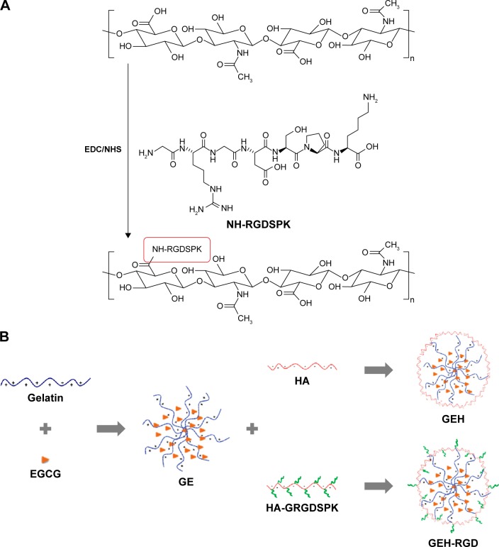
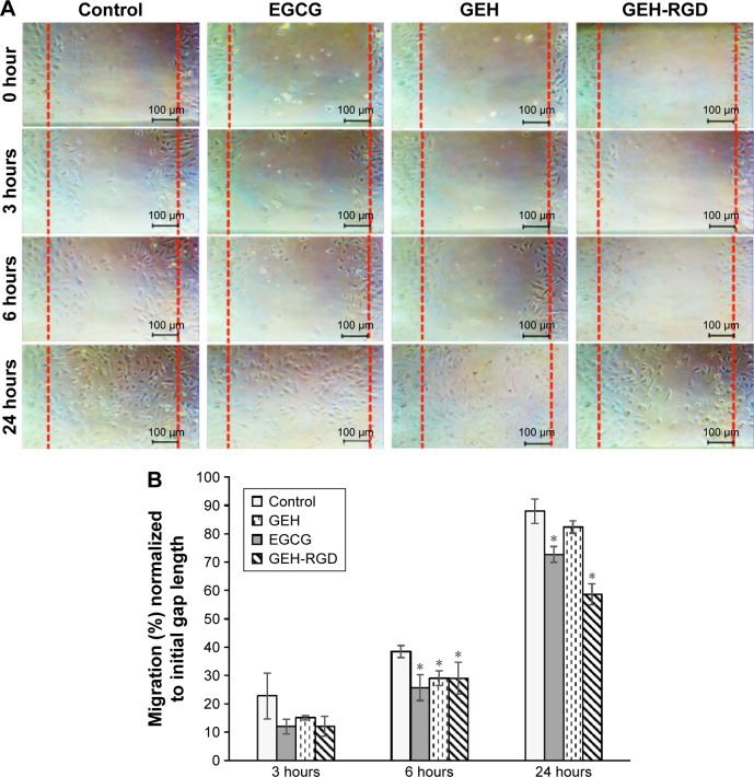
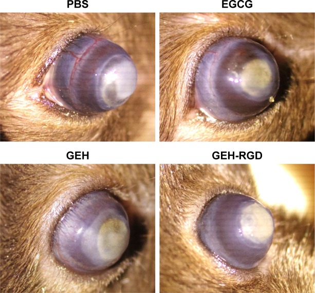
Neovascularization (NV) of the cornea can disrupt visual function, causing ocular diseases, including blindness. Therefore, treatment of corneal NV has a high public health impact. Epigalloccatechin-3-gallate (EGCG), presenting antiangiogenesis effects, was chosen as an inhibitor to treat human vascular endothelial cells for corneal NV treatment. An arginine-glycine-aspartic acid (RGD) peptide-hyaluronic acid (HA)-conjugated complex coating on the gelatin/EGCG self-assembly nanoparticles (GEH-RGD NPs) was synthesized for targeting the αvβ3 integrin on human umbilical vein endothelial cells (HUVECs) in this study, and a corneal NV mouse model was used to evaluate the therapeutic effect of this nanomedicine used as eyedrops. HA-RGD conjugation via COOH and amine groups was confirmed by 1H-nuclear magnetic resonance and Fourier-transform infrared spectroscopy. The average diameter of GEH-RGD NPs was 168.87±22.5 nm with positive charge (19.7±2 mV), with an EGCG-loading efficiency up to 95%. Images of GEH-RGD NPs acquired from transmission electron microscopy showed a spherical shape and shell structure of about 200 nm. A slow-release pattern was observed in the nanoformulation at about 30% after 30 hours. Surface plasmon resonance confirmed that GEH-RGD NPs specifically bound to the integrin αvβ3. In vitro cell-viability assay showed that GEH-RGD efficiently inhibited HUVEC proliferation at low EGCG concentrations (20 μg/mL) when compared with EGCG or non-RGD-modified NPs. Furthermore, GEH-RGD NPs significantly inhibited HUVEC migration down to 58%, lasting for 24 hours. In the corneal NV mouse model, fewer and thinner vessels were observed in the alkali-burned cornea after treatment with GEH-RGD NP eyedrops. Overall, this study indicates that GEH-RGD NPs were successfully developed and synthesized as an inhibitor of vascular endothelial cells with specific targeting capacity. Moreover, they can be used in eyedrops to inhibit angiogenesis in corneal NV mice.

Keywords: RGD peptide; antiangiogenesis; corneal neovascularization; epigallocatechin gallate (EGCG); hyaluronic acid (HA); vascular endothelial cells.


Conflict of interest statement

The authors report no conflicts of interest in this work.

Figures

Figure 1
Figure 1
Schematic representations. Notes: (A) HA-RGD peptide-conjugation reaction; (B) step for variant NP preparation with/without RGD modification. First, gelatin–EGCG self-assembling NPs were prepared (GE), followed by surface decoration with HA or HA-RGD (GEH and GEH-RGD, respectively). Abbreviations: HA, hyaluronic acid; RGD, arginine–glycine–aspartic acid; NP, nanoparticle; EGCG, epigallocatechin-3-gallate; EDC, 1-ethyl-3-(3-dimethylaminopropyl) carbodiimide hydrochloride; NHS, N-hydroxysuccinimide; GRGDSPK, H-Gly-Arg-Asp-Ser-Pro-Lys-OH. NH-RGDSPK, RGDSPK peptide conjugated with HA via amide bond formation.
Figure 2
Figure 2
(A) FT-IR patterns of HA, RGD peptide, their mixture, and the conjugated product (HA-RGD); (B) 1H-NMR spectra of HA and HA-RGD conjugates. Note: The arrows indicate the acetamido moiety of the N-acetyl-d-glucosamine residue of HA and proline in GRGDSPK. Abbreviations: FT-IR, Fourier-transform infrared; HA, hyaluronic acid; RGD, arginine–glycine–aspartic acid; NMR, nuclear magnetic resonance; GRGDSPK, H-Gly-Arg-Asp-Ser-Pro-Lys-OH.
Figure 3
Figure 3
(A) DLS size distribution of NPs; (B) TEM of GEH-RGD; (C) release pattern of variant NPs in PBS (pH 4) at 37°C±0.5°C. Abbreviations: DLS, dynamic light scattering; NPs, nanoparticles; TEM, transmission electron microscopy; GEH, gelatin–epigallocatechin-3-gallate–hyaluronic acid; RGD, arginine–glycine–aspartic acid; PBS, phosphate-buffered saline.
Figure 4
Figure 4
SPR analysis of the binding affinity of NPs with the integrin αvβ3, grafted on a chip. Notes: (A) GEH-RGD presented a stronger signal than GE; (B) GEH-RGD signal was also stronger than that of GEH. Abbreviations: SPR, surface plasmon resonance; NPs, nanoparticles; GEH, gelatin–epigallocatechin-3-gallate–hyaluronic acid; RGD, arginine–glycine–aspartic acid; GE, gelatin–EGCG; EGCG, epigallocatechin-3-gallate.
Figure 5
Figure 5
Results of cell viability and live/dead stain assays. Notes: (A) Cell viability of HUVECs treated with various NP formulations at different EGCG concentrations; (B) live/dead cells identified by fluorescence (scale bars 100 μm) of HUVECs cultured in medium (control), EGCG, GE, GEH, and GEH-RGD NPs at an EGCG concentration of 200 μg/mL or 20 μg/mL at day 1. Results expressed as mean ± standard deviation (n=6); *P<0.05. Abbreviations: HUVECs, human umbilical vein endothelial cells; NP, nanoparticle; EGCG, epigallocatechin-3-gallate; GE, gelatin–EGCG; GEH, GE–hyaluronic acid; RGD, arginine–glycine–aspartic acid; BF, bright field; L/D, live/dead.
Figure 6
Figure 6
(A) TAMRA, GEH, and GEH-RGD taken up by HUVECs after 2 hours of coculture; (B) quantification of NPs in cells from A. Notes: Vertical bars represent standard error of mean (n=3); *P<0.05. Abbreviations: TAMRA, carboxytetramethylrhodamine; GEH, gelatin–epigallocatechin-3-gallate–hyaluronic acid; RGD, arginine–glycine–aspartic acid; HUVECs, human umbilical vein endothelial cells; NPs, nanoparticles.
Figure 7
Figure 7
(A) Migration assay: photomicrographs of the wound closure. (B) Migration rate of HUVECs treated with EGCG, GEH, and GEH-RGD. Notes: All groups were treated with the same EGCG concentration (20 μg/mL); *P<0.05. Abbreviations: HUVECs, human umbilical vein endothelial cells; EGCG, epigallocatechin-3-gallate; GEH, gelatin–EGCG–hyaluronic acid; RGD, arginine–glycine–aspartic acid.
Figure 8
Figure 8
GEH-RGD NPs used as eyedrops prevent corneal NV formation. Notes: The ocular images show angiogenesis in the PBS group. Vessel reduction was observed in corneas treated with free EGCG and EGCG NPs, especially when treated with GEH-RGD (EGCG 30 μg/mL twice daily). Abbreviations: GEH, gelatin–epigallocatechin-3-gallate–hyaluronic acid; RGD, arginine–glycine–aspartic acid; NPs, nanoparticles; NV, neovascularization; PBS, phosphate-buffered saline; EGCG, epigallocatechin-3-gallate.

References

    1. Menzel-Severing J. Emerging techniques to treat corneal neovascularisation. Eye (Lond) 2012;26:2–12. - PMC - PubMed
    1. Huang TL, Hsu SY, Tsai RK, Sheu MM. Etiology of ocular diseases in elderly Amis aborigines in Eastern Taiwan (the Amis Eye Study) Jpn J Ophthalmol. 2010;54:266–271. - PubMed
    1. Bachmann BO, Bock F, Wiegand SJ, et al. Promotion of graft survival by vascular endothelial growth factor: a neutralization after high-risk corneal transplantation. Arch Ophthalmol. 2008;126:71–77. - PubMed
    1. Azar DT. Corneal angiogenic privilege: angiogenic and antiangiogenic factors in corneal avascularity, vasculogenesis, and wound healing (an American Ophthalmological Society thesis) Trans Am Ophthalmol Soc. 2006;104:264–302. - PMC - PubMed
    1. Bock F, König Y, Kruse F, Baier M, Cursiefen C. Bevacizumab (Avastin) eye drops inhibit corneal neovascularization. Graefes Arch Clin Exp Ophthalmol. 2008;246:281–284. - PubMed

MeSH terms

LinkOut - more resources