. 2017 Jan 4:10:79-88.
doi: 10.2147/JPR.S123828. eCollection 2017.

Sonographic tracking of trunk nerves: essential for ultrasound-guided pain management and research

Affiliations

Sonographic tracking of trunk nerves: essential for ultrasound-guided pain management and research

Ke-Vin Chang et al. J Pain Res. .

Abstract

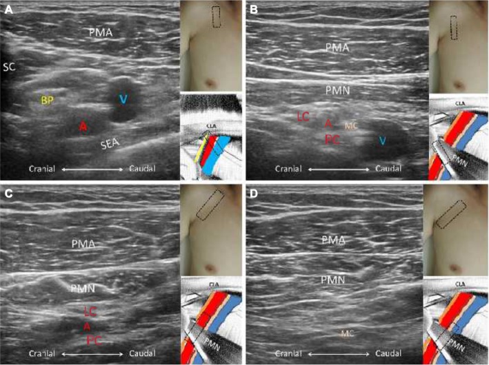
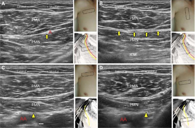
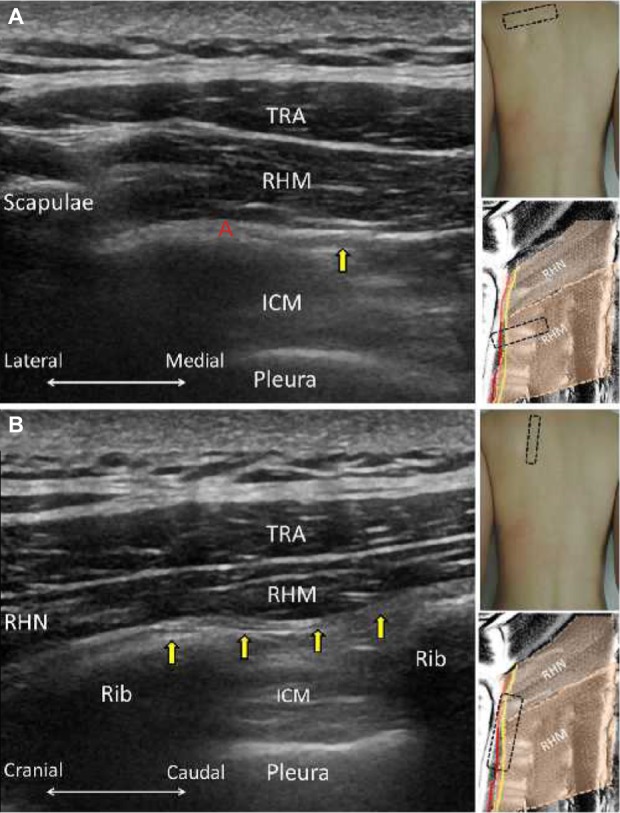
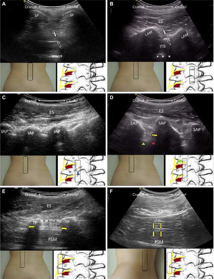
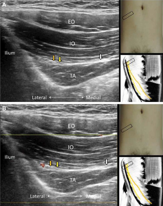
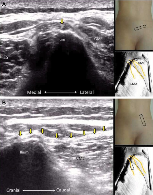
Delineation of architecture of peripheral nerves can be successfully achieved by high-resolution ultrasound (US), which is essential for US-guided pain management. There are numerous musculoskeletal pain syndromes involving the trunk nerves necessitating US for evaluation and guided interventions. The most common peripheral nerve disorders at the trunk region include thoracic outlet syndrome (brachial plexus), scapular winging (long thoracic nerve), interscapular pain (dorsal scapular nerve), and lumbar facet joint syndrome (medial branches of spinal nerves). Until now, there is no single article systematically summarizing the anatomy, sonographic pictures, and video demonstration of scanning techniques regarding trunk nerves. In this review, the authors have incorporated serial figures of transducer placement, US images, and videos for scanning the nerves in the trunk region and hope this paper helps physicians familiarize themselves with nerve sonoanatomy and further apply this technique for US-guided pain medicine and research.

Keywords: lumbar; nerve; pain; trunk; ultrasound.


Conflict of interest statement

The current research was supported by Ministry of Science and Technology, Taiwan, Republic of China (MOST 105-2314-B-002-008). The authors report no other conflicts of interest in this work.

Figures

Figure 1
Figure 1
The short axis views of the brachial plexus just distal to the clavicle (A) and underneath the pectoralis minor muscle (B); the long axis views of the lateral and posterior cords (C) and the medial cord (D). Note: The red A indicates axillary artery. Abbreviations: BP, brachial plexus; LC, lateral cord; PC, posterior cord; MC, medial cord; PMA, pectoralis major; PMN, pectoralis minor; SC, subclavius; SEA, serratus anterior; V, axillary vein; CLA, clavicle.
Figure 2
Figure 2
The short axis view of the long thoracic nerve at the junction between pectoralis major and serratus anterior muscles (A) and overlying the serratus anterior muscle (B); the short axis (C) and long axis (D) views of the thoracodorsal nerve. Notes: Yellow arrows indicate long thoracic nerve; yellow arrowheads indicate thoracodorsal nerve. The red A in (A) and (B) indicates lateral thoracic artery and thoracodorsal artery in (C). Abbreviations: PMA, pectoralis major; LD, latissimus dorsi; SEA, serratus anterior; ICM, intercostal muscle.
Figure 3
Figure 3
The short axis (A) and long axis (B) views of the lateral pectoral nerve; the short axis views (C) of the medial pectoral nerve at its origin near the medial cord and descending beneath the pectoralis minor muscle (D). Notes: Yellow arrows indicate the lateral pectoral nerve; yellow arrowheads indicate medial pectoral nerve; white arrow indicates medial cord; red A indicates pectoral branch of the thoracoacromial artery; red AA indicates axillary artery. Abbreviations: PMA, pectoralis major; PMN, pectoralis minor; ICM, intercostal muscle.
Figure 4
Figure 4
The short axis (A) and long axis (B) views of the dorsal scapular nerve (yellow arrows). Note: The red A indicates dorsal scapular artery. Abbreviations: TRA, trapezius; RHM, rhomboid major; RHN, rhomboid minor; ICM, intercostal muscle.
Figure 5
Figure 5
The sagittal planes for the ribs (A), transverse processes (B), laminae (C), and spinous processes of the thoracic vertebrae (D). The horizontal planes for the transverse processes (E) and the intercostal space (F) of the thoracic vertebrae. Notes: Yellow arrows indicate spinal nerve roots; purple arrows indicate interlaminar spaces; orange arrows indicate lateral costal transverse ligament; green arrows indicate superior costal transverse ligament. Abbreviations: TRA, trapezius; ES, erector spinae; TP, transverse process; SP, spinous process; LAM, lamina.
Figure 6
Figure 6
The horizontal planes for the cranial sacrum (A), alae of the sacrum (B), bilateral L4/L5 facet joints (C), and waist of the L4 lamina (D). Notes: Green arrowheads indicate ala of the sacrum; red circles indicate medial branch; yellow arrows indicate nerve roots; white arrows indicate the ligamentum flavum; black arrows indicate the dura. The asterisks indicate the posterior longitudinal ligament. Abbreviations: ES, erector spinae; AP, articular process; SP, spinous process; TP, transverse process; SAP, superior articular process; IAP, inferior articular process; ITS, intrathecal space; LAM, lamina; PSM, psoas major.
Figure 7
Figure 7
The sagittal planes for the spinous processes (A), laminae (B), inferior articular processes (C), superior articular processes (D), and transverse processes (E) of the lumbar spine; the oblique sagittal plane for the lumbar plexus (F). Notes: Green arrowheads indicate vertebral bodies; brown arrowheads indicate intervertebral discs; yellow arrows indicate nerve roots or lumbar plexus; black asterisks indicate intertransverse ligaments; white asterisks indicate anterior complex containing the posterior longitudinal ligament and anterior dura. Abbreviations: ES, erector spinae; SP, spinous process; TP, transverse process; SAP, superior articular process; IAP, inferior articular process; ITS, intrathecal space; LAM, lamina; PSM, psoas major; QL, quadratus lumborum; ITL, intertransversalis lumborum.
Figure 8
Figure 8
The sagittal plane for the origins (A) and middle portions (B) of the ribs. Notes: Yellow arrow indicates intercostal nerve; the red A indicates intercostal artery. Abbreviations: EIM, external intercostal muscle; IIM, internal intercostal muscle; IMM, innermost intercostal muscle; V, intercostal vein.
Figure 9
Figure 9
Gray scale (A) and power Doppler imaging (B) for the ilioinguinal, iliohypogastric, and subcostal nerves. Notes: Orange arrows indicate ilioinguinal nerve; yellow arrows indicate iliohypogastric nerve; white arrows indicate subcostal nerve. Abbreviations: EO, external oblique muscle; IO, internal oblique muscle; TA, transverse abdominis muscle.
Figure 10
Figure 10
The short axis (A) and long axis (B) views of the superior cluneal nerve. Note: Yellow arrows indicate superior cluneal nerve. Abbreviations: GME, gluteus medius; ES, erector spinae.

References

    1. Wu CH, Chang KV, Ozcakar L, et al. Sonographic tracking of the upper limb peripheral nerves: a pictorial essay and video demonstration. Am J Phys Med Rehabil. 2015;94(9):740–747. - PubMed
    1. Hung CY, Hsiao MY, Ozcakar L, et al. Sonographic tracking of the lower limb peripheral nerves: a pictorial essay and video demonstration. Am J Phys Med Rehabil. 2016;95(9):698–708. - PubMed
    1. Chang KV, Lin CP, Hung CY, Ozcakar L, Wang TG, Chen WS. Sonographic nerve tracking in the cervical region: a pictorial essay and video demonstration. Am J Phys Med Rehabil. 2016 Jun 26; Epub. - PubMed
    1. Demondion X, Herbinet P, Van Sint Jan S, Boutry N, Chantelot C, Cotten A. Imaging assessment of thoracic outlet syndrome. Radio-Graphics. 2006;26(6):1735–1750. - PubMed
    1. Martinoli C, Gandolfo N, Perez MM, et al. Brachial plexus and nerves about the shoulder. Semin Musculoskelet Radiol. 2010;14(5):523–546. - PubMed

LinkOut - more resources