

Analysis of the clinical effects of transforaminal endoscopic discectomy on lumbar disk herniation combined with common peroneal nerve paralysis: a 2-year follow-up retrospective study on 32 patients
- PMID: 28115870
- PMCID: PMC5221719
- DOI: 10.2147/JPR.S120463
Analysis of the clinical effects of transforaminal endoscopic discectomy on lumbar disk herniation combined with common peroneal nerve paralysis: a 2-year follow-up retrospective study on 32 patients
Abstract
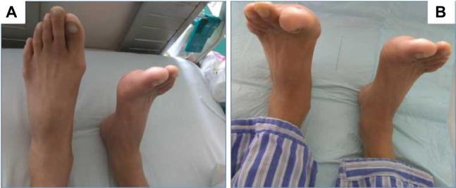
Background: Very few studies have discussed transforaminal endoscopic discectomy (TED) in the treatment of common peroneal nerve paralysis induced by lumbar disk herniation (LDH). This study aimed to evaluate the efficacy of TED in the treatment of LDH combined with common peroneal nerve paralysis.
Materials and methods: The clinical and follow-up data of 32 patients with common peroneal nerve paralysis induced by LDH undergoing TED from March 2011 to April 2014 were retrospectively analyzed in this study. Follow-up was conducted immediately after the surgery, as well as 3, 12, and 24 months postoperatively. The parameters (including muscle strength recovery of the anterior tibial muscle, leg pain visual analog scale score, neurological function Japanese Orthopaedic Association [JOA] score, MacNab scores in the last follow-up, and the intraoperative and postoperative complications) were recorded.
Results: Three patients (9.4%) had the anterior tibial muscle strength recovered to ≥ grade 4 immediately after the surgery. The anterior tibial muscle strength of patients recovered to basically stable form in the 6-month postoperative follow-up and that in the last follow-up were as follows: one case of grade 1, one case of grade 2, 28 cases of grade 4, and two cases of grade 5. The visual analog scale scores of leg pain were significantly reduced immediately after the surgery and also on 3, 12, and 24 months compared with preoperative period (all P<0.05). The postoperative JOA scores in the last follow-up were significantly higher than the preoperative JOA scores (P<0.05), and there were nine excellent cases (28.2%), 21 good cases (65.6%), one fair case (3.1%) and one poor case (3.1%) in the last follow-up, with an overall excellent and good rate of 93.8%.
Conclusion: TED, which can offer sufficient decompression of the nerve root, has excellent overall clinical effects in treating common peroneal nerve paralysis induced by LDH.
Keywords: common peroneal nerve paralysis; lumbar disk herniation; minimally invasive surgery; transforaminal endoscopic discectomy.
Conflict of interest statement
The authors report no conflicts of interest in this work.
Figures


References
-
- Aono H, Iwasaki M, Ohwada T, et al. Surgical outcome of drop foot caused by degenerative lumbar diseases. Spine (Phila Pa 1976) 2007;32(8):E262–E266. - PubMed
-
- Ghahreman A, Ferch RD, Rao PJ, Bogduk N. Minimal access versus open posterior lumbar interbody fusion in the treatment of spondylolisthesis. Neurosurgery. 2010;66(2):296–304. - PubMed
-
- Girardi FP, Cammisa FJ, Huang RC, Parvataneni HK, Tsairis P. Improvement of preoperative foot drop after lumbar surgery. J Spinal Disord Tech. 2002;15(6):490–494. - PubMed
-
- Iizuka Y, Iizuka H, Tsutsumi S, et al. Foot drop due to lumbar degenerative conditions: mechanism and prognostic factors in herniated nucleus pulposus and lumbar spinal stenosis. J Neurosurg Spine. 2009;10(3):260–264. - PubMed
-
- Rodriguez-Martinez NG, Perez-Orribo L, Kalb S, et al. The role of obesity in the biomechanics and radiological changes of the spine: an in vitro study. J Neurosurg Spine. 2016;24(4):615–623. - PubMed
LinkOut - more resources
Full Text Sources
Other Literature Sources
Miscellaneous
